During swallowing food moves from the mouth to the ___________.
- laryngopharynx
- esophagus
- nasopharynx
- oropharynx
oropharynx
Ex.
During swallowing, food from the mouth enters the oropharynx next.
To send food on its way from the mouth, it is first compacted by the tongue into a bolus and is then swallowed. This complicated process involves the coordinated activity of over 22 separate muscle groups. There are two major phases involved in deglutition, or swallowing.
The buccal phase occurs in the mouth and is voluntary. It ends when a food bolus or a “bit of saliva” leaves the mouth and stimulates tactile receptors in the posterior pharynx, initiating the next phase.
The pharyngeal-esophageal phase is involuntary and is controlled by the swallowing center in the brain stem (medulla and lower pons). Various cranial nerves, most importantly the vagus nerves, transmit motor impulses from the swallowing center to the muscles of the pharynx and esophagus. Once food enters the pharynx, respiration is momentarily inhibited and all routes except the desired one into the digestive tract are blocked off. Solid foods pass from the oropharynx to the stomach in about 8 seconds, and fluids, aided by gravity, pass in 1 to 2 seconds.
Match the following part of a tooth with its description: Periodontal ligament.
- Forms the support of the gomphosis
- Calcified connective tissue around the bottom of the tooth
- Embedded in the jawbone
- Exposed and covered in enamel
Forms the support of the gomphosis
Ex.
The periodontal ligament forms the support of the gomphosis.
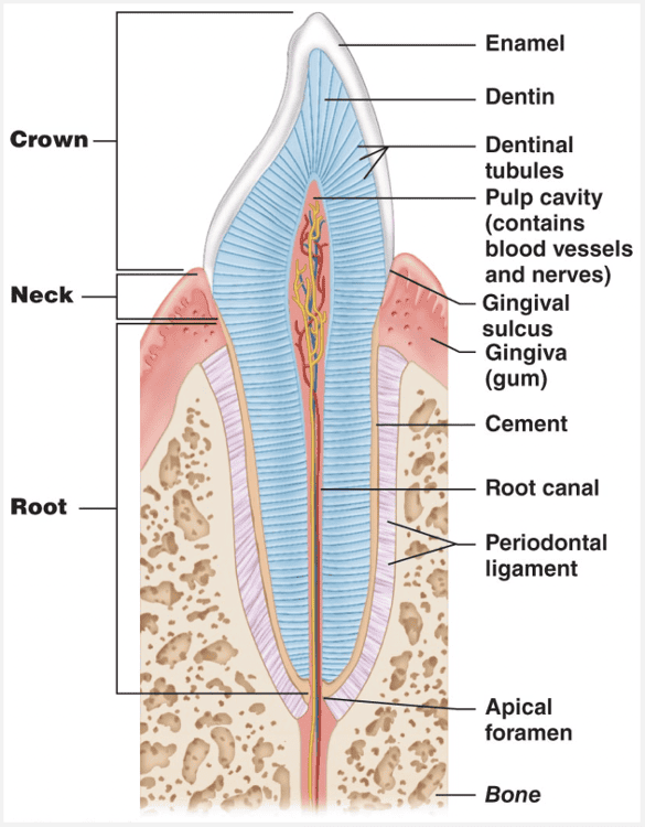
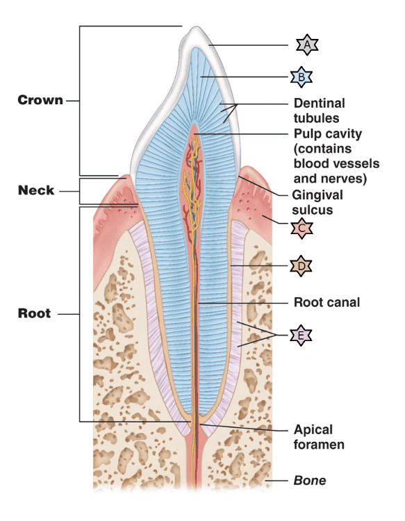
A constricted tooth region called the neck connects the crown and root. Cement, a calcified connective tissue, covers the outer surface of the root and attaches the tooth to the thin periodontal ligament (per”e-o-don’tal; “around the tooth”). This ligament anchors the tooth in the bony socket (alveolus) of the jaw, forming a fibrous joint called a gomphosis. Where the gingiva borders on a tooth, it dips downward to form a shallow groove called the gingival sulcus.
Select the description for the structure labeled 'B.'
- Surrounds the tooth like a collar
- Forms a gomphosis
- Covers the root of the tooth
- Shock absorber during biting
- Brittle and thick as a dime
Shock absorber during biting
Ex.
'B' is denting which is a shock absorber during biting.
Each tooth has two major regions: the crown and the root. The enamel-covered crown is the exposed part of the tooth above the gingiva (jin’jĭ-vah), or gum, which surrounds the tooth like a tight collar. Enamel, a brittle ceramic-like material thick as a dime, directly bears the force of chewing. The hardest substance in the body, it is heavily mineralized with calcium salts, and its densely packed hydroxyapatite (mineral) crystals are oriented in force-resisting columns perpendicular to the tooth’s surface. The cells that produce enamel degenerate when the tooth erupts; consequently, decayed or cracked areas of enamel will not heal and must be artificially filled.
The root is the portion of the tooth embedded in the jawbone. Canine teeth, incisors, and premolars have one root, although the first upper premolars commonly have two. The first two upper molars have three roots, while the corresponding lower molars have two. The root pattern of the third molar varies, but a fused single root is most common.
A constricted tooth region called the neck connects the crown and root. Cement, a calcified connective tissue, covers the outer surface of the root and attaches the tooth to the thin periodontal ligament (per”e-o-don’tal; “around the tooth”). This ligament anchors the tooth in the bony socket (alveolus) of the jaw, forming a fibrous joint called a gomphosis. Where the gingiva borders on a tooth, it dips downward to form a shallow groove called the gingival sulcus.
Dentin, a protein-rich bone-like material, underlies the enamel cap and forms the bulk of a tooth. More resilient than enamel, dentin acts as a shock absorber during biting and chewing. Dentin surrounds a central pulp cavity containing a number of soft tissue structures (connective tissue, blood vessels, and nerve fibers) collectively called pulp. Pulp supplies nutrients to the tooth tissues and provides tooth sensation. Where the pulp cavity extends into the root, it becomes the root canal.
Match the following term with its correct description: In direct contact with ingested food.
- Submucosa
- Serosa
- Mucosa
- Muscularis
Mucosa
Ex.
The mucosa is in direct contact with ingested food.
The innermost layer is the mucosa, or mucous membrane, a moist epithelial membrane that lines the alimentary canal lumen from mouth to anus. Its major functions are to:
- Secrete mucus, digestive enzymes, and hormones
- Absorb the end products of digestion into the blood
- Protect against infectious disease
The mucosa in a particular region of the GI tract may perform one or all three of these functions. More complex than most other mucosae in the body, the typical digestive mucosa consists of three sublayers: (1) a lining epithelium, (2) a lamina propria, and (3) a muscularis mucosae. Except for that of the mouth, esophagus, and anus where it is stratified squamous, the epithelium of the mucosa is a simple columnar epithelium rich in mucus-secreting cells. The slippery mucus it produces protects certain digestive organs from being digested by enzymes working within their cavities and eases food passage along the tract. In the stomach and small intestine, the mucosa also contains both enzyme-synthesizing and hormone-secreting cells. In such sites, the mucosa is a diffuse endocrine organ as well as part of the digestive organ.
Select the description for the structure labeled 'D.'
- Brittle and thick as a dime
- Shock absorber during biting
- Surrounds the tooth like a collar
- Covers the root of the tooth
- Forms a gomphosis
Covers the root of the tooth
Ex.
“D” is the cement which covers the root of the tooth.
Each tooth has two major regions: the crown and the root. The enamel-covered crown is the exposed part of the tooth above the gingiva (jin’jĭ-vah), or gum, which surrounds the tooth like a tight collar. Enamel, a brittle ceramic-like material thick as a dime, directly bears the force of chewing. The hardest substance in the body, it is heavily mineralized with calcium salts, and its densely packed hydroxyapatite (mineral) crystals are oriented in force-resisting columns perpendicular to the tooth’s surface. The cells that produce enamel degenerate when the tooth erupts; consequently, decayed or cracked areas of enamel will not heal and must be artificially filled.
The root is the portion of the tooth embedded in the jawbone. Canine teeth, incisors, and premolars have one root, although the first upper premolars commonly have two. The first two upper molars have three roots, while the corresponding lower molars have two. The root pattern of the third molar varies, but a fused single root is most common.
A constricted tooth region called the neck connects the crown and root. Cement, a calcified connective tissue, covers the outer surface of the root and attaches the tooth to the thin periodontal ligament (per”e-o-don’tal; “around the tooth”). This ligament anchors the tooth in the bony socket (alveolus) of the jaw, forming a fibrous joint called a gomphosis. Where the gingiva borders on a tooth, it dips downward to form a shallow groove called the gingival sulcus.
Dentin, a protein-rich bone-like material, underlies the enamel cap and forms the bulk of a tooth. More resilient than enamel, dentin acts as a shock absorber during biting and chewing. Dentin surrounds a central pulp cavity containing a number of soft tissue structures (connective tissue, blood vessels, and nerve fibers) collectively called pulp. Pulp supplies nutrients to the tooth tissues and provides tooth sensation. Where the pulp cavity extends into the root, it becomes the root canal.
Which of the following teeth are most commonly impacted?
- Deciduous teeth
- Baby teeth
- Premolars
- Wisdom teeth
Wisdom teeth
Ex.
Wisdom teeth are the most commonly impacted.
When a tooth remains trapped in the jawbone, it is said to be impacted. Impacted teeth can cause a good deal of pressure and pain and must be removed surgically. Wisdom teeth are most commonly involved.
Which phase of swallowing is voluntary?
- Pharyngeal-esophageal phase
- All phases of swallowing are voluntary
- Gastric phase
- Buccal phase
Buccal phase
Ex.
The buccal phase of swallowing is the only voluntary phase of swallowing.
To send food on its way from the mouth, it is first compacted by the tongue into a bolus and is then swallowed. This complicated process involves the coordinated activity of over 22 separate muscle groups. There are two major phases involved in deglutition, or swallowing.
The buccal phase occurs in the mouth and is voluntary. It ends when a food bolus or a “bit of saliva” leaves the mouth and stimulates tactile receptors in the posterior pharynx, initiating the next phase.
The pharyngeal-esophageal phase is involuntary and is controlled by the swallowing center in the brain stem (medulla and lower pons). Various cranial nerves, most importantly the vagus nerves, transmit motor impulses from the swallowing center to the muscles of the pharynx and esophagus. Once food enters the pharynx, respiration is momentarily inhibited and all routes except the desired one into the digestive tract are blocked off. Solid foods pass from the oropharynx to the stomach in about 8 seconds, and fluids, aided by gravity, pass in 1 to 2 seconds.
Match the following substance involved in organic molecule digestion with its description: Salivary amylase
- Emulsifies fats for digestion
- Begins carbohydrate digestion in the mouth
- Digests proteins in the small intestines
- Brush border enzymes that act on disaccharides
Begins carbohydrate digestion in the mouth
Ex.
Salivary amylase begins carbohydrate digestion in the mouth.
A number of glands associated with the oral cavity secrete saliva. Saliva:
- Cleanses the mouth
- Dissolves food chemicals so they can be tasted
- Moistens food and helps compact it into a bolus
- Contains the enzyme amylase that begins the digestion of starchy foods
Pepsin is an enzyme that digests proteins in the stomach.
Bile emulsifies fats for digestion in the small intestine.
Brush border enzymes of the small intestine break olig- and disaccharides into monosaccharides.
What type of epithelium is the esophageal mucosa lined with?
- Keratinized stratified squamous
- Nonkeratinized stratified squamous
- Simple columnar
- Simple cuboidal
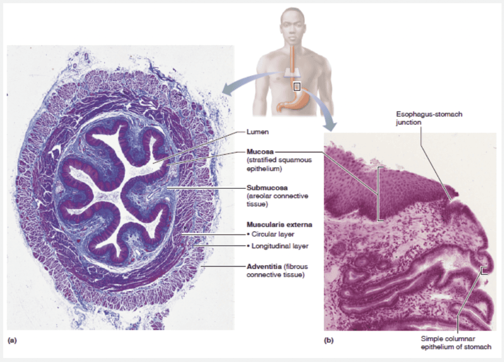
Nonkeratinized stratified squamous
Ex.
The esophageal mucosa lined with nonkeratinized stratified squamous epithelium.
At the esophagus-stomach junction, that abrasion-resistant epithelium changes abruptly to the simple columnar epithelium of the stomach, which is specialized for secretion.
The calcified form of dental plaque is called __________.
- calculus or tartar
- cement
- dentin
- enamel
calculus or tartar
Ex.
The calcified form of dental plaque is called calculus or tartar.
Dental caries ((kerēz;“rottenness”), or cavities, result from bacterial action that gradually demineralizes enamel and underlying dentin. Decay begins when dental plaque (a film of sugar, bacteria, and other mouth debris) adheres to the teeth.
More serious than tooth decay is the effect of unremoved plaque on the gums. As dental plaque accumulates, it calcifies, forming calculus (kalkyələs; “stone”) or tartar. These stonyhard deposits disrupt the seal between gingivae and teeth, deepening the sulcus and putting the gums at risk for infection by pathogenic anaerobic bacteria. In the early stages of such an infection, called gingivitis (jinjəˈvīdəs), the gums are red, sore, swollen, and may bleed.
Enamel, dentin, and cement are all parts of healthy teeth.
Match the following digestive process with the correct description: Mechanical digestion
- Enzymatic degradation of foodstuffs into simpler molecules
- Taking food into the digestive system
- Passage of digested materials from the lumen of the GI tract into the blood or lymph
- Chewing, mixing, churning, and segmenting food
- Elimination of indigestible solids
Chewing, mixing, churning, and segmenting food
Ex.
Mechanical digestion includes chewing, mixing, churning, and segmenting food.
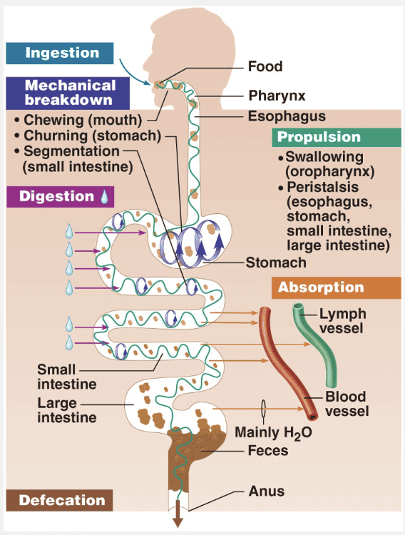
Mechanical breakdown increases the surface area of ingested food, physically preparing it for digestion by enzymes. Mechanical processes include chewing, mixing food with saliva by the tongue, churning food in the stomach, and segmentation (rhythmic local constrictions of the small intestine). Segmentation mixes food with digestive juices and makes absorption more efficient by repeatedly moving different parts of the food mass over the intestinal wall.
Absorption is the passage of digested end products (plus vitamins, minerals, and water) from the lumen of the GI tract through the mucosal cells by active or passive transport into the blood or lymph.
Taking food into the digestive system is called ingestion.
Enzymatic degradation of foodstuffs into simpler molecules is chemical digestion.
Elimination of indigestible solids is the process of defecation.
Match the following: GERD
- The gums are red, sore, swollen, and may bleed.
- Tartar or calculus is formed as dental plaque accumulates and calcifies.
- Neutrophils and other immune cells attack body tissues, carving deep pockets around the teeth.
- Results from bacterial action that gradually demineralizes enamel.
- Causes a burning radiating substernal pain due to regurgitation of stomach acid into the esophagus.
- Inflammation of the parotid salivary gland that carries a 25% risk of leading to sterility in adult males.
Causes a burning radiating substernal pain due to regurgitation of stomach acid into the esophagus.
Ex.
GERD causes a burning radiating substernal pain due to regurgitation of stomach acid into the esophagus.
Heartburn, the first symptom of gastroesophageal reflux disease (GERD), is the burning, radiating substernal pain that occurs when stomach acid regurgitates into the esophagus. Symptoms are so similar to those of a heart attack that many first-time sufferers of heartburn are rushed to the emergency room. Heartburn is most likely when a person has eaten or drunk to excess, and in conditions that force abdominal contents superiorly, such as extreme obesity, pregnancy, and running, which splashes stomach contents upward with each step.
In 25% of mumps cases in adult males, the testes are also infected, which can lead to sterility.
Dental caries (kār’ēz; “rottenness”), or cavities, result from bacterial action that gradually demineralizes enamel and underlying dentin.
In periodontitis, neutrophils and other immune cells attack body tissues, carving deep pockets around the teeth.
As dental plaque accumulates, it calcifies, forming calculus (kal’ku-lus; “stone”) or tartar. These stony hard deposits disrupt the seal between gingivae and teeth, deepening the sulcus and putting the gums at risk for infection by pathogenic anaerobic bacteria. In the early stages of such an infection, called gingivitis (jin”jĭ-vi’tis), the gums are red, sore, swollen, and may bleed.
The myxovirus causes __________.
- irritable bowel syndrome
- infectious mononucleosis
- mumps
- measles
mumps
Ex.
The myxovirus causes mumps.
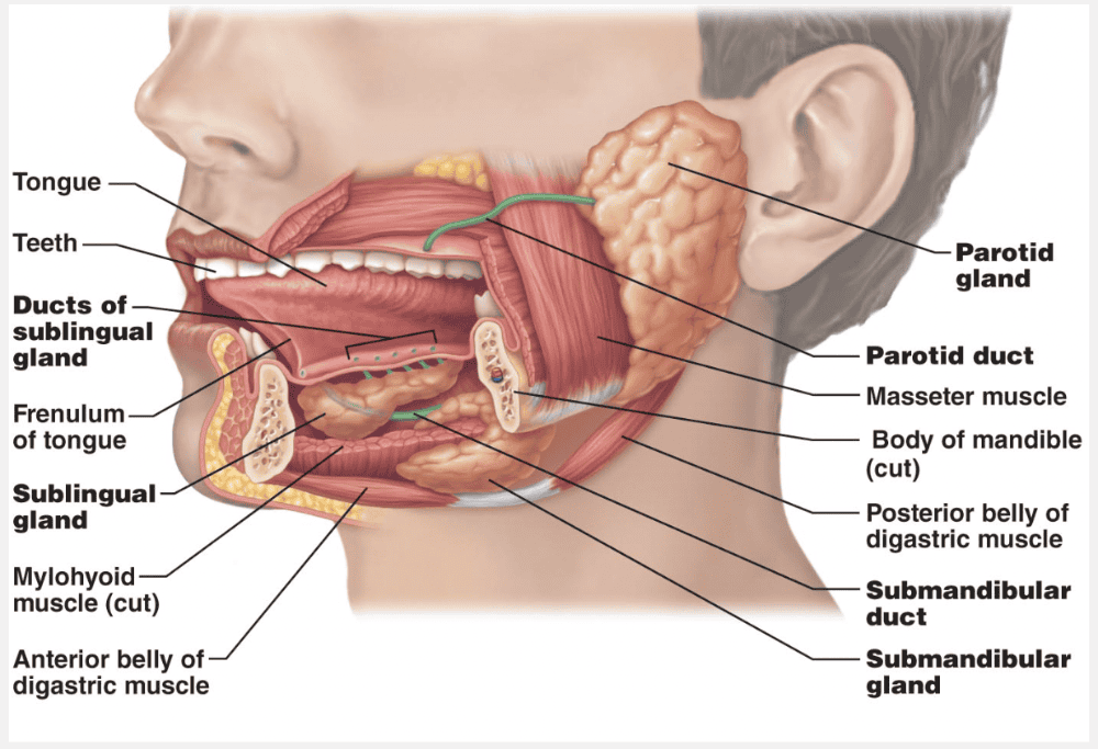
Mumps, a common children’s disease, is an inflammation of the parotid glands caused by the mumps virus (myxovirus), which spreads from person to person in saliva. If you check the location of the parotid glands in the figure below, you can understand why people with mumps complain that it hurts to open their mouth or chew. Other signs and symptoms include moderate fever and pain when swallowing acidic foods (pickles, grapefruit juice, etc.). Mumps in adult males carries a 25% risk of infecting the testes too, leading to sterility.
Select the description for the structure labeled 'A.'
Brittle and thick as a dime
Ex.
“A” is the enamel, which is brittle and thick as a dime.
Each tooth has two major regions: the crown and the root. The enamel-covered crown is the exposed part of the tooth above the gingiva (jin’jĭ-vah), or gum, which surrounds the tooth like a tight collar. Enamel, a brittle ceramic-like material thick as a dime, directly bears the force of chewing. The hardest substance in the body, it is heavily mineralized with calcium salts, and its densely packed hydroxyapatite (mineral) crystals are oriented in force-resisting columns perpendicular to the tooth’s surface. The cells that produce enamel degenerate when the tooth erupts; consequently, decayed or cracked areas of enamel will not heal and must be artificially filled.
The root is the portion of the tooth embedded in the jawbone. Canine teeth, incisors, and premolars have one root, although the first upper premolars commonly have two. The first two upper molars have three roots, while the corresponding lower molars have two. The root pattern of the third molar varies, but a fused single root is most common.
A constricted tooth region called the neck connects the crown and root. Cement, a calcified connective tissue, covers the outer surface of the root and attaches the tooth to the thin periodontal ligament (per”e-o-don’tal; “around the tooth”). This ligament anchors the tooth in the bony socket (alveolus) of the jaw, forming a fibrous joint called a gomphosis. Where the gingiva borders on a tooth, it dips downward to form a shallow groove called the gingival sulcus.
Dentin, a protein-rich bonelike material, underlies the enamel cap and forms the bulk of a tooth. More resilient than enamel, dentin acts as a shock absorber during biting and chewing. Dentin surrounds a central pulp cavity containing a number of soft tissue structures (connective tissue, blood vessels, and nerve fibers) collectively called pulp. Pulp supplies nutrients to the tooth tissues and provides tooth sensation. Where the pulp cavity extends into the root, it becomes the root canal.
Match the following: Gingivitis
- The gums are red, sore, swollen, and may bleed.
- Tartar or calculus is formed as dental plaque accumulates and calcifies.
- Results from bacterial action that gradually demineralizes enamel.
- Causes a burning radiating substernal pain due to regurgitation of stomach acid into the esophagus.
- Inflammation of the parotid salivary gland that carries a 25% risk of leading to sterility in adult males.
- Neutrophils and other immune cells attack body tissues, carving deep pockets around the teeth.
The gums are red, sore, swollen, and may bleed.
Ex.
In ginigivits, the gums are red, sore, swollen, and may bleed.
As dental plaque accumulates, it calcifies, forming calculus (kal’ku-lus; “stone”) or tartar. These stony hard deposits disrupt the seal between gingivae and teeth, deepening the sulcus and putting the gums at risk for infection by pathogenic anaerobic bacteria. In the early stages of such an infection, called gingivitis (jin”jĭ-vi’tis), the gums are red, sore, swollen, and may bleed.
A burning, radiating substernal pain may be caused by gastroesophageal disease (GERD), in which stomach acid regurgitates into the esophagus.
In 25% of mumps cases in adult males, the testes are also infected, which can lead to sterility.
Match the following: Caries
- Neutrophils and other immune cells attack body tissues, carving deep pockets around the teeth.
- The gums are red, sore, swollen, and may bleed.
- Causes a burning radiating substernal pain due to regurgitation of stomach acid into the esophagus.
- Results from bacterial action that gradually demineralizes enamel.
- Tartar or calculus is formed as dental plaque accumulates and calcifies.
- Inflammation of the parotid salivary gland that carries a 25% risk of leading to sterility in adult males.
Results from bacterial action that gradually demineralizes enamel.
Ex.
Dental caries (kār’ēz; “rottenness”), or cavities, result from bacterial action that gradually demineralizes enamel and underlying dentin.
Decay begins when dental plaque (a film of sugar, bacteria, and other mouth debris) adheres to the teeth. Bacterial metabolism of the trapped sugars produces acids, which dissolve the calcium salts of the teeth. Once the salts are leached out, enzymes released by the bacteria readily digest the remaining organic matrix of the tooth. Frequent brushing and daily flossing help prevent caries by removing plaque.
As dental plaque accumulates, it calcifies, forming calculus (kal’ku-lus; “stone”) or tartar. These stony hard deposits disrupt the seal between gingivae and teeth, deepening the sulcus and putting the gums at risk for infection by pathogenic anaerobic bacteria. In the early stages of such an infection, called gingivitis (jin”jĭ-vi’tis), the gums are red, sore, swollen, and may bleed.
A burning, radiating substernal pain may be caused by gastroesophageal disease (GERD), in which stomach acid regurgitates into the esophagus.
In 25% of mumps cases in adult males, the testes are also infected, which can lead to sterility.
Select the description for the structure labeled 'C.'
- Shock absorber during biting
- Covers the root of the tooth
- Brittle and thick as a dime
- Forms a gomphosis
- Surrounds the tooth like a collar
Surrounds the tooth like a collar
Ex.
“C” is the gingiva, which surrounds a tooth like a collar.
Each tooth has two major regions: the crown and the root. The enamel-covered crown is the exposed part of the tooth above the gingiva (jin’jĭ-vah), or gum, which surrounds the tooth like a tight collar. Enamel, a brittle ceramic-like material thick as a dime, directly bears the force of chewing. The hardest substance in the body, it is heavily mineralized with calcium salts, and its densely packed hydroxyapatite (mineral) crystals are oriented in force-resisting columns perpendicular to the tooth’s surface. The cells that produce enamel degenerate when the tooth erupts; consequently, decayed or cracked areas of enamel will not heal and must be artificially filled.
The root is the portion of the tooth embedded in the jawbone. Canine teeth, incisors, and premolars have one root, although the first upper premolars commonly have two. The first two upper molars have three roots, while the corresponding lower molars have two. The root pattern of the third molar varies, but a fused single root is most common.
A constricted tooth region called the neck connects the crown and root. Cement, a calcified connective tissue, covers the outer surface of the root and attaches the tooth to the thin periodontal ligament (per”e-o-don’tal; “around the tooth”). This ligament anchors the tooth in the bony socket (alveolus) of the jaw, forming a fibrous joint called a gomphosis. Where the gingiva borders on a tooth, it dips downward to form a shallow groove called the gingival sulcus.
Dentin, a protein-rich bone-like material, underlies the enamel cap and forms the bulk of a tooth. More resilient than enamel, dentin acts as a shock absorber during biting and chewing. Dentin surrounds a central pulp cavity containing a number of soft tissue structures (connective tissue, blood vessels, and nerve fibers) collectively called pulp. Pulp supplies nutrients to the tooth tissues and provides tooth sensation. Where the pulp cavity extends into the root, it becomes the root canal.
Match the structure in or around the oral cavity with its description: Parotid gland.
- Lingual frenulum
- Large salivary gland lying near the ear
- Salivary gland below the tongue
- Smaller salivary gland located under the jaw
Large salivary gland lying near the ear
Ex.
The parotid gland is the large salivary gland lying near the ear.
The major salivary glands are paired compound tubuloalveolar glands that develop from the oral mucosa and remain connected to it by ducts. The large, roughly triangular parotid gland (pah-rot’id; par = near, oto = the ear) lies anterior to the ear between the masseter muscle and the skin. Its prominent duct parallels the zygomatic arch, pierces the buccinator muscle, and opens into the vestibule next to the second upper molar.
The permanent dentition consists of __________ teeth in a full set.
- 16
- 32
- 10
- 20
32
Ex.
The permanent dentition consists of 32 teeth in a full set.
As the deep-lying permanent teeth enlarge and develop, the roots of the milk teeth are resorbed from below, causing them to loosen and fall out between ages 6 and 12. Generally, all the permanent teeth but the third molars have erupted by the end of adolescence. The third molars, also called wisdom teeth, emerge between ages 17 and 25. There are usually 32 permanent teeth in a full set, but sometimes the wisdom teeth never erupt or are completely absent.
The dental formula is a shorthand way of indicating the numbers and relative positions of the different types of teeth. This formula is written as a ratio, uppers over lowers, for one-half of the mouth. Since the other side is a mirror image, we obtain total dentition by multiplying the dental formula by 2. The primary dentition consists of two incisors (I), one canine (C), and two molars (M) on each side of each jaw, and its dental formula is written as
2I, 1C, 2M (upper jaw)
2I, 1C, 2M (lower jaw) × 2 (20 teeth)
Similarly, the permanent dentition [two incisors, one canine, two premolars (PM), and three molars] is
2I, 1C, 2PM, 3M
2I, 1C, 2PM, 3M × 2 (32 teeth)
Match the following: Mumps in males
- As dental plaque accumulates, it calcifies, forming calculus.
- Neutrophils and other immune cells attack body tissues, carving deep pockets around the teeth.
- Carries a 25% risk of leading to sterility.
- The gums are red, sore, swollen, and may bleed.
- This results from bacterial action that gradually demineralizes enamel.
Carries a 25% risk of leading to sterility.
Ex.
Mumps in males carries a 25% risk of infecting the testes too, leading to sterility.
Mumps, a common children’s disease, is an inflammation of the parotid glands caused by the mumps virus (myxovirus), which spreads from person to person in saliva. If you check the location of the parotid glands in the figure below, you can understand why people with mumps complain that it hurts to open their mouth or chew. Other signs and symptoms include moderate fever and pain when swallowing acidic foods (pickles, grapefruit juice, etc.). Mumps in adult males carries a 25% risk of infecting the testes too, leading to sterility.
In ginigivits, the gums are red, sore, swollen, and may bleed.
As dental plaque accumulates, it calcifies, forming calculus (kal’ku-lus; “stone”) or tartar. These stony hard deposits disrupt the seal between gingivae and teeth, deepening the sulcus and putting the gums at risk for infection by pathogenic anaerobic bacteria. In the early stages of such an infection, called gingivitis (jin”jĭ-vi’tis), the gums are red, sore, swollen, and may bleed.
The point at which the esophagus passes through the diaphragm is called the __________.
- esophageal sphincter
- esophageal hiatus
- cardial orifice
- central canal
esophageal hiatus
Ex.
The point at which the esophagus passes through the diaphragm is called the esophageal hiatus.
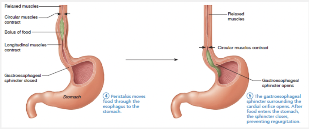
The esophagus takes a fairly straight course through the mediastinum of the thorax. It pierces the diaphragm at the esophageal hiatus (hi-a’tus; “gap”) to enter the abdomen. It joins the stomach at the cardial orifice within the abdominal cavity. The cardial orifice is surrounded by the gastroesophageal or cardiac sphincter (ga-strō-i-ˌsä-fə-ˈjē-əl), which is a physiological sphincter.
Select the description for the structure labeled 'E.'
- Surrounds the tooth like a collar
- Forms a gomphosis
- Brittle and thick as a dime
- Covers the root of the tooth
- Shock absorber during biting
Forms a gomphosis
Ex.
“E” is the periodontal ligament which forms a gomphosis.
Each tooth has two major regions: the crown and the root. The enamel-covered crown is the exposed part of the tooth above the gingiva jin’ji-vah), or gum, which surrounds the tooth like a tight collar. Enamel, a brittle ceramic-like material thick as a dime, directly bears the force of chewing. The hardest substance in the body, it is heavily mineralized with calcium salts, and its densely packed hydroxyapatite (mineral) crystals are oriented in force-reisting columns perpendicular to the tooth’s surface. The cells that produce enamel degenerate when the tooth erupts; consequently, decayed or cracked areas of enamel will not heal and must be artificially filled.
The root is the portion of the tooth embedded in the jawbone. Canine teeth, incisors, and premolars have one root, althrough the first upper premolars commonly have two. The first two upper molars have three roots, while the corresponding lower molars have two. The root patern of the third molar varies, but a fused single root is most common.
A constricted tooth region called the neck connects the crown and root. Cement, a calcified connective tissue, covers the outer surface of the root and attaches the tooth to the thin periodontal ligament (per”e-o-don’tal; “around the tooth”). This ligament anchors the tooth in the bony socket (alveolus) of the jaw, forming fibrous joint called a gomphosis. Where the gingiva borders on a tooth, it dips downward to form a shallow groove called the gingival sulcus.
Dentin, a protetin-rich bonelike material, underlies the enamel cap and forms the bulk of a tooth. More resilient than enamel, dentin acts as a shock absorber during biting and chewing. Dentin surround central pulp cavity containing a numberof soft tissue structures (connective tissue, blood vessels, and nerve fibers) collectively called pulp. Pulp supplies nutrients to the tooth tissues and provides tooth sensation. Where the pulp cavity extends into the root, it becomes the root canal.
Match the structure in or around the oral cavity with its description: Sublingual gland.
- Large salivary gland lying near the ear
- Smaller salivary gland located under the jaw
- Lingual frenulum
- Salivary gland below the tongue
Salivary gland below the tongue
Ex.
The sublingual gland is the salivary gland below the tongue.
The small, almond-shaped sublingual gland lies anterior to the submandibular gland under the tongue and opens via 10–20 ducts into the floor of the mouth. About the size of a walnut, the submandibular gland lies along the medial aspect of the mandibular body. Its duct runs beneath the mucosa of the oral cavity floor and opens at the base of the lingual frenulum.
Match the following part of a tooth with its description: Crown.
- Embedded in the jawbone
- Exposed and covered in enamel
- Forms the support of the gomphosis
- Calcified connective tissue around the bottom of the tooth
Exposed and covered in enamel
Ex.
The crown is exposed and covered in enamel.
Each tooth has two major regions: the crown and the root. The enamel-covered crown is the exposed part of the tooth above the gingiva (jin’jĭ-vah), or gum, which surrounds the tooth like a tight collar. Enamel, a brittle ceramic-like material thick as a dime, directly bears the force of chewing. The hardest substance in the body, it is heavily mineralized with calcium salts, and its densely packed hydroxyapatite (mineral) crystals are oriented in force-resisting columns perpendicular to the tooth’s surface. The cells that produce enamel degenerate when the tooth erupts; consequently, decayed or cracked areas of enamel will not heal and must be artificially filled.
Match the following: Periodontitis.
- The gums are red, sore, swollen, and may bleed.
- Inflammation of the parotid salivary gland that carries a 25% risk of leading to sterility in adult males.
- Causes a burning radiating substernal pain due to regurgitation of stomach acid into the esophagus.
- Tartar or calculus is formed as dental plaque accumulates and calcifies.
- Results from bacterial action that gradually demineralizes enamel.
- Neutrophils and other immune cells attack body tissues, carving deep pockets around the teeth.
Neutrophils and other immune cells attack body tissues, carving deep pockets around the teeth.
Ex.
In periodontitis, neutrophils and other immune cells attack body tissues, carving deep pockets around the teeth.
Gingivitis is reversible if the calculus is removed, but if it is neglected the bacteria eventually form pockets of infection which become inflamed. Neutrophils and other immune cells attack not only the intruders but also body tissues, carving deep pockets around the teeth, destroying the periodontal ligament, and activating osteoclasts which dissolve the bone. This serious condition, periodontal disease or periodontitis, affects up to 95% of all people over age 35 and accounts for 80–90% of tooth loss in adults.
Tooth loss from periodontitis is not inevitable. Various treatments can alleviate the bacterial infestations and encourage the surrounding tissues to reattach to the teeth and bone.
How does food move through the esophagus to the stomach?
- Peristalsis
- Bulk Flow
- Gravity
- Segmentation
Peristalsis
Ex.
After swallowing, food moves through the esophagus and to the stomach by peristalsis.
The pharyngeal-esophageal phase is involuntary and is controlled by the swallowing center in the brain stem (medulla and lower pons). Various cranial nerves, most importantly the vagus nerves, transmit motor impulses from the swallowing center to the muscles of the pharynx and esophagus. Once food enters the pharynx, respiration is momentarily inhibited and all routes except the desired one into the digestive tract are blocked off. Solid foods pass from the oropharynx to the stomach in about 8 seconds, and fluids, aided by gravity, pass in 1 to 2 seconds.
Match the following digestive process with the correct description: Ingestion
- Chewing, mixing, churning, and segmentation of food
- Passage of digested materials from the lumen of the GI tract into the blood or lymph
- Elimination of indigestible solids
- Taking food into the digestive system
- Enzymatic degradation of foodstuffs into simpler molecules
Taking food into the digestive system
Ex.
Ingestion is taking food into the digestive tract (eating).
Absorption is the passage of digested end products (plus vitamins, minerals, and water) from the lumen of the GI tract through the mucosal cells by active or passive transport into the blood or lymph.
Chewing, mixing, churning, and segmentation are part of mechanical digestion.
Enzymatic degradation of foodstuffs into simpler molecules is chemical digestion.
Elimination of indigestible solids is the process of defecation.