Match the following term to its correct description: Muscularis externa
- Protective outermost layer of the alimentary canal
- areolar connective tissue that has a rich supply of blood, lymphatic vessels, and nerve fibers
- Main site of nutrient absorption
- Responsible for segmentation and peristalsis
Responsible for segmentation and peristalsis
Ex.
The muscularis externa is responsible for segmentation and peristalsis.
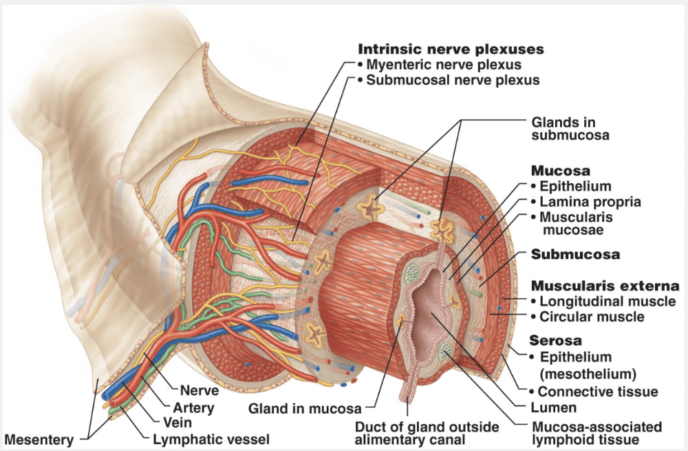
Surrounding the submucosa is the muscularis externa, also simply called the muscularis. This layer is responsible for segmentation and peristalsis. It typically has an inner circular layer and an outer longitudinal layer of smooth muscle cells. In several places along the tract, the circular layer thickens, forming sphincters that act as valves to control food passage from one organ to the next and prevent backflow.
The mucosa is the main site of nutrient absorption.
The protective outer layer of the alimentary canal is the serosa.
Areolar connective tissue that has a rich supply of blood, lymphatic vessels, and nerve fibers is the submucosa.
Identify the organ indicated by 'D.'
- Pancreas
- Stomach
- Large intestine
- Pharynx
- Salivary gland
Pancreas
Identify the organ indicated by 'D'
- Tongue
- Liver
- Small intestine
- Mouth
- Gallbladder
- Esophagus
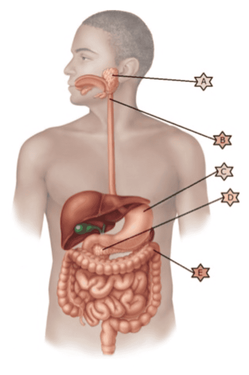
Liver
Identify the organ indicated by 'B.'
- Pharynx
- Pancreas
- Stomach
- Large intestine
- Salivary gland
Pharynx
Which sequence below represents the correct layering of the wall of the GI tract, starting from the layer next to the lumen?
- Serosa, mucosa, muscularis externa, submucosa
- Mucosa, muscularis externa, submucosa, serosa
- Muscularis externa, serosa, submucosa, mucosa
- Mucosa, submucosa, muscularis externa, serosa
- Submucosa, mucosa, muscularis externa, serosa
Mucosa, submucosa, muscularis externa, serosa
Ex.
The correct layering of the wall of the GI tract, starting from the layer next to the lumen is mucosa, submucosa, muscularis externa, serosa.
From the esophagus to the anal canal, the walls of the alimentary canal have the same four basic layers, or tunics—mucosa, submucosa, muscularis externa, and serosa. Each layer contains a predominant tissue type that plays a specific role in food breakdown.
Match the following term to its correct description: Serosa
- Responsible for segmentation and peristalsis
- Areolar connective tissue that has a rich supply of blood, lymphatic vessels, and nerve fibers
- Main site of nutrient absorption
- Protective outermost layer of the alimentary canal
Protective outermost layer of the alimentary canal
Ex.
The serosa is the protective outermost layer of the alimentary canal.
The serosa, the outermost layer of the intraperitoneal organs, is the visceral peritoneum. In most alimentary canal organs, it is formed of areolar connective tissue covered with mesothelium, a single layer of squamous epithelial cells.
In the esophagus, which is located in the thoracic instead of the abdominopelvic cavity, the serosa is replaced by an adventitia (ad”ven-tish’e-ah), ordinary dense connective tissue that binds the esophagus to surrounding structures. Retroperitoneal organs have both an adventitia (on the side facing the dorsal body wall) and a serosa (on the side facing the peritoneal cavity).
The mucosa is the main site of nutrient absorption.
Areolar connective tissue that has a rich supply of blood, lymphatic vessels, and nerve fibers is the submucosa.
The muscularis externa is responsible for segmentation and peristalsis.
__________ is the major means of propulsion in the digestive system.
- Defecation
- Ingestion
- Mechanical digestion
- Peristalsis
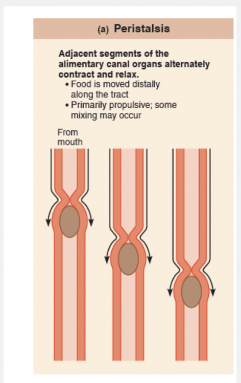
Peristalsis
Ex.
Peristalsis is the major means of propulsion in the digestive system.
Propulsion, which moves food through the alimentary canal, includes swallowing, which is initiated voluntarily, and peristalsis (per”ĭ-stal’sis), an involuntary process. Peristalsis (peri = around; stalsis = constriction), the major means of propulsion, involves alternating waves of contraction and relaxation of muscles in the organ walls. Its main effect is to squeeze food along the tract, but some mixing occurs as well. In fact, peristaltic waves are so powerful that, once swallowed, food and fluids will reach your stomach even if you stand on your head.
Which of the following is not an accessory organ of the digestive system?
- Tongue
- Teeth
- Intestines
- Salivary glands
Intestines
Ex.
The intestines are not accessory digestive organs.
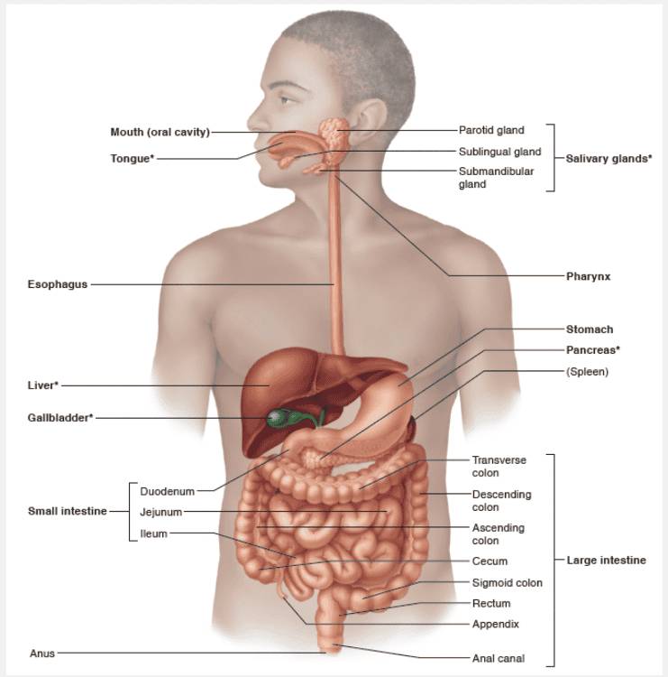
The accessory digestive organs are the teeth, tongue, gallbladder, and a number of large digestive glands—the salivary glands, liver, and pancreas. The teeth and tongue are in the mouth, or oral cavity, while the digestive glands and gallbladder lie outside the GI tract and connect to it by ducts. The accessory digestive glands produce a variety of secretions that help break down foodstuffs.
Which of the following is the most common cause of peritonitis?
- A burst appendix
- A perforating abdominal wound
- A ruptured pancreatic cyst
- A perforating ulcer of the stomach
A burst appendix
Ex.
The most common cause of peritonitis is a burst appendix.
Peritonitis is inflammation of the peritoneum. It can arise from a piercing abdominal wound, a perforating ulcer that leaks stomach juices into the peritoneal cavity, or poor sterile technique during abdominal surgery. However, most commonly it results from a burst appendix that sprays bacteria-containing feces all over the peritoneum. In peritonitis, the peritoneal coverings tend to stick together around the infection site. This localizes the infection, providing time for macrophages to prevent the inflammation from spreading. If peritonitis becomes widespread within the peritoneal cavity, it is dangerous and often lethal. Treatment includes removing as much infectious debris as possible and administering mega-doses of antibiotics.
Identify the organ indicated by 'E'
- Tongue
- Esophagus
- Liver
- Small intestine
- Mouth
- Gallbladder
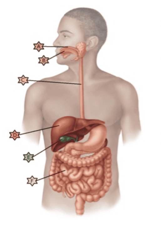
Gallbladder
The serous membrane that covers the external surface of most digestive organs is called the __________.
- visceral peritoneum
- mesentery
- parietal peritoneum
- omentum
visceral peritoneum
Ex.
The serous membrane that covers the external surface of most digestive organs is called the visceral peritoneum.
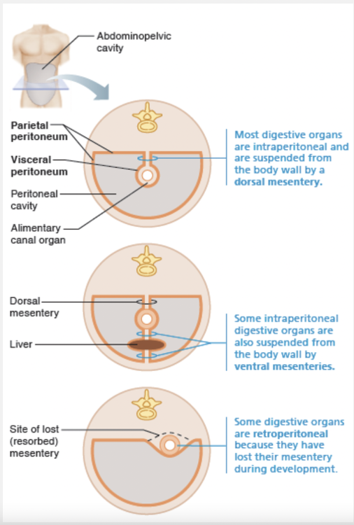
Most digestive system organs reside in the abdomino pelvic cavity. All ventral body cavities contain slippery serous membranes. The peritoneum of the abdomino pelvic cavity is the most extensive of these membranes. The visceral peritoneum covers the external surfaces of most digestive organs and is continuous with the parietal peritoneum that lines the body wall. Between the two peritoneums is the peritoneal cavity, a slit like potential space containing a slippery fluid secreted by the serous membranes.
The omenta are two mesenteries that help tether the stomach to other digestive organs and the body wall.
Identify the organ indicated by 'F'
- Tongue
- Liver
- Small intestine
- Mouth
- Gallbladder
- Esophagus
Small intestine
The alimentary canal in a cadaver is longer than in a living person because, in a cadaver, there is no __________.
- hormonal influence
- digestion taking place
- muscle tone
- food in the tube
muscle tone
Ex.
The alimentary canal in a cadaver is longer than in a living person because, in a cadaver, there is no muscle tone.
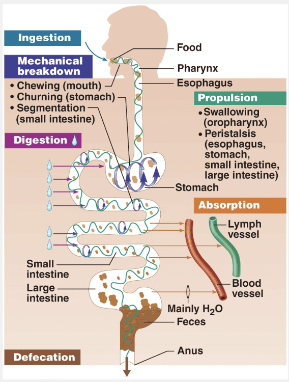
The organs of the alimentary canal are the mouth, pharynx, esophagus, stomach, small intestine, and large intestine. The large intestine leads to the terminal opening, or anus. In a cadaver, the alimentary canal is approximately 9 m (about 30 ft) long, but in a living person, it is considerably shorter because of its muscle tone. Food material in this tube is technically outside the body because the canal is open to the external environment at both ends.
Identify the organ indicated by 'A'.
- Stomach
- Pharynx
- Pancreas
- Large intestine
- Salivary gland
Salivary gland
Match the following digestive process with the correct description: Chemical digestion
- Chewing, mixing, churning, and segmentation of food
- Enzymatic degradation of foodstuffs into simpler molecules
- Elimination of indigestible solids
- Passage of digested materials from the lumen of the GI tract into the blood or lymph
- Taking food into the digestive system
Enzymatic degradation of foodstuffs into simpler molecules
Ex.
Chemical digestion is described as the enzymatic degradation of foodstuffs into simpler molecules.
Digestion involves a series of catabolic steps in which enzymes secreted into the lumen (cavity) of the alimentary canal break down complex food molecules to their chemical building blocks.
Absorption is the passage of digested end products (plus vitamins, minerals, and water) from the lumen of the GI tract through the mucosal cells by active or passive transport into the blood or lymph.
Taking food into the digestive system is called ingestion.
Chewing, mixing, churning, and segmentation are part of mechanical digestion.
Elimination of indigestible solids is the process of defecation.
Which of the following statements about the mesentery is incorrect?
- It provides a route for blood vessels, lymphatics, and nerves to reach the digestive viscera.
- It is composed of a layer of serous membrane fused with a layer of mucus membrane.
- Omenta is the special name for part of the mesentery.
- It holds the organs of the abdomen in place.
It is composed of a layer of serous membrane fused with a layer of mucus membrane.
Ex.
It is incorrect that the mesentery is composed of a layer of serous membrane fused with a layer of mucus membrane.
A mesentery (mes’en-ter”e) is a double layer of peritoneum—a sheet of two serous membranes fused back to back—that extends to the digestive organs from the body wall. Mesenteries provide routes for blood vessels, lymphatics, and nerves to reach the digestive viscera, hold organs in place, and store fat. In most places the mesentery is dorsal and attaches to the posterior abdominal wall, but there are ventral mesenteries too, such as the one that extends from the liver to the anterior abdominal wall digestive organ mesenteries have specific names (such as the omenta), or are called “ligaments” (even though these peritoneal folds are nothing like the fibrous ligaments that connect bones).
Match the following term to its correct description: Mucosa
- Protective outermost layer of the alimentary canal
- Main site of nutrient absorption
- Areolar connective tissue that has a rich supply of blood, lymphatic vessels, and nerve fibers
- Responsible for segmentation and peristalsis
Main site of nutrient absorption
Ex.
The mucosa is the main site of nutrient absorption.
The innermost layer is the mucosa, or mucous membrane, a moist epithelial membrane that lines the alimentary canal lumen from mouth to anus. Its major functions are to:
- Secrete mucus, digestive enzymes, and hormones
- Absorb the end products of digestion into the blood
- Protect against infectious disease
The mucosa in a particular region of the GI tract may perform one or all three of these functions.
The protective outer layer of the alimentary canal is the serosa.
Areolar connective tissue that has a rich supply of blood, lymphatic vessels, and nerve fibers is the submucosa.
The muscularis externa is responsible for segmentation and peristalsis.
Identify the organ indicated by 'A'
- Mouth
- Small intestine
- Tongue
- Gallbladder
- Esophagus
- Liver
Mouth
Match the following digestive process with the correct description: Absorption
- Passage of digested materials from the lumen of the GI tract into the blood or lymph
- Chewing, mixing, churning, and segmentation of food
- Elimination of indigestible solids
- Taking food into the digestive system
- Enzymatic degradation of foodstuffs into simpler molecules
Passage of digested materials from the lumen of the GI tract into the blood or lymph
Ex.
Absorption is the passage of digested end products (plus vitamins, minerals, and water) from the lumen of the GI tract through the mucosal cells by active or passive transport into the blood or lymph.
Taking food into the digestive system is called ingestion.
Chewing, mixing, churning, and segmentation are part of mechanical digestion.
Enzymatic degradation of foodstuffs into simpler molecules is chemical digestion.
Elimination of indigestible solids is the process of defecation.
Identify the organ indicated by 'C.'
- Salivary gland
- Stomach
- Large intestine
- Pancreas
- Pharynx
Stomach
Identify the organ indicated by 'C'
- Small intestine
- Liver
- Mouth
- Esophagus
- Tongue
- Gallbladder
Esophagus
Hollow muscular organs, like the stomach act as reservoirs because of an intrinsic smooth muscle stress-relaxation response known as _________.
- receptive relaxation
- peristalsis
- accommodation
- positive feedback
accommodation
Ex.
Hollow muscular organs, like the stomach act as reservoirs because of an intrinsic smooth muscle stress-relaxation response known as accommodation.
Gastric accommodation is the intrinsic ability of visceral smooth muscle to exhibit the stress-relaxation response (usually discussed with smooth muscle). In other words, the stomach can stretch without greatly increasing its tension and contracting expulsively. This capability is very important in hollow organs, like the stomach, that must serve as temporary reservoirs.
Receptive relaxation of smooth muscle in the stomach fundus and body occurs both in anticipation of and in response to food moving through the esophagus and into the stomach. The swallowing center of the brain stem coordinates this process, which is mediated by the vagus nerves.
Peristalsis (peri = around; stalsis = constriction), the major means of propulsion, involves alternating waves of contraction and relaxation of muscles in the organ walls.
Match the following term with its correct description: Same structure as the visceral peritoneum.
- Muscularis
- Serosa
- Mucosa
- Submucosa
Serosa
Ex.
The serosa, the outermost layer of the intraperitoneal organs, is the visceral peritoneum. In most alimentary canal organs, it is formed of areolar connective tissue covered with mesothelium, a single layer of squamous epithelial cells.
In the esophagus, which is located in the thoracic instead of the abdominopelvic cavity, the serosa is replaced by an adventitia (ad”ven-tish’e-ah), ordinary dense connective tissue that binds the esophagus to surrounding structures. Retroperitoneal organs have both an adventitia (on the side facing the dorsal body wall) and a serosa (on the side facing the peritoneal cavity).
The short reflexes in the digestive system are mediated by __________.
- the sympathetic system
- the parasympathetic system
- the enteric nerve plexuses
- higher brain centers
the enteric nerve plexuses
Ex.
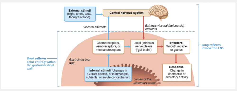
The short reflexes in the digestive system are mediated by the enteric nerve plexuses.
Short reflexes are mediated entirely by enteric nervous system plexuses in response to stimuli within the GI tract. Control of the patterns of segmentation and peristalsis is largely automatic, involving pacemaker cells and reflex arcs between enteric neurons in the same or different organs.
Identify the organ indicated by 'B'
- Liver
- Tongue
- Esophagus
- Gallbladder
- Mouth
- Small intestine
Tongue
Identify the organ indicated by 'E.'
- Pharynx
- Pancreas
- Small intestine
- Large intestine
- Stomach
Large intestine