Which of the following is not a function of the conducting zone?
- Warming of air
- Transport of air
- Mucus secretion
- Cleansing of air
- Gas exchange
Gas exchange
Ex.
Gas exchange is not a function of the conducting zone. Rather, it is a function of the respiratory zone where gas exchange occurs.
The conducting zone consists of all of the respiratory passageways from the nose to the respiratory bronchioles. These provide fairly rigid conduits for air to reach the gas exchange sites. The conducting zone organs also cleanse, humidify, and warm incoming air. As a result, air reaching the lungs has fewer irritants (dust, bacteria, etc.) than when it entered the body, and it is warm and damp, like the air of the tropics.
Match the following description to the image: “A.”
- Secretes a fluid to reduce the surface tension of alveolar fluid
- Cells that regularly get swept up and out of the lung
- Creates the blood air barrier in the lungs
- Creates a wall 15 times thinner than a piece of paper
Creates a wall 15 times thinner than a piece of paper
Ex.
“A” represents the type I alveolar cell that creates a wall 15 times thinner than a piece of paper.
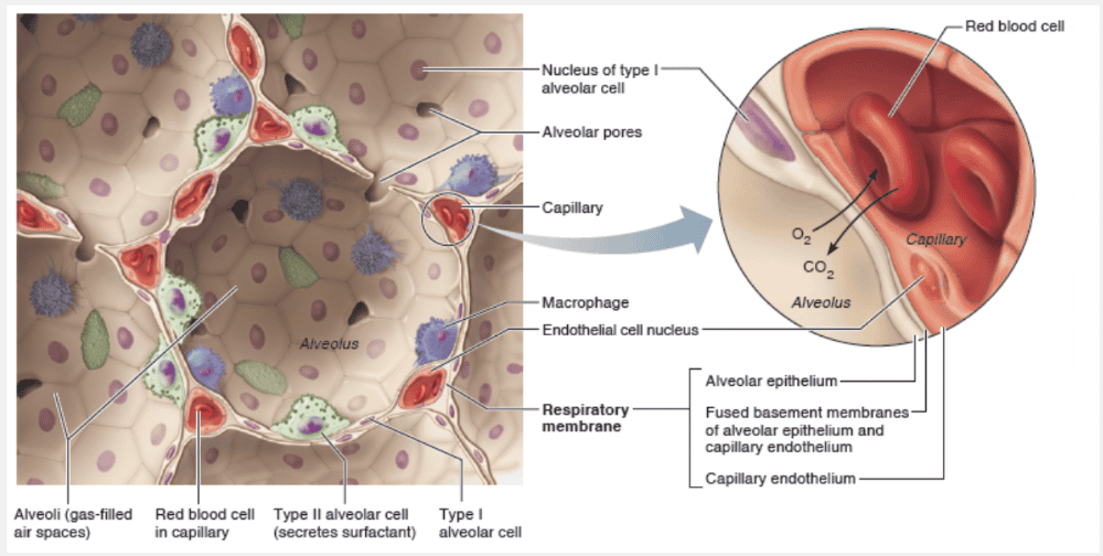
The walls of the alveoli are composed primarily of a single layer of squamous epithelial cells, called type I alveolar cells, surrounded by a flimsy basement membrane. The thinness of their walls is hard to imagine, but a sheet of tissue paper is 15 times thicker!
The external surfaces of the alveoli are densely covered with a “cobweb” of pulmonary capillaries. Together, the capillary and alveolar walls and their fused basement membranes form the respiratory membrane, a 0.5-μm-thick blood air barrier that has blood flowing past on one side and gas on the other. Gas exchanges occur readily by simple diffusion across the respiratory membrane—O2 passes from the alveolus into the blood, and CO2 leaves the blood to enter the gas-filled alveolus. Scattered amid the squamous type I alveolar cells that form the major part of the alveolar walls are cuboidal type II alveolar cells. Type II alveolar cells secrete a fluid containing a detergent-like substance called surfactant that coats the gas-exposed alveolar surfaces. Type II alveolar cells also secrete a number of antimicrobial proteins that are important elements of innate immunity.
The alveoli have three other significant features: (1) They are surrounded by fine elastic fibers of the same type that surround the entire bronchial tree. (2) Open alveolar pores connecting adjacent alveoli allow air pressure throughout the lung to be equalized and provide alternate air routes to any alveoli whose bronchi have collapsed due to disease. (3) Remarkably efficient alveolar macrophages crawl freely along the internal alveolar surfaces.
Match the following: The Heimlich maneuver.
- Inhibits and ultimately destroys cilia
- Often caused by viral infections, but may also be due to overusing the voice, very dry air, bacterial infections, tumors on the vocal folds, or inhalation of irritating chemicals
- Has saved many people from becoming victims of “cafe coronaries”
- A partial vacuum in openings in the skull
- May result in air that is not properly moistened, warmed, or filtered before reaching the lungs
Has saved many people from becoming victims of “cafe coronaries”
Ex.
The Heimlich maneuver has saved many people from becoming victims of “cafe coronaries.”
Tracheal obstruction is life-threatening. Many people have suffocated after choking on a piece of food that suddenly closed off their trachea. The Heimlich maneuver, a procedure in which air in the victim’s lungs is used to “pop out,” or expel, an obstructing piece of food, has saved many people from becoming victims of “cafe coronaries.” The maneuver is simple to learn and easy to do. However, it is best learned by demonstration because cracked ribs are a distinct possibility when it is done incorrectly.
Smoking inhibits and ultimately destroys cilia.
Without ciliary activity, coughing is the only way to prevent mucus from accumulating in the lungs. For this reason, smokers with respiratory congestion should avoid medications that inhibit the cough reflex.
Laryngitis is often caused by viral infections, but may also be due to overusing the voice, very dry air, bacterial infections, tumors on the vocal folds, or inhalation of irritating chemicals.
Sinus headaches are caused by a partial vacuum in openings of the skull.
Swelling of adenoids may result in air that is not properly moistened, warmed, or filtered before reaching the lungs.
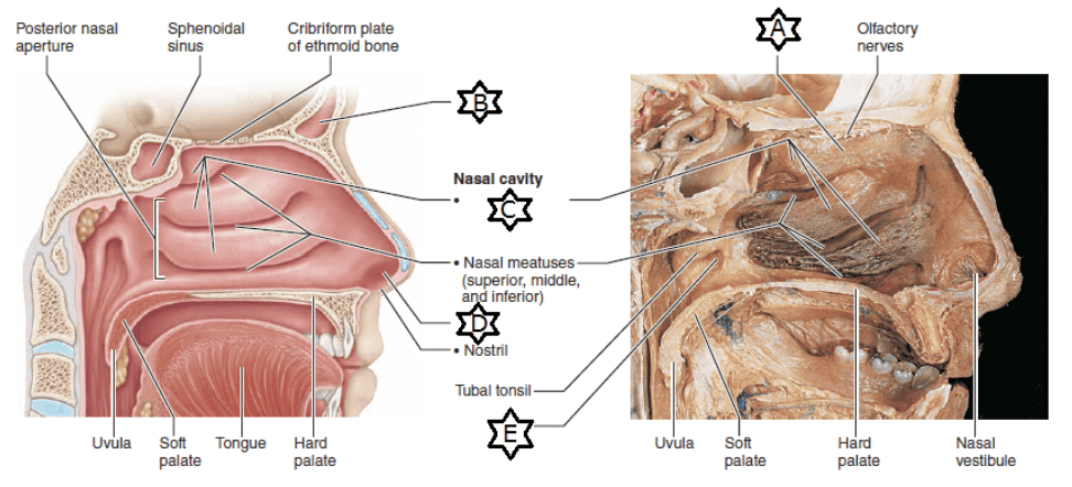
What is the function of the structure labeled “E?”
- Equalizes pressure between atmosphere and middle ear
- Structure that allows heavier particles to be deflected into nasal mucosa
- Region of nasal cavities that contains smell receptors
- Helps warm and moisten inspired air
- Lined with sebaceous and sweat glands and filters coarse particles
Equalizes pressure between atmosphere and middle ear
Ex.
“E” represents the the pharyngotympanic (auditory) tubes, which drain the middle ear cavities and allow middle ear pressure to equalize with atmospheric pressure, by opening into the lateral walls of the nasopharynx. A ridge of pharyngeal mucosa posterior to each of these openings constitutes the tubal tonsil. Its strategic location helps protect the middle ear against infections likely to spread from the nasopharynx.
Match the following description with its corresponding term: About ten in each lung; small amounts of cartilage; smooth muscle dominates.
- Lobar (secondary) bronchi
- Trachea
- Segmental (tertiary) bronchi
- Bronchioles
- Main (primary) bronchi
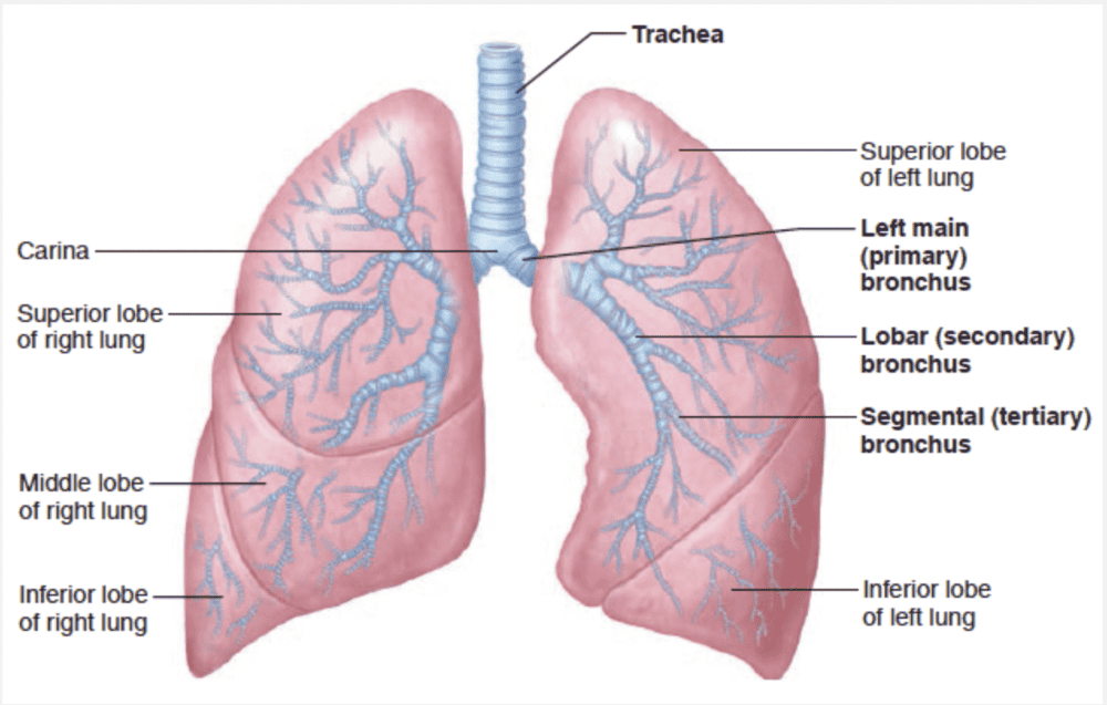
Segmental (tertiary) bronchi
Ex.
The structure described as having about ten in each lung, small amounts of cartilage and dominated by smooth muscle are segmental (tertiary) bronchi.
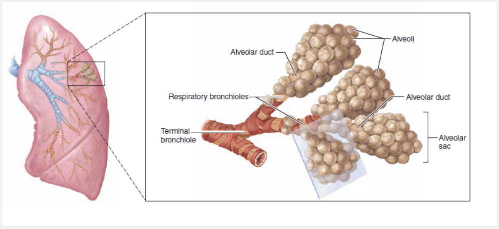
Once inside the lungs, each main bronchus subdivides into lobar (secondary) bronchi—three on the right and two on the left—each supplying one lung lobe. The lobar bronchi branch into third-order segmental (tertiary) bronchi, which divide repeatedly into smaller and smaller bronchi (fourth-order, fifth-order, etc.). Passages smaller than 1 mm in diameter are called bronchioles (“little bronchi”), and the tiniest of these, the terminal bronchioles, are less than 0.5 mm in diameter.
Match the following: Smoking.
- Inhibits and ultimately destroys cilia
- Has saved many people from becoming victims of “cafe coronaries”
- A partial vacuum in openings in the skull
- Often caused by viral infections, but may also be due to overusing the voice, very dry air, bacterial infections, tumors on the vocal folds, or inhalation of irritating chemicals
- May result in air that is not properly moistened, warmed, or filtered before reaching the lungs
Inhibits and ultimately destroys cilia
Ex.
Smoking inhibits and ultimately destroys cilia.
Without ciliary activity, coughing is the only way to prevent mucus from accumulating in the lungs. For this reason, smokers with respiratory congestion should avoid medications that inhibit the cough reflex.
The Heimlich maneuver has saved many people from becoming victims of “cafe coronaries.”
Laryngitis is often caused by viral infections, but may also be due to overusing the voice, very dry air, bacterial infections, tumors on the vocal folds, or inhalation of irritating chemicals.
Sinus headaches are caused by a partial vacuum in openings of the skull.
Swelling of adenoids may result in air that is not properly moistened, warmed, or filtered before reaching the lungs.
Which of the following is not a process in respiration?
- Internal respiration
- Pulmonary ventilation
- External ventilation
- External respiration

External ventilation
Ex.
External ventilation is not a process in respiration.
The major function of the respiratory system is to supply the body with oxygen and dispose of carbon dioxide. To accomplish this function, at least four processes, collectively called respiration, must happen:
- Pulmonary ventilation (commonly called breathing): Air is moved into and out of the lungs (during inspiration and expiration) so the gases there are continuously changed and refreshed.
- External respiration: Oxygen diffuses from the lungs to the blood, and carbon dioxide diffuses from the blood to the lungs.
- Transport of respiratory gases: Oxygen is transported from the lungs to the tissue cells of the body, and carbon dioxide is transported from the tissue cells to the lungs. The cardiovascular system accomplishes this transport using blood as the transporting fluid.
- Internal respiration: Oxygen diffuses from blood to tissue cells, and carbon dioxide diffuses from tissue cells to blood.
The elastic cartilage that shields the opening to the larynx during swallowing is the __________.
- cricoid cartilage
- cuneiform cartilage
- thyroid cartilage
- corniculate cartilage
- epiglottis
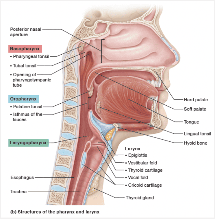
epiglottis
Ex.
The elastic cartilage that shields the opening to the larynx during swallowing is the epiglottis.
The ninth cartilage of the larynx, the flexible, spoon-shaped epiglottis (ep”ĭ-glot’is; “above the glottis”), is composed of elastic cartilage and is almost entirely covered by a taste bud–containing mucosa. The epiglottis extends from the posterior aspect of the tongue to its anchoring point on the anterior rim of the thyroid cartilage. When only air is flowing into the larynx, the inlet to the larynx is wide open and the free edge of the epiglottis projects upward. During swallowing, the larynx is pulled superiorly and the epiglottis tips to cover the laryngeal inlet. Because this action keeps food out of the lower respiratory passages, the epiglottis has been called the guardian of the airways. Anything other than air entering the larynx initiates the cough reflex to expel the substance. This protective reflex does not work when we are unconscious, so it is never a good idea to administer liquids when attempting to revive an unconscious person.
The thyroid, cricoid, cuneiform, and corniculate cartilages are made of hyaline cartilage and form the major structures of the larynx.
The __________ is covered by mucosa containing taste buds and keeps food out of the lower respiratory passages.
- glottis
- epiglottis
- vestibular folds
- uvula
epiglottis
Ex.
The epiglottis is covered by mucosa containing taste buds and keeps food out of the lower respiratory passages.
The ninth cartilage of the larynx, the flexible, spoon-shaped epiglottis (ep”ĭ-glot’is; “above the glottis”), is composed of elastic cartilage and is almost entirely covered by a taste bud– containing mucosa. The epiglottis extends from the posterior aspect of the tongue to its anchoring point on the anterior rim of the thyroid cartilage. When only air is flowing into the larynx, the inlet to the larynx is wide open and the free edge of the epiglottis projects upward. During swallowing, the larynx is pulled superiorly and the epiglottis tips to cover the laryngeal inlet. Because this action keeps food out of the lower respiratory passages, the epiglottis has been called the guardian of the airways. Anything other than air entering the larynx initiates the cough reflex to expel the substance. This protective reflex does not work when we are unconscious, so it is never a good idea to administer liquids when attempting to revive an unconscious person.
The glottis is the opening between the vocal folds (true vocal cords).
Vestibular folds are the false vocal cords within the larynx that help to close the glottis when we swallow.
The uvula is the piece of soft tissue suspended from the back of the soft palate, and closes off the nasopharynx to prevent food from entering the nasal cavity.
What is the function of the structure labeled “A?”
- Structure that allows heavier particles to be deflected into nasal mucosa
- Equalizes pressure between atmosphere and middle ear
- Region of nasal cavities that contains smell receptors
- Lined with sebaceous and sweat glands and filters coarse particles
- Helps warm and moisten inspired air
Region of nasal cavities that contains smell receptors
Ex.
“A” represents the olfactory epithelium, the region of nasal cavities that contains smell receptors.
The small patch of olfactory mucosa lines the slitlike superior region of the nasal cavity and contains smell receptors in its olfactory epithelium.
Which of the following statements is false?
- Remarkably efficient alveolar macrophages crawl freely along the internal and external alveolar surfaces.
- We clear and swallow over 2 million alveolar macrophages per hour.
- Huge numbers of infectious microorganisms are continuously carried into the alveoli.
- Alveolar surfaces are usually sterile.
Remarkably efficient alveolar macrophages crawl freely along the internal and external alveolar surfaces.
Ex.
It is false to say that remarkably efficient alveolar macrophages crawl freely along the internal and external alveolar surfaces. They are located only on the internal, not the external, alveolar surfaces.
Although huge numbers of infectious microorganisms are continuously carried into the alveoli, alveolar surfaces are usually sterile. Because the alveoli are “dead ends,” aged and dead macrophages must be prevented from accumulating in them. Most macrophages simply get swept up by the ciliary current of superior regions and carried to the pharynx. In this manner, we clear and swallow over 2 million alveolar macrophages per hour!
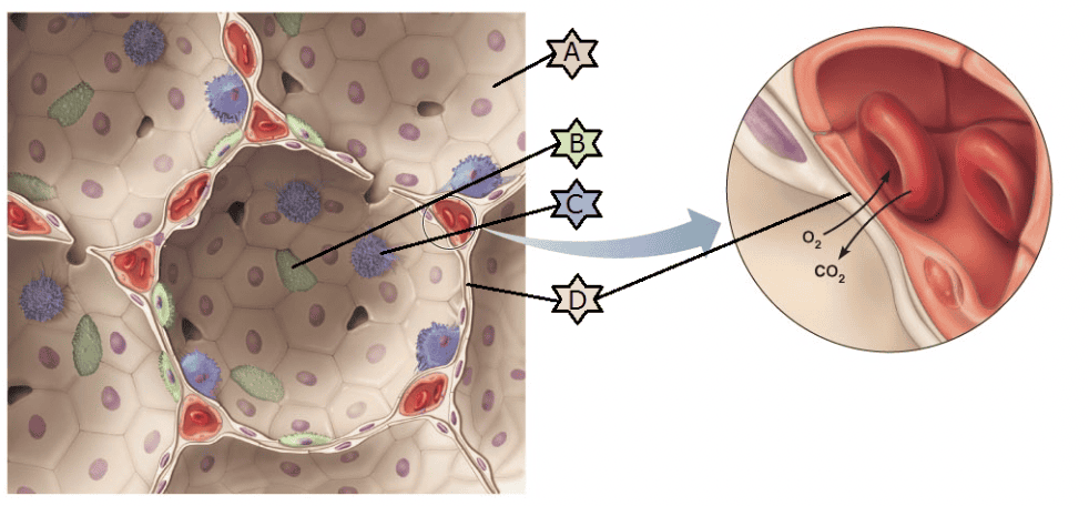
Match the following description to the image: “D.”
- Cells that regularly get swept up and out of the lung
- Creates the blood air barrier in the lungs
- Secretes a fluid to reduce the surface tension of alveolar fluid
- Creates a wall 15 times thinner than a piece of paper
Creates the blood air barrier in the lungs
Ex.
“D” represents the respiratory membrane that creates the blood air barrier in the lungs.
The walls of the alveoli are composed primarily of a single layer of squamous epithelial cells, called type I alveolar cells, surrounded by a flimsy basement membrane. The thinness of their walls is hard to imagine, but a sheet of tissue paper is 15 times thicker!
The external surfaces of the alveoli are densely covered with a “cobweb” of pulmonary capillaries. Together, the capillary and alveolar walls and their fused basement membranes form the respiratory membrane, a 0.5-μm-thick blood air barrier that has blood flowing past on one side and gas on the other. Gas exchanges occur readily by simple diffusion across the respiratory membrane—O2 passes from the alveolus into the blood, and CO2 leaves the blood to enter the gas-filled alveolus. Scattered amid the squamous type I alveolar cells that form the major part of the alveolar walls are cuboidal type II alveolar cells. Type II alveolar cells secrete a fluid containing a detergent-like substance called surfactant that coats the gas-exposed alveolar surfaces. Type II alveolar cells also secrete a number of antimicrobial proteins that are important elements of innate immunity.
The alveoli have three other significant features: (1) They are surrounded by fine elastic fibers of the same type that surround the entire bronchial tree. (2) Open alveolar pores connecting adjacent alveoli allow air pressure throughout the lung to be equalized and provide alternate air routes to any alveoli whose bronchi have collapsed due to disease. (3) Remarkably efficient alveolar macrophages crawl freely along the internal alveolar surfaces.
Match the following description to the image: “C.”
- Creates the blood air barrier in the lungs
- Creates a wall 15 times thinner than a piece of paper
- Cells that regularly get swept up and out of the lung
- Secretes a fluid to reduce the surface tension of alveolar fluid
Cells that regularly get swept up and out of the lung
Ex.
“C” represents macrophages, cells that regularly get swept up and out of the lung.
The walls of the alveoli are composed primarily of a single layer of squamous epithelial cells, called type I alveolar cells, surrounded by a flimsy basement membrane. The thinness of their walls is hard to imagine, but a sheet of tissue paper is 15 times thicker!
The external surfaces of the alveoli are densely covered with a “cobweb” of pulmonary capillaries. Together, the capillary and alveolar walls and their fused basement membranes form the respiratory membrane, a 0.5-μm-thick blood air barrier that has blood flowing past on one side and gas on the other. Gas exchanges occur readily by simple diffusion across the respiratory membrane—O2 passes from the alveolus into the blood, and CO2 leaves the blood to enter the gas-filled alveolus. Scattered amid the squamous type I alveolar cells that form the major part of the alveolar walls are cuboidal type II alveolar cells. Type II alveolar cells secrete a fluid containing a detergent-like substance called surfactant that coats the gas-exposed alveolar surfaces. Type II alveolar cells also secrete a number of antimicrobial proteins that are important elements of innate immunity.
The alveoli have three other significant features: (1) They are surrounded by fine elastic fibers of the same type that surround the entire bronchial tree. (2) Open alveolar pores connecting adjacent alveoli allow air pressure throughout the lung to be equalized and provide alternate air routes to any alveoli whose bronchi have collapsed due to disease. (3) Remarkably efficient alveolar macrophages crawl freely along the internal alveolar surfaces.
Match the following: Swelling of adenoids.
- Often caused by viral infections, but may also be due to overusing the voice, very dry air, bacterial infections, tumors on the vocal folds, or inhalation of irritating chemicals
- May result in air that is not properly moistened, warmed, or filtered before reaching the lungs
- Inhibits and ultimately destroys cilia
- Has saved many people from becoming victims of “cafe coronaries”
- A partial vacuum in openings in the skull
May result in air that is not properly moistened, warmed, or filtered before reaching the lungs
Ex.
Swelling of adenoids may result in air that is not properly moistened, warmed, or filtered before reaching the lungs.
Infected and swollen adenoids block air passage in the nasopharynx, making it necessary to breathe through the mouth. As a result, the air is not properly moistened, warmed, or filtered before reaching the lungs. When the adenoids are chronically enlarged, both speech and sleep may be disturbed.
Smoking inhibits and ultimately destroys cilia.
Without ciliary activity, coughing is the only way to prevent mucus from accumulating in the lungs. For this reason, smokers with respiratory congestion should avoid medications that inhibit the cough reflex.
The Heimlich maneuver has saved many people from becoming victims of “cafe coronaries.”
Laryngitis is often caused by viral infections, but may also be due to overusing the voice, very dry air, bacterial infections, tumors on the vocal folds, or inhalation of irritating chemicals.
Sinus headaches are caused by a partial vacuum in openings of the skull.
What is the function of the structure labeled “B?”
- Structure that allows heavier particles to be deflected into nasal mucosa
- Lightens the skull and helps warm and moisten inspired air
- Lined with sebaceous and sweat glands and filters coarse particles
- Equalizes pressure between atmosphere and middle ear
- Region of nasal cavities that contains smell receptors
Lightens the skull and helps warm and moisten inspired air
Ex.
“B” represents the paranasal sinuses that lighten the skull and help warm and moisten inspired air.
The nasal cavity is surrounded by a ring of paranasal sinuses. They are located in the frontal, sphenoid, ethmoid, and maxillary bones. The sinuses lighten the skull, and they may help warm and moisten the air. The mucus they produce ultimately flows into the nasal cavity, and the suctioning effect created by nose blowing helps drain the sinuses.
The __________ is covered by mucosa containing taste buds and keeps food out of the lower respiratory passages.
- glottis
- uvula
- vestibular folds
- epiglottis
epiglottis
Ex.
The epiglottis is covered by mucosa containing taste buds and keeps food out of the lower respiratory passages.
The ninth cartilage of the larynx, the flexible, spoon-shaped epiglottis (ep”ĭ-glot’is; “above the glottis”), is composed of elastic cartilage and is almost entirely covered by a taste bud– containing mucosa. The epiglottis extends from the posterior aspect of the tongue to its anchoring point on the anterior rim of the thyroid cartilage. When only air is flowing into the larynx, the inlet to the larynx is wide open and the free edge of the epiglottis projects upward. During swallowing, the larynx is pulled superiorly and the epiglottis tips to cover the laryngeal inlet. Because this action keeps food out of the lower respiratory passages, the epiglottis has been called the guardian of the airways. Anything other than air entering the larynx initiates the cough reflex to expel the substance. This protective reflex does not work when we are unconscious, so it is never a good idea to administer liquids when attempting to revive an unconscious person.
The glottis is the opening between the vocal folds (true vocal cords).
Vestibular folds are the false vocal cords within the larynx that help to close the glottis when we swallow.
The uvula is the piece of soft tissue suspended from the back of the soft palate, and closes off the nasopharynx to prevent food from entering the nasal cavity.
Which of the following are the hairs within the nasal cavity that filter coarse particles, such as pollen and dust, from inspired air?
- Alveoli
- Microvilli
- Cilia
- Vibrissae
Vibrissae
Ex.
Vibrissae are the hairs within the nasal cavity that filter coarse particles, such as pollen and dust, from inspired air.
The part of the nasal cavity just superior to the nostrils, called the nasal vestibule, is lined with skin containing sebaceous and sweat glands and numerous hair follicles. The hairs, or vibrissae (vi-bris’e; vibro = to quiver), filter coarse particles (dust, pollen) from inspired air.
Cilia are found in much of the respiratory system to help move mucus and particulate matter, but are not coarse hairs.
Microvilli are found in areas of the body which require cells to increase the surface area in contact with substances to which cells are exposed. They are extensions of the cell membrane, but are not coarse hairs.
Alveoli are the simple squamous air sacs in the respiratory portion of the respiratory system.
Match the following description with its corresponding term: Many small tubes of less than 1 mm in diameter; smooth muscle only in the walls; no cartilage.
- Tertiary bronchi
- Bronchioles
- Trachea
- Primary bronchi
Bronchioles
Ex.
The structures with many small tubes of less than 1 mm in diameter, smooth muscle only in the walls and no cartilage are bronchioles.
Once inside the lungs, each main bronchus subdivides into lobar (secondary) bronchi—three on the right and two on the left—each supplying one lung lobe. The lobar bronchi branch into third-order segmental (tertiary) bronchi, which divide repeatedly into smaller and smaller bronchi (fourth-order, fifth-order, etc.). Passages smaller than 1 mm in diameter are called bronchioles (“little bronchi”), and the tiniest of these, the terminal bronchioles, are less than 0.5 mm in diameter.
Match the following description to the image: “B.”
- Creates a wall 15 times thinner than a piece of paper
- Secretes a fluid to reduce the surface tension of alveolar fluid
- Creates the blood air barrier in the lungs
- Cells that regularly get swept up and out of the lung
Secretes a fluid to reduce the surface tension of alveolar fluid
Which respiratory structure has the smallest diameter?
- Larynx
- Pharynx
- Trachea
- Bronchiole
- Secondary bronchus
Bronchiole
Ex.
The respiratory structure that has the smallest diameter is the bronchiole.
Once inside the lungs, each main bronchus subdivides into lobar (secondary) bronchi—three on the right and two on the left—each supplying one lung lobe. The lobar bronchi branch into third-order segmental (tertiary) bronchi, which divide repeatedly into smaller and smaller bronchi (fourth-order, fifthorder, etc.). Passages smaller than 1 mm in diameter are called bronchioles (“little bronchi”), and the tiniest of these, the terminal bronchioles, are less than 0.5 mm in diameter.
Match the following: Laryngitis.
- Has saved many people from becoming victims of “cafe coronaries”
- Often caused by viral infections, but may also be due to overusing the voice, very dry air, bacterial infections, tumors on the vocal folds, or inhalation of irritating chemicals
- May result in air that is not properly moistened, warmed, or filtered before reaching the lungs
- A partial vacuum in openings in the skull
- Inhibits and ultimately destroys cilia
Often caused by viral infections, but may also be due to overusing the voice, very dry air, bacterial infections, tumors on the vocal folds, or inhalation of irritating chemicals
Ex.
Laryngitis is often caused by viral infections, but may also be due to overusing the voice, very dry air, bacterial infections, tumors on the vocal folds, or inhalation of irritating chemicals.
Inflammation of the vocal folds, or laryngitis, causes the vocal folds to swell, interfering with their vibration. This changes the vocal tone, causing hoarseness, or in severe cases limiting us to a whisper. Laryngitis is most often caused by viral infections, but may also be due to overusing the voice, very dry air, bacterial infections, tumors on the vocal folds, or inhalation of irritating chemicals.
Smoking inhibits and ultimately destroys cilia.
Without ciliary activity, coughing is the only way to prevent mucus from accumulating in the lungs. For this reason, smokers with respiratory congestion should avoid medications that inhibit the cough reflex.
The Heimlich maneuver has saved many people from becoming victims of “cafe coronaries.”
Sinus headaches are caused by a partial vacuum in openings of the skull.
Swelling of adenoids may result in air that is not properly moistened, warmed, or filtered before reaching the lungs.
Which tissue lines the trachea?
- Areolar connective tissue
- Simple squamous epithelium
- Pseudostratified columnar epithelium
- Hyaline cartilage
- Stratified squamous epithelium
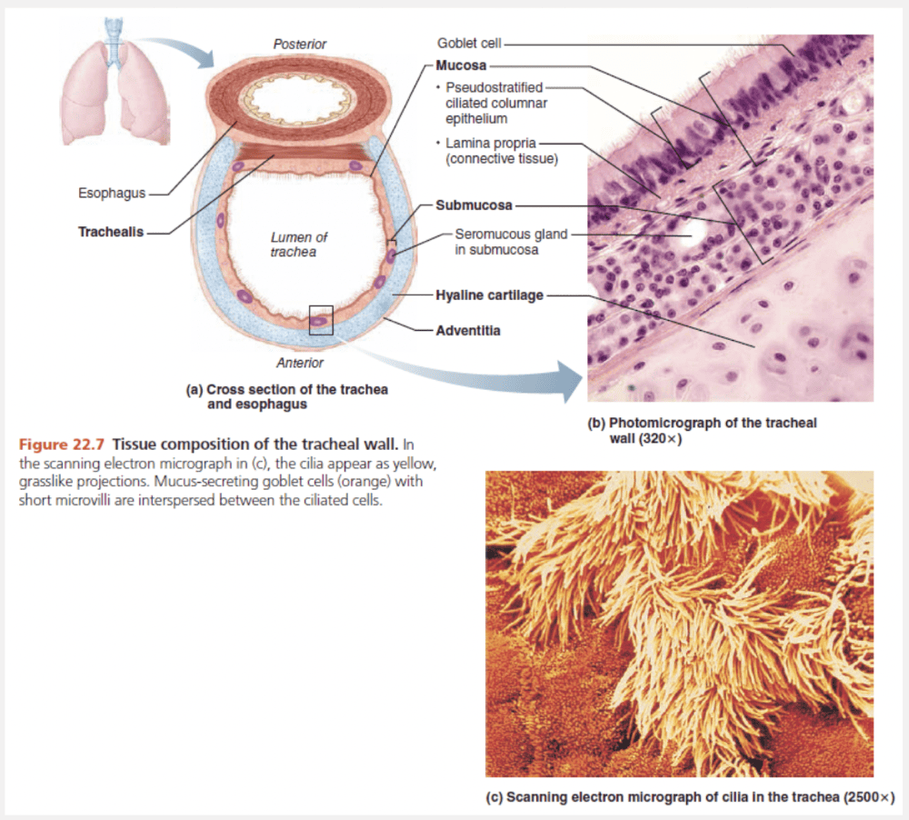
Pseudostratified columnar epithelium
Ex.
Pseudostratified columnar epithelium is the tissue that lines the trachea.
The tracheal wall consists of several layers that are common to many tubular body organs—the mucosa, submucosa, and adventitia—plus a layer of hyaline cartilage. The mucosa has the same goblet cell-containing pseudostratified epithelium that occurs throughout most of the respiratory tract. Its cilia continually propel debris-laden mucus toward the pharynx. This epithelium rests on a fairly thick lamina propria that has a rich supply of elastic fibers.
Which of the following lists the regions of the pharynx from superior to inferior?
- Nasopharynx, oropharynx, laryngopharynx
- Oropharynx, laryngopharynx, nasopharynx
- Nasopharynx, laryngopharynx, oropharynx
- Laryngopharynx, oropharynx, nasopharynx
Nasopharynx, oropharynx, laryngopharynx
Ex.
The regions of the pharynx from superior to inferior are nasopharynx, oropharynx, laryngopharynx.
The funnel-shaped pharynx (far’ingks) connects the nasal cavity and mouth superiorly to the larynx and esophagus inferiorly. Commonly called the throat, the pharynx vaguely resembles a short length of garden hose as it extends for about 13 cm (5 inches) from the base of the skull to the level of the sixth cervical vertebra. From superior to inferior, the pharynx is divided into three regions—the nasopharynx, oropharynx, and laryngopharynx. The muscular pharynx wall is composed of skeletal muscle throughout its length. However, the cellular composition of its mucosa varies from one pharyngeal region to another.
What is the function of the structure labeled “D?”
- Structure that allows heavier particles to be deflected into nasal mucosa
- Helps warm and moisten inspired air
- Lined with sebaceous and sweat glands that filter coarse particles
- Equalizes pressure between atmosphere and middle ear
- Region of nasal cavities that contains smell receptors
Lined with sebaceous and sweat glands that filter coarse particles
Ex.
“D” represents the nasal vestibule, a structure lined with sebaceous and sweat glands that filter coarse particles.
The part of the nasal cavity just superior to the nostrils, called the nasal vestibule, is lined with skin containing sebaceous and sweat glands and numerous hair follicles. The hairs, or vibrissae (vi-bris e; vibro = to quiver), filter coarse particles (dust, pollen) from inspired air. The rest of the nasal cavity is lined with two types of mucous membranes.
_________ is the inflammation of the nasal mucosa accompanied by excessive mucus production, nasal congestion, and postnasal drip.
- Bronchitis
- Laryngitis
- Rhinitis
- Sinusitis
Rhinitis
Ex.
Rhinitis is the inflammation of the nasal mucosa accompanied by excessive mucus production, nasal congestion, and postnasal drip.
Cold viruses, streptococcal bacteria, and various allergens can cause rhinitis (ri-ni’tis), inflammation of the nasal mucosa accompanied by excessive mucus production, nasal congestion, and postnasal drip. The nasal mucosa is continuous with the mucosa of the other respiratory passageways, explaining the typical nose to throat to chest progression of colds.
Because the mucosa extends tentacle-like into the nasolacrimal (tear) ducts and paranasal sinuses, nasal cavity infections often spread to those regions, causing sinusitis (inflamed sinuses). When mucus or infectious materials block the passageways connecting the sinuses to the nasal cavity, the air in the sinus cavities is absorbed. The result is a partial vacuum and a sinus headache localized over the inflamed areas.
Laryngitis is inflammation of the larynx.
Bronchitis is an inflammation of the lower respiratory passages.
Match the following: Sinus headache.
- May result in air that is not properly moistened, warmed, or filtered before reaching the lungs
- Has saved many people from becoming victims of “cafe coronaries”
- A partial vacuum in openings in the skull
- Often caused by viral infections, but may also be due to overusing the voice, very dry air, bacterial infections, tumors on the vocal folds, or inhalation of irritating chemicals
- Inhibits and ultimately destroys cilia
A partial vacuum in openings in the skull
Ex.
A sinus headache results from a partial vacuum in openings in the skull.
When mucus or infectious materials block the passageways connecting the sinuses to the nasal cavity, the air in the sinus cavities is absorbed. The result is a partial vacuum and a sinus headache localized over the inflamed areas.
Smoking inhibits and ultimately destroys cilia.
Without ciliary activity, coughing is the only way to prevent mucus from accumulating in the lungs. For this reason, smokers with respiratory congestion should avoid medications that inhibit the cough reflex.
The Heimlich maneuver has saved many people from becoming victims of “cafe coronaries.”
Laryngitis is often caused by viral infections, but may also be due to overusing the voice, very dry air, bacterial infections, tumors on the vocal folds, or inhalation of irritating chemicals.
Swelling of adenoids may result in air that is not properly moistened, warmed, or filtered before reaching the lungs.
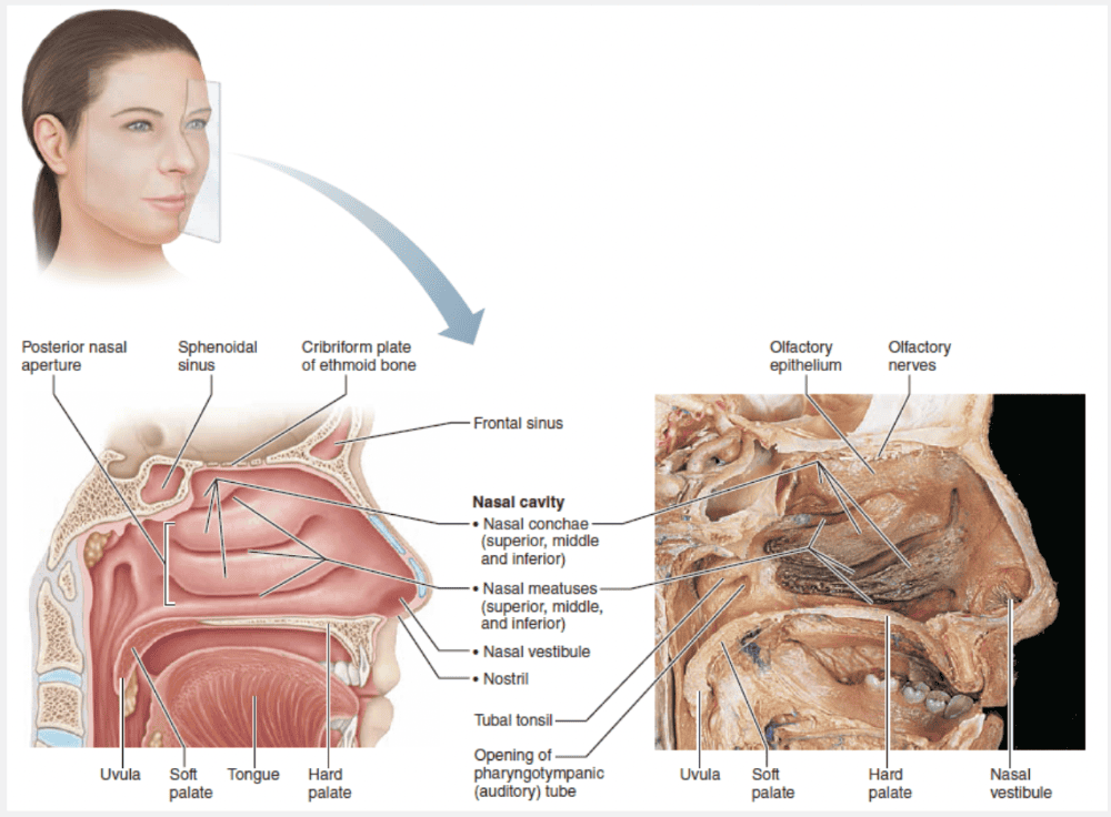
What is the function of the structure labeled “C?”
- Lined with sebaceous and sweat glands and filters coarse particles
- Structures that allows heavier particles to be deflected into nasal mucosa
- Region of nasal cavities that contains smell receptors
- Helps warm and moisten inspired air
- Equalizes pressure between atmosphere and middle ear
Structures that allows heavier particles to be deflected into nasal mucosa
Ex.
“C” represent the nasal conchae, structures that allow heavier particles to be deflected into nasal mucosa.
Protruding medially from each lateral wall of the nasal cavity are three scroll-like mucosa-covered projections, the superior, middle, and inferior nasal conchae (kong’ke). The groove inferior to each concha is a nasal meatus (me-a’tus).
The curved conchae greatly increase the mucosal surface area exposed to air and enhance air turbulence in the cavity. The gases in inhaled air swirl through the twists and turns, deflecting heavier, nongaseous particles onto the mucus-coated surfaces, where they become trapped. As a result, few particles larger than 6 μm make it past the nasal cavity.
The conchae and nasal mucosa not only function during inhalation to filter, heat, and moisten the air, but also act during exhalation to reclaim this heat and moisture. In other words, inspired air cools the conchae, then during exhalation these cooled conchae precipitate moisture and extract heat from the humid air flowing over them. This reclamation process minimizes the amount of moisture and heat lost from the body through breathing, helping us to survive in dry and cold climates.