Lymphangitis presents itself as __________.
- enlarged tender tonsils
- red lines under the skin that are sensitive to touch
- tender lymph nodes that are filled with pus
- severe localized edema
red lines under the skin that are sensitive to touch
Ex.
Lymphangitis presents itself as red lines under the skin that are sensitive to touch.
Like the larger blood vessels, the larger lymphatics receive their nutrient blood supply from a branching vasa vasorum. When lymphatic vessels are severely inflamed, the related vessels of the vasa vasorum become congested with blood. As a result, the pathway of the associated superficial lymphatics becomes visible through the skin as red lines that are tender to the touch. This unpleasant condition is called lymphangitis (lim”fan-ji’tis; angi = vessel).
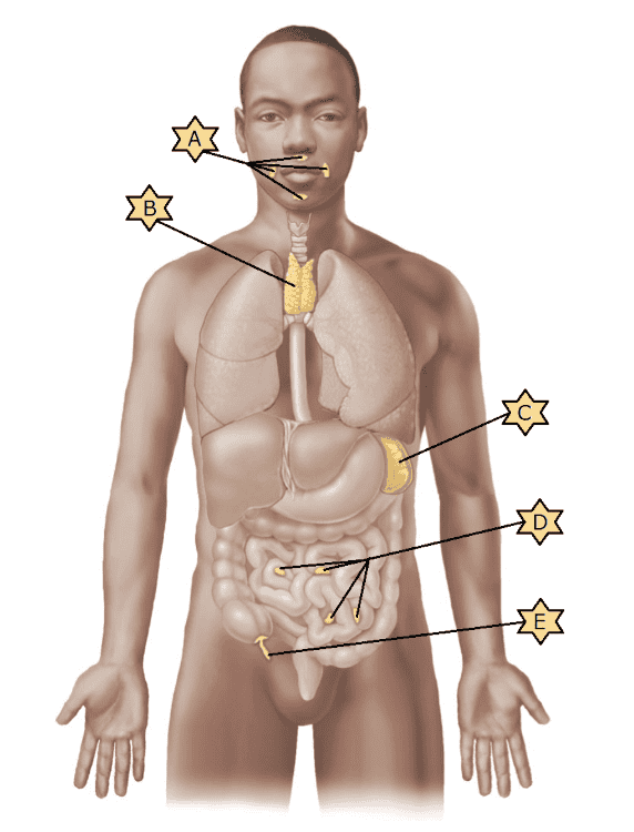
Identify the lymphoid organs indicated by “A.”
- Appendix
- Peyer's patches
- Spleen
- Tonsils
- Thymus
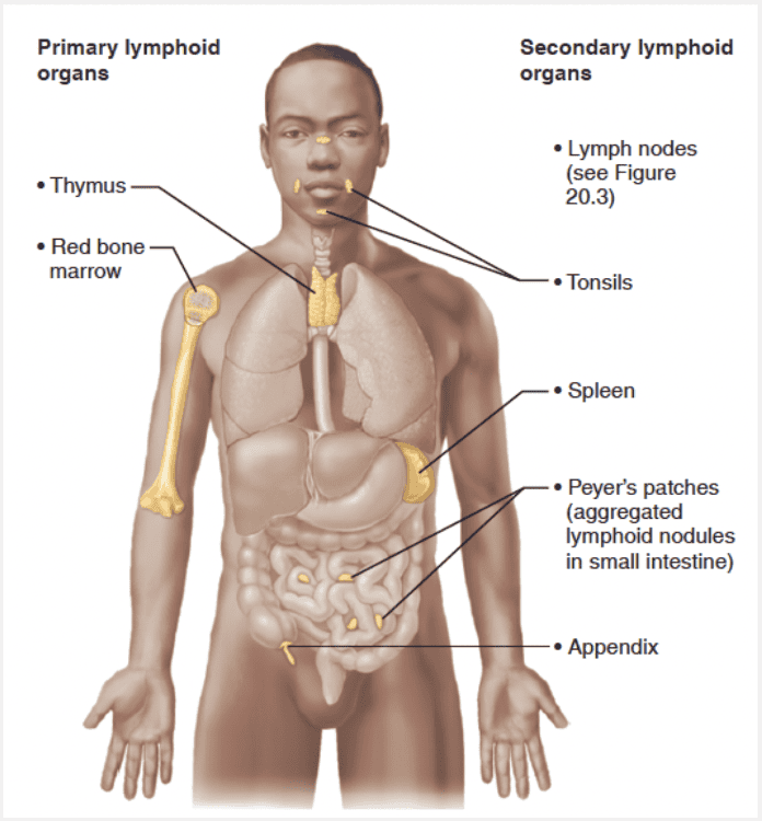
Tonsils
Match the following with the appropriate description: Collecting lymphatic vessels.
- Same three tunics as veins; the largest of lymph vessels
- Bean-shaped structure surrounded by a dense fibrous capsule with fibrous strands that divide it into compartments
- Same three tunics as veins; the second-smallest lymph vessels
- Begin as blind-ended tubes that weave between tissue cells and blood capillaries in loose connective tissues; the smallest vessels
- A type of loose connective tissue called reticular connective tissue; dominates all lymphoid organs except the thymus
Same three tunics as veins; the second-smallest lymph vessels
Ex.
Collecting lymphatic vessels contain the same three tunics as veins and are the second smallest of lymph vessels.
From the lymphatic capillaries, lymph flows through successively larger and thicker-walled channels—first collecting vessels, then trunks, and finally the largest of all, the ducts (figure below). The collecting lymphatic vessels have the same three tunics as veins, but the collecting vessels have thinner walls and more internal valves, and they anastomose more. In general, lymphatics in the skin travel along with superficial veins, while the deep lymphatic vessels of the trunk and digestive viscera travel with the deep arteries. The exact anatomical distribution of lymphatic vessels varies greatly between individuals, even more than it does for veins.
Match the lymphatic structure with the correct characteristic: Lymphatic capillary.
- Drains lymph from specific body areas
- Larger of the two lymphatic ducts receiving lymph from all but the right head and neck region
- Smallest lymphatic vessel that collects excess tissue fluid
- Drains right head and neck region
- Houses lymphatic cells and filters lymph
Smallest lymphatic vessel that collects excess tissue fluid
Ex.
Lymphatic capillaries are the s mallest lymphatic vessel that collects excess tissue fluid.
The transport of lymph begins in microscopic blind-ended lymphatic capillaries. These capillaries weave between the tissue cells and blood capillaries in the loose connective tissues of the body. Lymphatic capillaries are widespread, but they are absent from bones and teeth, bone marrow, and most of the central nervous system (where the excess tissue fluid drains into the cerebrospinal fluid).
As blood circulates through the body, nutrients, wastes, and gases are exchanged between the blood and the interstitial fluid. The hydrostatic and colloid osmotic pressures operating at capillary beds force fluid out of the blood at the arterial ends of the beds (“upstream”) and cause most of it to be reabsorbed at the venous ends (“downstream”). The fluid that remains behind in the tissue spaces, as much as 3L daily, becomes part of the interstitial fluid.
This leaked fluid, plus any plasma proteins that escape from the bloodstream, must somehow be returned to the blood to ensure that the cardiovascular system has sufficient blood volume. This problem of circulatory dynamics is resolved by the lymphatic vessels, or lymphatics, elaborate networks of drainage vessels that collect the excess protein-containing interstitial fluid and return it to the bloodstream.
Match the following with the appropriate description: Lymphoid tissue.
- Same three tunics as veins; the second-smallest lymph vessels
- Same three tunics as veins; the largest of lymph vessels
- Begin as blind-ended tubes that weave between tissue cells and blood capillaries in loose connective tissues; the smallest vessels
- Bean-shaped structure surrounded by a dense fibrous capsule with fibrous strands that divide it into compartments
- A type of loose connective tissue called reticular connective tissue; dominates all lymphoid organs except the thymus
A type of loose connective tissue called reticular connective tissue; dominates all lymphoid organs except the thymus
Ex.
Lymphoid tissue is a type of loose connective tissue called reticular connective tissue. It dominates all lymphoid organs except the thymus.
Lymphoid tissue is an important component of the immune system, mainly because it:
- Houses and provides a proliferation site for lymphocytes
- Furnishes an ideal surveillance vantage point for lymphocytes and macrophages
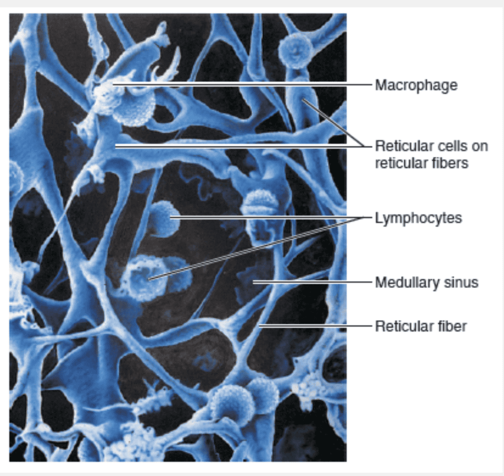
Lymphoid tissue, largely composed of loose connective tissue called reticular connective tissue, dominates all the lymphoid organs except the thymus. Macrophages live on the fibers of the reticular connective tissue network. Huge numbers of lymphocytes squeeze through the walls of postcapillary venules coursing through this network. The lymphocytes temporarily occupy the spaces in the network before leaving to patrol the body again (figure below). The cycling of lymphocytes between the circulatory vessels, lymphoid tissues, and loose connective tissues of the body ensures that lymphocytes reach infected or damaged sites quickly.
Which of the following features is not common to both lymphatic vessels and veins?
- Transport of chyle
- The presence of valves
- A wall made of three tunic layers
- Anatomical distributions of each vary between individuals
Transport of chyle
Ex.
It is not common to both lymphatic vessels and veins to transport chyle.
A special set of lymphatic capillaries called lacteals (lak’tealz) transports absorbed fat from the small intestine to the bloodstream. Lacteals are so called because of the milky white lymph that drains through them (lact = milk). This fatty lymph, called chyle (“juice”), drains from the fingerlike villi of the intestinal mucosa.
A major function of the lymphatic system is __________.
- gas distribution
- return of tissue fluid to the cardiovascular system
- circulation of blood
- distribution of nutrients
return of tissue fluid to the cardiovascular system
Ex.
A major function of the lymphatic system is to return tissue fluid to the cardiovascular system.
As blood circulates through the body, nutrients, wastes, and gases are exchanged between the blood and the interstitial fluid. The hydrostatic and colloid osmotic pressures operating at capillary beds force fluid out of the blood at the arterial ends of the beds (“upstream”) and cause most of it to be reabsorbed at the venous ends (“downstream”). The fluid that remains behind in the tissue spaces, as much as 3L daily, becomes part of the interstitial fluid.
This leaked fluid, plus any plasma proteins that escape from the bloodstream, must somehow be returned to the blood to ensure that the cardiovascular system has sufficient blood volume. This problem of circulatory dynamics is resolved by the lymphatic vessels, or lymphatics, elaborate networks of drainage vessels that collect the excess protein-containing interstitial fluid and return it to the bloodstream.
Which statement is true of the thoracic duct?
- It drains the lymph from the right head, neck, shoulder, arm, and upper right chest.
- It drains the lymph from the entire left side of the body and also the right abdomen and leg.
- It forms from the merging of collecting vessels from the left upper limb and neck.
- It forms from the merging of collecting vessels on the right side of the body.
- It drains lymph only from the arms.
It drains the lymph from the entire left side of the body and also the right abdomen and leg.
Ex.
The thoracic duct drains the lymph from the entire left side of the body and also the right abdomen and leg.
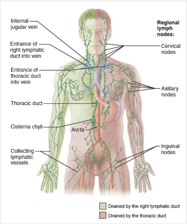
Lymph is eventually delivered to one of two large ducts in the thoracic region. The right lymphatic duct drains lymph from the right upper limb and the right side of the head and thorax (figure below). The much larger thoracic duct receives lymph from the rest of the body. It collects lymph from the two large lumbar trunks that drain the lower limbs and from the intestinal trunk that drains the digestive organs. In about half of individuals, the thoracic duct begins as an enlarged sac, the cisterna chyli (sis-ter’nah ki’li), located in the region between the last thoracic and second lumbar vertebrae. As the thoracic duct runs superiorly, it receives lymphatic drainage from the left side of the thorax, left upper limb, and the left side of the head. Each terminal duct empties its lymph into the venous circulation at the junction of the internal jugular vein and subclavian vein on its own side of the body.
Match the lymphatic structure with the correct characteristic: Right lymphatic duct.
- Larger of the two lymphatic ducts
- Directly drains the left and right lumbar regions
- Houses lymphatic cells and filters lymph
- Drains right side of the head, upper limb, and neck region
- Empties into the left subclavian vein
Drains right side of the head, upper limb, and neck region
Ex.
The right lymphatic duct drains the right side of the head, upper limb, and neck region.
Lymph is eventually delivered to one of two large ducts in the thoracic region. The right lymphatic duct drains lymph from the right upper limb and the right side of the head and thorax (figure below). The much larger thoracic duct receives lymph from the rest of the body. It collects lymph from the two large lumbar trunks that drain the lower limbs and from the intestinal trunk that drains the digestive organs. In about half of individuals, the thoracic duct begins as an enlarged sac, the cisterna chyli (sis-ter’nah ki’li), located in the region between the last thoracic and second lumbar vertebrae. As the thoracic duct runs superiorly, it receives lymphatic drainage from the left side of the thorax, left upper limb, and the left side of the head. Each terminal duct empties its lymph into the venous circulation at the junction of the internal jugular vein and subclavian vein on its own side of the body.
Match the following structure with its function: Lymphatic capillaries.
- Provide a proliferation site for lymphocytes and furnishes surveillance vantage point for lymphocytes and macrophages
- Smallest lymphatic vessels that collect tissue fluid
- Collect lymph fluid draining from lymphatic vessels
- Largest vessels; carry lymph fluid to subclavian veins
- Distinct bean-shaped structures that "filter" lymph fluid as it is moved toward the circulatory system
Smallest lymphatic vessels that collect tissue fluid
Ex.
Lymphatic capillaries are the s mallest lymphatic vessels that collect tissue fluid.
As blood circulates through the body, nutrients, wastes, and gases are exchanged between the blood and the interstitial fluid. The hydrostatic and colloid osmotic pressures operating at capillary beds force fluid out of the blood at the arterial ends of the beds (“upstream”) and cause most of it to be reabsorbed at the venous ends (“downstream”). The fluid that remains behind in the tissue spaces, as much as 3L daily, becomes part of the interstitial fluid.
This leaked fluid, plus any plasma proteins that escape from the bloodstream, must somehow be returned to the blood to ensure that the cardiovascular system has sufficient blood volume. This problem of circulatory dynamics is resolved by the lymphatic vessels, or lymphatics, elaborate networks of drainage vessels that collect the excess protein-containing interstitial fluid and return it to the bloodstream.
From the lymphatic capillaries, lymph flows through successively larger and thicker-walled channels:
Lymphatic capillaries → Collecting lymphatic vessels → Lymphatic trunks → Lymphatic ducts
What part of the lymphatic system is most closely associated with capillary beds?
- Lymphatic capillaries
- Lymphatic trunks
- Lymph nodes
- Lymph ducts
Lymphatic capillaries
Ex.
Lymphatic capillaries are closely associated with capillary beds.
The transport of lymph begins in microscopic blind-ended lymphatic capillaries. From the lymphatic capillaries, lymph flows through successively larger and thicker-walled channels—first collecting vessels, then trunks, and finally the largest of all, the ducts.
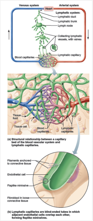
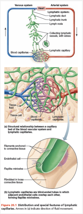
These capillaries weave between the tissue cells and blood capillaries in the loose connective tissues of the body. Lymphatic capillaries are widespread, but they are absent from bones and teeth, bone marrow, and most of the central nervous system (where the excess tissue fluid drains into the cerebrospinal fluid). Although similar to blood capillaries, lymphatic capillaries are so remarkably permeable that they were once thought to be open at one end like a straw.
Select the lymphoid organ that cleanses the lymph.
- MALT
- Lymph nodes
- Tonsils
- Peyer's patches
- Spleen
Lymph nodes
Ex.
The lymph nodes cleanse the lymph.
Although all lymphoid organs help protect the body, only the lymph nodes cleanse the lymph. The other secondary lymphoid organs typically have efferent lymphatics draining them, but lack afferent lymphatics.
Identify the lymphatic system structures indicated by “C.”
- Axillary nodes
- Inguinal nodes
- Cervical nodes
- Thoracic duct
- Cisterna chyli
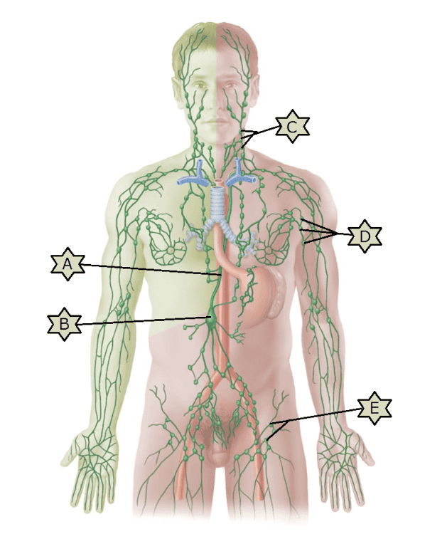
Cervical nodes
Identify the lymphatic system structure indicated by “A.”
- Axillary nodes
- Inguinal nodes
- Cervical nodes
- Cisterna chyli
- Thoracic duct
Thoracic duct
How are lymphatic capillaries different from blood capillaries?
- Lymphatic capillaries have fewer tunics than blood capillaries.
- Lymphatic capillaries have endothelial flap valves, but blood capillaries do not.
- Lymphatic capillaries have thicker walls than do blood capillaries.
- Lymphatic capillaries do not form branches; blood capillaries do form branches.
Lymphatic capillaries have endothelial flap valves, but blood capillaries do not.
Ex.
Lymphatic capillaries are different from veins in that lymphatic capillaries have endothelial flap valves, but blood capillaries do not.
Although similar to blood capillaries, lymphatic capillaries are so remarkably permeable that they were once thought to be open at one end like a straw. We now know that they owe their permeability to two unique structural modifications:
- The endothelial cells forming the walls of lymphatic capillaries are not tightly joined. Instead, the edges of adjacent cells overlap each other loosely, forming easily opened, flaplike minivalves (figure below).
- Collagen filaments anchor the endothelial cells to surrounding structures so that any increase in interstitial fluid volume opens the minivalves, rather than causing the lymphatic capillaries to collapse.
Identify the lymphoid organ indicated by “E.”
- Peyer's patches
- Appendix
- Thymus
- Tonsils
- Spleen
Appendix
Which sequence best describes the flow of lymph through the lymphatic system?
- Capillaries, trunks, vessels, ducts
- Ducts, trunks, capillaries, vessels
- Capillaries, vessels, trunks, ducts
- Trunks, capillaries, vessels, ducts
- Ducts, vessels, trunks, capillaries
Capillaries, vessels, trunks, ducts
Ex.
The sequence that best describes the flow of lymph through the lymphatic system is capillaries, vessels, trunks, ducts.
The transport of lymph begins in microscopic blind-ended lymphatic capillaries. From the lymphatic capillaries, lymph flows through successively larger and thicker-walled channels—first collecting vessels, then trunks, and finally the largest of all, the ducts.
Identify the lymphoid organ indicated by “B.”
- Tonsils
- Appendix
- Thymus
- Spleen
- Peyer's patches
Thymus
Match the following structure with its function: Lymphatic ducts.
- Provide a proliferation site for lymphocytes and furnishes surveillance vantage point for lymphocytes and macrophages
- Collect lymph fluid draining from lymphatic capillaries
- Weave between the tissue cells and blood capillaries
- Distinct bean-shaped structures that "filter" lymph fluid as it is moved toward the circulatory system
- Largest vessels; carry lymph fluid to subclavian veins
Largest vessels; carry lymph fluid to subclavian veins
Ex.
Lymphatic ducts are the largest vessles and carry lymph fluid to the subclavian veins.
Lymph is eventually delivered to one of two large ducts in the thoracic region. The right lymphatic duct drains lymph from the right upper limb and the right side of the head and thorax (figure below). The much larger thoracic duct receives lymph from the rest of the body. It collects lymph from the two large lumbar trunks that drain the lower limbs and from the intestinal trunk that drains the digestive organs. In about half of individuals, the thoracic duct begins as an enlarged sac, the cisterna chyli (sis-ter’nah ki’li), located in the region between the last thoracic and second lumbar vertebrae. As the thoracic duct runs superiorly, it receives lymphatic drainage from the left side of the thorax, left upper limb, and the left side of the head. Each terminal duct empties its lymph into the venous circulation at the junction of the internal jugular vein and subclavian vein on its own side of the body.
Which of the following vessels transport fluid back into the blood that leaks from the vascular system?
- Blood capillaries
- Lymphatics
- Veins
- Sinusoids
Lymphatics
Ex.
Lymphatics are vessels that transport fluid that leaks from the vascular system back into the blood.
As blood circulates through the body, nutrients, wastes, and gases are exchanged between the blood and the interstitial fluid. The hydrostatic and colloid osmotic pressures operating at capillary beds force fluid out of the blood at the arterial ends of the beds (“upstream”) and cause most of it to be reabsorbed at the venous ends (“downstream”). The fluid that remains behind in the tissue spaces, as much as 3L daily, becomes part of the interstitial fluid.
This leaked fluid, plus any plasma proteins that escape from the bloodstream, must somehow be returned to the blood to ensure that the cardiovascular system has sufficient blood volume. This problem of circulatory dynamics is resolved by the lymphatic vessels, or lymphatics, elaborate networks of drainage vessels that collect the excess protein-containing interstitial fluid and return it to the bloodstream.
Identify the lymphatic system structures indicated by “E.”
- Thoracic duct
- Cisterna chyli
- Axillary nodes
- Inguinal nodes
- Cervical nodes
Inguinal nodes
Match the lymphatic structure with the correct characteristic: Thoracic duct.
- Smaller of the two lymphatic ducts
- Empties into the right subclavian vein
- Houses lymphatic cells and filters lymph
- Directly drains the left and right lumbar regions
- Receives lymph from all but the right upper limb and right side of the head and thorax
Receives lymph from all but the right upper limb and right side of the head and thorax
Ex.
The thoracic ducts receives lymph from all but the right upper limb and right side of the head and thorax.
Lymph is eventually delivered to one of two large ducts in the thoracic region. The right lymphatic duct drains lymph from the right upper limb and the right side of the head and thorax (figure below). The much larger thoracic duct receives lymph from the rest of the body. It collects lymph from the two large lumbar trunks that drain the lower limbs and from the intestinal trunk that drains the digestive organs. In about half of individuals, the thoracic duct begins as an enlarged sac, the cisterna chyli (sis-ter’nah ki’li), located in the region between the last thoracic and second lumbar vertebrae. As the thoracic duct runs superiorly, it receives lymphatic drainage from the left side of the thorax, left upper limb, and the left side of the head. Each terminal duct empties its lymph into the venous circulation at the junction of the internal jugular vein and subclavian vein on its own side of the body.
Which statement below describes the lymphatic system's role in relation to the cardiovascular system?
- It maintains blood volume and, hence, pressure.
- It serves as a pathway for distribution of neutrophils, eosinophils, and basophils.
- It is the primary source for regulation of blood pressure.
- It is the major source for distribution of all hormones.
- It helps regulate cardiac activity.
It maintains blood volume and, hence, pressure.
Ex.
The lymphatic system's role in relation to the cardiovascular system is to maintain blood volume and, hence, pressure.
As blood circulates through the body, nutrients, wastes, and gases are exchanged between the blood and the interstitial fluid. The hydrostatic and colloid osmotic pressures operating at capillary beds force fluid out of the blood at the arterial ends of the beds (“upstream”) and cause most of it to be reabsorbed at the venous ends (“downstream”). The fluid that remains behind in the tissue spaces, as much as 3 L daily, becomes part of the interstitial fluid.
This leaked fluid, plus any plasma proteins that escape from the bloodstream, must somehow be returned to the blood to ensure that the cardiovascular system has sufficient blood volume. This problem of circulatory dynamics is resolved by the lymphatic vessels, or lymphatics, elaborate networks of drainage vessels that collect the excess protein-containing interstitial fluid and return it to the bloodstream.
Which of the following best describes the arrangement of lymphatic vessels?
- A system of large vessels designed to fill quickly with lymph as the heart pushes blood through the coronary sinus
- A system that carries lymph through lymphatic arteries, lymphatic capillaries, and lymphatic veins
- A system that pumps lymph through lymphatic ventricles to the lymphatic capillaries and through lymph veins back to the atria
- A one-way system of vessels beginning with blind-ended lymphatic capillaries
- A system that collects fluid from arteries and veins and takes it into lymphatic arteries to be pumped back to the blood circulation
A one-way system of vessels beginning with blind-ended lymphatic capillaries
Ex.
The lymphatic vessels form a one-way system in which lymph flows only toward the heart.
The transport of lymph begins in microscopic blind-ended lymphatic capillaries. These capillaries weave between the tissue cells and blood capillaries in the loose connective tissues of the body. Lymphatic capillaries are widespread, but they are absent from bones and teeth, bone marrow, and most of the central nervous system (where the excess tissue fluid drains into the cerebrospinal fluid).
Which of the following is not a function of lymphatic vessels?
- Transportation of absorbed fat from the intestines to the blood
- Delivery of nutrients to tissues
- The return of tissue fluid to the bloodstream
- The return of leaked proteins to the blood
Delivery of nutrients to tissues
Ex.
Lymphatic vessels do not deliver nutrients to tissues.
As blood circulates through the body, nutrients, wastes, and gases are exchanged between the blood and the interstitial fluid. The hydrostatic and colloid osmotic pressures operating at capillary beds force fluid out of the blood at the arterial ends of the beds (“upstream”) and cause most of it to be reabsorbed at the venous ends (“downstream”). The fluid that remains behind in the tissue spaces, as much as 3L daily, becomes part of the interstitial fluid.
This leaked fluid, plus any plasma proteins that escape from the bloodstream, must somehow be returned to the blood to ensure that the cardiovascular system has sufficient blood volume. This problem of circulatory dynamics is resolved by the lymphatic vessels, or lymphatics, elaborate networks of drainage vessels that collect the excess protein-containing interstitial fluid and return it to the bloodstream.
A special set of lymphatic capillaries called lacteals (lak’tealz) transports absorbed fat from the small intestine to the bloodstream. Lacteals are so called because of the milky white lymph that drains through them (lact = milk). This fatty lymph, called chyle (“juice”), drains from the fingerlike villi of the intestinal mucosa.
Tumors that block the lymphatics or lymphatics are removed during cancer surgery may result in what condition?
- Lymphangitis
- Lymphoma
- Splenomegaly
- Lymphedema
Lymphedema
Ex.
Tumors that block the lymphatics or lymphatics are removed during cancer surgery may result in what condition termed lymphedema.
Anything that prevents the normal return of lymph to the blood—such as when tumors block the lymphatics or lymphatics are removed during cancer surgery—results in shortterm but severe localized edema (lymphedema). In some cases, the lymphedema improves if some lymphatic pathways remain and can enlarge.
Identify the lymphatic system structures indicated by “D.”
- Inguinal nodes
- Axillary nodes
- Cisterna chyli
- Thoracic duct
- Cervical nodes
Axillary nodes
Match the following structure with its function: Lymphatic collecting vessels.
- Collect lymph fluid draining from lymphatic capillaries
- The smallest lymphatic vessels that collect fluid that leaks from blood capillaries into tissue fluid
- Distinct bean-shaped structures that "filter" lymph fluid as it is moved toward the circulatory system
- Largest vessels; carry lymph fluid to subclavian veins
- Provide a proliferation site for lymphocytes and furnishes surveillance vantage point for lymphocytes and macrophages
Collect lymph fluid draining from lymphatic capillaries
Ex.
Lymphatic collecting vessels collect lymph fluid draining from lymphatic capillaries.
From the lymphatic capillaries, lymph flows through successively larger and thicker-walled channels—first collecting vessels, then trunks, and finally the largest of all, the ducts. The collecting lymphatic vessels have the same three tunics as veins, but the collecting vessels have thinner walls and more internal valves, and they anastomose more. In general, lymphatics in the skin travel along with superficial veins, while the deep lymphatic vessels of the trunk and digestive viscera travel with the deep arteries. The exact anatomical distribution of lymphatic vessels varies greatly between individuals, even more than it does for veins.
Identify the lymphoid organs indicated by “D.”
- Peyer's patches
- Thymus
- Spleen
- Tonsils
- Appendix
Peyer's patches
Identify the lymphatic system structure indicated by “B.”
- Inguinal nodes
- Cervical nodes
- Axillary nodes
- Cisterna chyli
- Thoracic duct
Cisterna chyli