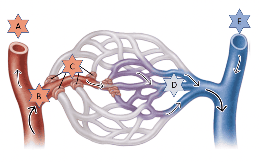
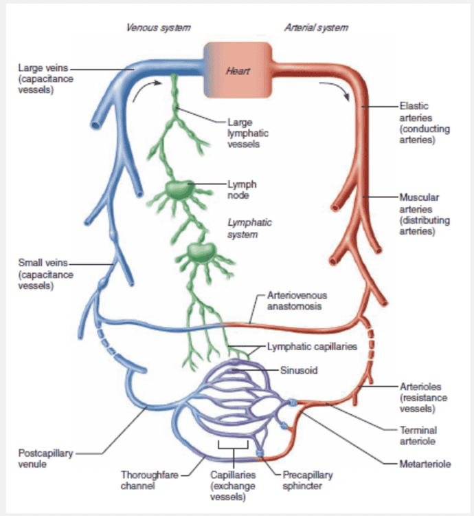
Identify the part of the special (mesenteric) capillary bed indicated by “D.”
- Terminal arteriole
- Postcapillary venule
- Metarteriole
- Thoroughfare channel
- Precapillary sphincter
Thoroughfare channel
Identify the part of a special (mesenteric) capillary bed indicated by “B.”
- Precapillary sphincters
- Postcapillary venule
- Thoroughfare channel
- Terminal arteriole
- Metarteriole
Terminal arteriole
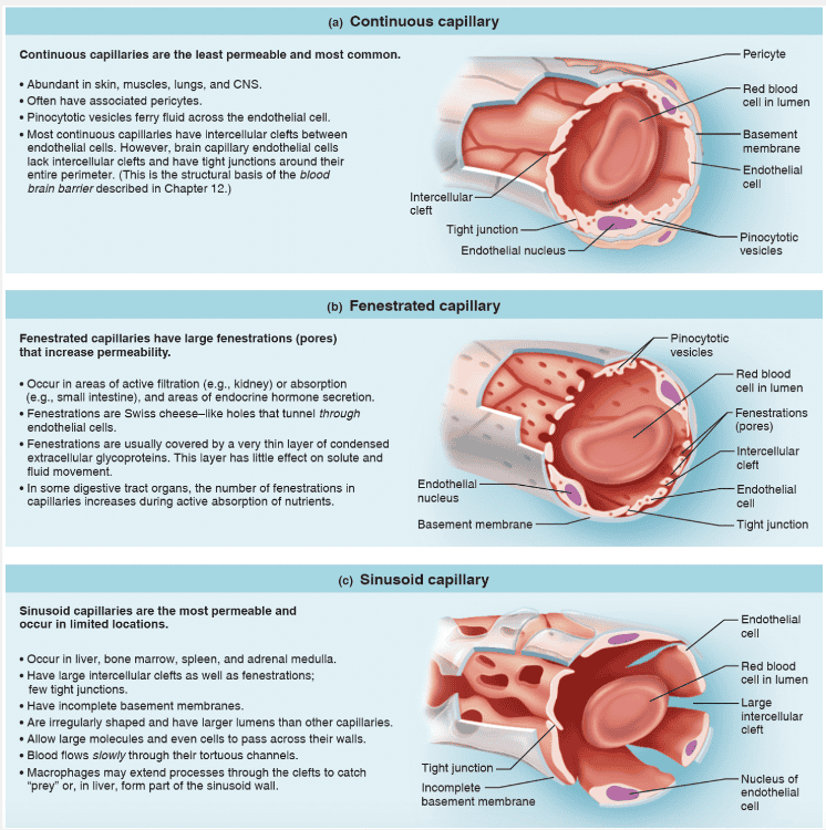
The vessels that exhibit the lowest level of permeability are the __________.
- fenestrated capillaries
- sinusoid capillaries
- continuous capillaries
- capillaries found in the glomerulus of the kidney
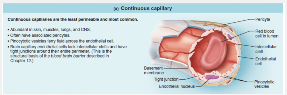
continuous capillaries
Ex.
The vessels that exhibit the lowest level of permeability are the continuous capillaries .
Continuous capillaries are the least permeable and most common.
Continuous capillaries are:
- Abundant in skin, muscles, lungs, and CNS.
- Often have associated pericytes.
- Pinocytotic vesicles ferry fluid across the endothelial cell.
- Brain capillary endothelial cells lack intercellular clefts and have tight junctions around their entire perimeter.
Match the following type of vessel with its structure: Venules.
- Smallest blood vessels with thin walls that allow exchange between blood and tissue cells
- Smallest of the vessels that lead into capillary beds
- Smallest vessels leading away from capillaries
- Contain valves to assist blood flow back toward heart
- Smaller vessels that distribute blood to specific body organs
- Thick-walled, large vessels near the heart that conduct blood continuously away from the heart
Smallest vessels leading away from capillaries
Ex.
Venules are the smallest vessels leading away from capillaries .
Capillaries unite to form venules, which range from 8 to 100 μm in diameter. The smallest venules, the postcapillary venules , consist entirely of endothelium around which pericytes congregate. Postcapillary venules are extremely porous (more like capillaries than veins in this way), and fluid and white blood cells move easily from the bloodstream through their walls. Indeed, a well recognized sign of inflammation is adhesion of white blood cells to the postcapillary venule endothelium, followed by their migration through the wall into the inflamed tissue. Larger venules have one or two layers of smooth muscle cells (a scanty tunica media) and a thin tunica externa as well.
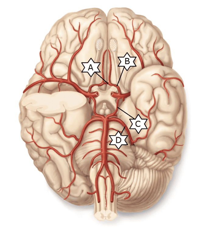
Identify the vessel of the cerebral arterial circle (of Willis) indicated by “A.”
- Posterior communicating artery
- Anterior communicating artery
- Anterior cerebral artery
- Posterior cerebral artery
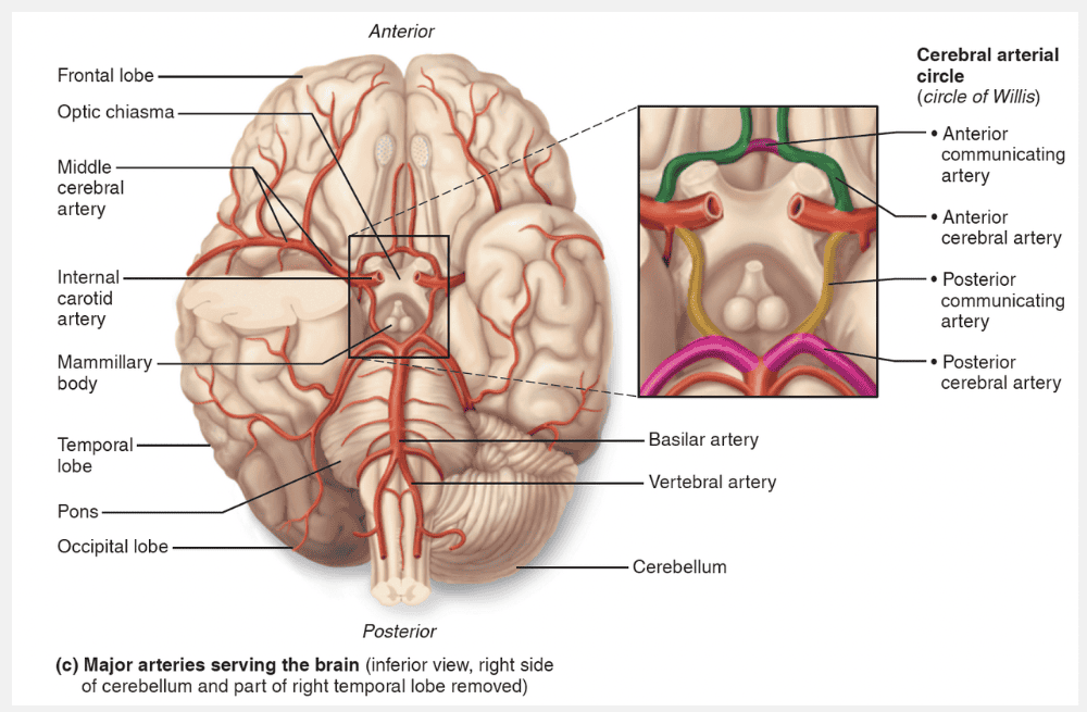
Anterior communicating artery
Match the following type of vessel with its structure: Veins.
- Smallest blood vessels with thin walls that allow exchange between blood and tissue cells
- Smallest of the vessels that lead into capillary beds
- Contain valves to assist blood flow back toward heart
- Smallest vessels leading away from capillaries
- Thick-walled, large vessels near the heart that conduct blood continuously away from the heart
- Smaller vessels that distribute blood to specific body organs
Contain valves to assist blood flow back toward heart
Ex.
Veins contain valves to assist blood flow back toward heart.
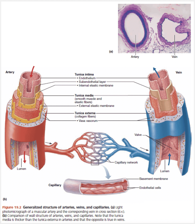
Venous valves prevent blood from flowing backward in veins just as valves do in the heart, and represent another adaptation to compensate for low venous pressure. They are formed from folds of the tunica intima and resemble the semilunar valves of the heart (see Figure 19.2). Venous valves are most abundant in the veins of the limbs, where gravity opposes the upward flow of blood. They are usually absent in veins of the thoracic and abdominal body cavities.
The effectiveness of venous valves is demonstrated by this simple experiment: Hang one hand by your side until the blood vessels on its dorsal aspect distend with blood. Next place two fingertips against one of the distended veins, and pressing firmly, move the superior finger proximally along the vein and then release that finger. The vein will remain collapsed (flat) despite the pull of gravity. Finally, remove your distal fingertip and watch the vein refill with blood.
Which of the following conditions would not increase the chances of developing varicose veins?
- Running in place
- A potbelly in an obese person
- Pregnancy
- Standing still in one position for long periods of time
Running in place
Ex.
Running in place would not increase the chances of developing varicose veins.
Varicose veins are veins that are tortuous and dilated because of incompetent (leaky) valves. More than 15% of adults suffer from varicose veins, usually in the lower limbs.
Several factors contribute, including heredity and conditions that hinder venous return, such as prolonged standing in one position, obesity, or pregnancy. Both the “potbelly” of an overweight person and the enlarged uterus of a pregnant woman exert downward pressure on vessels of the groin, restricting return of blood to the heart. Consequently, blood pools in the lower limbs, and with time, the valves weaken and the venous walls stretch. Superficial veins, which receive little support from surrounding tissues, are especially susceptible.
Elevated venous pressure can also cause varicose veins. For example, straining to deliver a baby or have a bowel movement raises intra-abdominal pressure, preventing blood from draining from anal veins. The resulting varicosities in the anal veins are called hemorrhoids (hem’ŏ-roidz).
Up to 65% of the body's blood supply is found in __________.
- capillaries
- arterioles
- arteries
- veins
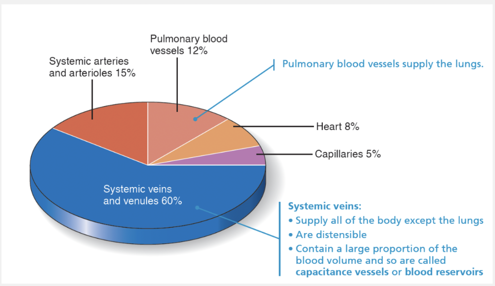
veins
Ex.
Up to 65% of the body's blood supply is found in veins.
With their large lumens and thin walls, veins can accommodate a fairly large blood volume. Veins are called capacitance vessels and blood reservoirs because they can hold up to 65% of the body’s blood supply at any time. Even so, these distensible vessels are usually not filled to capacity.
Which of the following blood vessels do not contain intercellular clefts?
- Arterioles
- Continuous capillaries
- Sinusoidal capillaries
- Fenestrated capillaries
Arterioles
Ex.
Arterioles do not contain intercellular clefts.
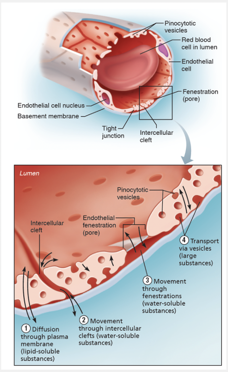
Structurally, there are three types of capillaries—continuous, fenestrated, and sinusoid. As you study their properties in the figure below, notice that all three types have tight junctions that join their endothelial cells together. However, these junctions are usually incomplete and leave gaps of unjoined membrane called intercellular clefts, which allow limited passage of fluids and small solutes. Leakier capillaries have specialized passageways that increase fluid movement.
Identify the part of a special (mesenteric) capillary bed indicated by “E.”
- Terminal arteriole
- Metarteriole
- Thoroughfare channel
- Venule
- Precapillary sphincters
Venule
The minute-to-minute blood flow through the capillary beds is determined by the __________.
- number of elastic fibers in conducting arteries
- diameter of arterioles
- elastic membranes on both sides of the tunica media in muscular arteries
- constriction of elastic arteries
diameter of arterioles
Ex.
Minute-to-minute blood flow into the capillary beds is determined by arteriolar diameter , which varies in response to changing neural, hormonal, and local chemical influences.
Changing diameter changes resistance to blood flow, and so arterioles are called resistance vessels . When arterioles constrict, the tissues served are largely bypassed. When arterioles dilate, blood flow into the local capillaries increases dramatically.
Match the following type of vessel with its structure: Capillaries.
- Smaller vessels that distribute blood to specific body organs
- Smallest of the vessels that lead into capillary beds
- Contain valves to assist blood flow back toward heart
- Smallest blood vessels with thin walls that allow exchange between blood and tissue cells
- Smallest vessels leading away from capillaries
- Thick-walled, large vessels near the heart that conduct blood continuously away from the heart
Smallest blood vessels with thin walls that allow exchange between blood and tissue cells
Ex.
Capillaries are the smallest blood vessels with thin walls that allow exchange between blood and tissue cells.
If we compare arteries and arterioles to expressways and roads, capillaries are the back alleys and driveways that provide direct access to nearly every cell in the body. Given their location and thin walls, capillaries are ideally suited for their role—exchange of materials (gases, nutrients, hormones, and so on) between the blood and the interstitial fluid.
Identify the vessel of the cerebral arterial circle (of Willis) indicated by “C.”
- Anterior cerebral artery
- Anterior communicating artery
- Posterior communicating artery
- Posterior cerebral artery
Posterior communicating artery
Identify the part of a special (mesenteric) capillary bed indicated by “C.”
- Terminal arteriole
- Thoroughfare channel
- Metarteriole
- Precapillary sphincters
- Postcapillary venule
Precapillary sphincters
The presence of __________ stabilizes the wall of capillaries.
- gap junctions
- elastic fibers
- pericytes
- valves
pericytes
Ex.
The presence of pericytes stabilizes the wall of capillaries.
The microscopic capillaries are the smallest blood vessels. Their exceedingly thin walls consist of just a thin tunica intima. In some cases, one endothelial cell forms the entire circumference of the capillary wall. At strategic locations along the outer surface of some capillaries are spider-shaped pericytes, contractile stem cells that can generate new vessels or scar tissue, stabilize the capillary wall, and help control capillary permeability.
Identify the vessel of the cerebral arterial circle (of Willis) indicated by “B.”
Anterior cerebral artery
Exchange of gases and nutrients occurs by diffusion between the __________.
- arterioles and tissue cells
- capillaries and tissue cells
- artery walls and tissue cells
- arteries and veins
- arterioles and venules
capillaries and tissue cells
Ex.
Exchange of gases and nutrients occurs by diffusion between the capillaries and tissue cells.
Oxygen, carbon dioxide, most nutrients, and metabolic wastes pass between the blood and interstitial fluid by diffusion. Recall that in diffusion, net movement always occurs along a concentration gradient—each substance moving from an area of its higher concentration to an area of its lower concentration.
Hence, oxygen and nutrients pass from the blood, where their concentration is fairly high, through the interstitial fluid to the tissue cells. Carbon dioxide and metabolic wastes leave the cells, where their content is higher, and diffuse into the capillary blood.
The only vessels that provide direct access to nearly every cell in the body are the __________.
- arterioles
- capillaries
- arteries
- venules
- veins
capillaries
Ex.
The only vessels that provide direct access to nearly every cell in the body are the capillaries.
Of all the blood vessels, only the capillaries have intimate contact with tissue cells and directly serve cellular needs. Exchanges between the blood and tissue cells occur primarily through the gossamer-thin capillary walls.
Which of the following regulates blood flow through most capillary beds?
- Terminal arterioles
- Valves
- Tunica externa
- Lymph nodes
- Tunica intima
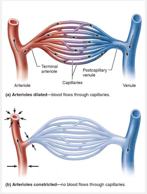
Terminal arterioles
Ex.
Terminal arterioles regulate blood flow through most capillary beds.
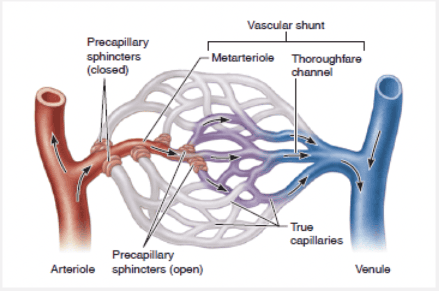
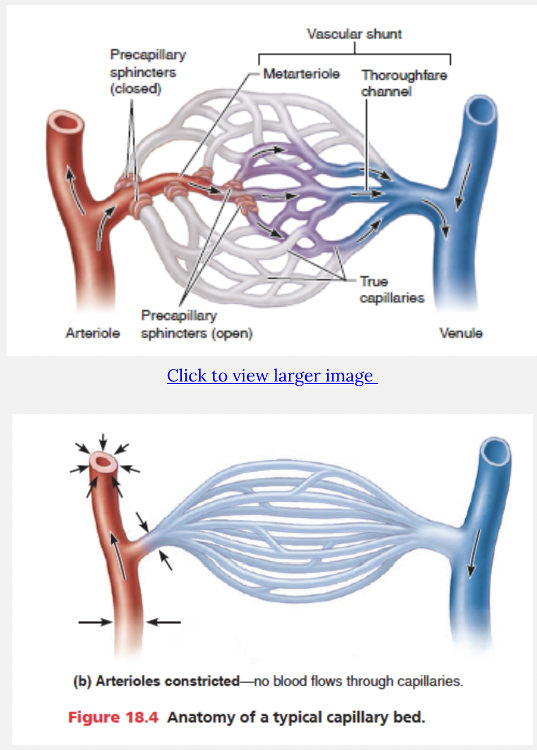
Capillaries do not function independently. Instead they form interweaving networks called capillary beds. The flow of blood from an arteriole to a venule is called the microcirculation. In most body regions, a terminal arteriole branches into 10 to 20 capillaries (exchange vessels) that form the capillary bed. These then drain into a postcapillary venule. Blood flow through the capillary bed is controlled by the diameter of the terminal arteriole as well as by all of the arterioles upstream from it.
Identify the vessel of the cerebral arterial circle (of Willis) indicated by “D.”
- Posterior cerebral artery
- Anterior cerebral artery
- Posterior communicating artery
- Anterior communicating artery
Posterior cerebral artery
Fluids would be likely to leave or filter out of the capillary if __________.
- net hydrostatic pressure (HP) is greater than net osmotic pressure (OP)
- net filtration pressure (NFP) is negative
- net hydrostatic pressure (HP) is less than net osmotic pressure (OP)
- osmotic pressure (OP) in the capillary is high
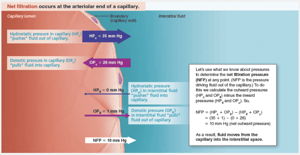
net hydrostatic pressure (HP) is greater than net osmotic pressure (OP)
Ex.
Fluids would be likely to leave or filter out of the capillary if net hydrostatic pressure (HP) is greater than net osmotic pressure (OP).
Identify the part of a capillary bed indicated by “A.”
- Postcapillary venule
- Precapillary sphincters
- Arteriole
- Thoroughfare channel
- Metarteriole
Arteriole
Ex.
The part of a capillary bed indicated by “A” is the terminal arteriole .