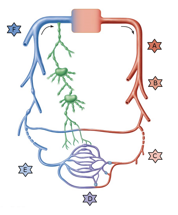
Identify the type of vessel indicated by “F.”
- Arterioles
- Postcapillary venule
- Muscular arteries
- Capillaries
- Elastic arteries
- Large veins
Large veins
Match the following type of vessel with its structure: Muscular arteries.
- Contain valves to assist blood flow back toward heart
- Thick-walled, large vessels near the heart that conduct blood continuously away from the heart
- Smallest of the vessels that lead into capillary beds
- Smallest vessels leading away from capillaries
- Smallest blood vessels with thin walls that allow exchange between blood and tissue cells
- Smaller vessels that distribute blood to specific body organs
Smaller vessels that distribute blood to specific body organs
Ex.
Muscular arteries are smaller vessels that distribute blood to specific body organs.
Distally the elastic arteries give way to the muscular arteries, which deliver blood to specific body organs (and so are sometimes called distributing arteries). Muscular arteries account for most of the named arteries studied in the anatomy laboratory. Their internal diameter ranges from that of a little finger to that of a pencil lead.
Proportionately, muscular arteries have the thickest tunica media of all vessels. Their tunica media contains relatively more smooth muscle and less elastic tissue than do elastic arteries. For this reason, they are more active in vasoconstriction and less capable of stretching. In muscular arteries, however, there is an elastic membrane on each face of the tunica media.
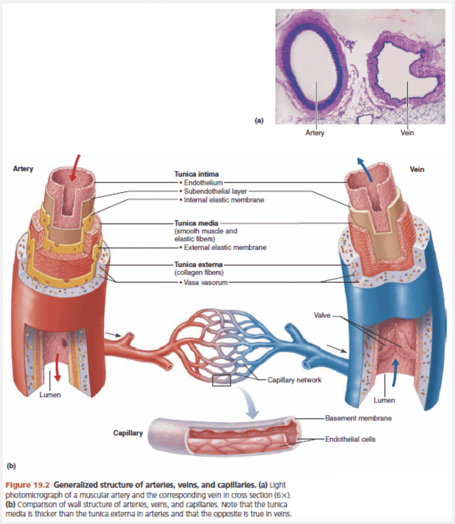
Match the following blood vessel layer with its description: Tunica intima.
- Blood-containing space in the center of the vessel
- Mostly circularly arranged smooth muscle cells and sheets of elastin
- Composed largely of loosely woven collagen fibers that protect and reinforce the vessel
- Contains the endothelium (made of simple squamous epithelium)
Contains the endothelium (made of simple squamous epithelium)
Ex.
The tunica intima contains the endothelium (made of simple squamous epithelium) .
The innermost tunic is the tunica intima (in′tĭ-mah). The name is easy to remember once you know that this tunic is in intimate contact with the blood in the lumen. The tunica intima contains the endothelium, the simple squamous epithelium that lines the lumen of all vessels. The endothelium is continuous with the endocardial lining of the heart, and its flat cells fit closely together, forming a slick surface that minimizes friction as blood moves through the lumen. In vessels larger than 1 mm in diameter, a subendothelial layer , consisting of a basement membrane and loose connective tissue, supports the endothelium.
The vessel layer that has a direct role in vasoconstriction is the __________.
- endothelium
- tunica externa
- tunica intima
- tunica media
tunica media
Ex.
The vessel layer that has a direct role in vasoconstriction is the tunica media.
The middle tunic, the tunica media (me’de-ah), is mostly circularly arranged smooth muscle cells and sheets of elastin. The activity of the smooth muscle is regulated by sympathetic vasomotor nerve fibers of the autonomic nervous system and a whole battery of chemicals. Depending on the body’s needs at any given moment, regulation causes either vasoconstriction (lumen diameter decreases as the smooth muscle contracts) or vasodilation (lumen diameter increases as the smooth muscle relaxes). The activities of the tunica media are critical in regulating circulatory dynamics because small changes in vessel diameter greatly influence blood flow and blood pressure. Generally, the tunica media is the bulkiest layer in arteries, which bear the chief responsibility for maintaining blood pressure and circulation.
Blood flows directly from __________ into capillary beds.
- venules
- arterioles
- elastic arteries
- muscular arteries
arterioles
Ex.
Blood flows directly from arterioles into capillary beds.
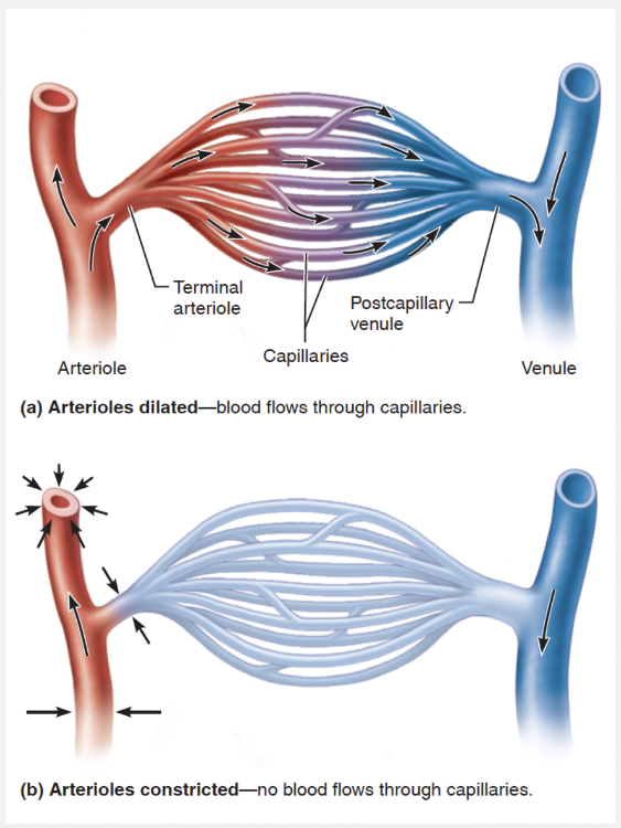
The smallest of the arteries, arterioles have a lumen diameter ranging from 0.3 mm down to 10 μm. Larger arterioles have all three tunics, but their tunica media is chiefly smooth muscle with a few scattered elastic fibers. Smaller arterioles, which lead into the capillary beds, are little more than a single layer of smooth muscle cells spiraling around the endothelial lining.
Minute-to-minute blood flow into the capillary beds is determined by arteriolar diameter, which varies in response to changing neural, hormonal, and local chemical influences. Changing diameter changes resistance to blood flow, and so arterioles are called resistance vessels. When arterioles constrict, the tissues served are largely bypassed. When arterioles dilate, blood flow into the local capillaries increases dramatically.
Which of the following is the correct sequence of layers in the vessel wall from outside to inside?
- It varies from vessel to vessel.
- Tunica media, tunica intima, tunica externa
- Tunica externa, tunica media, tunica intima
- Tunica intima, tunica externa, tunica media
- Tunica intima, tunica media, tunica externa
Tunica externa, tunica media, tunica intima
Ex.
The correct sequence of layers in the vessel wall from outside to inside is tunica externa, tunica media, tunica intima.
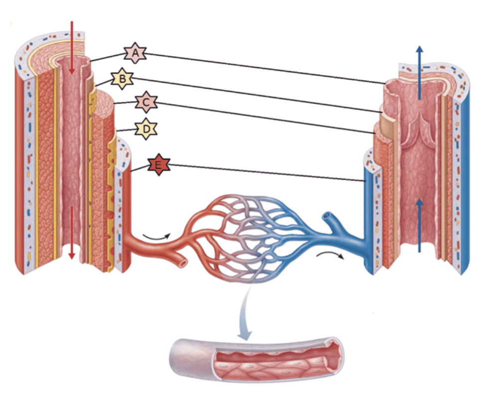
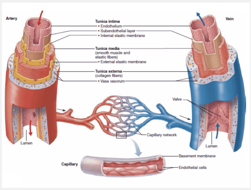
The walls of all blood vessels, except the very smallest, have three distinct layers, or tunics (“coverings”), that surround a central blood-containing space, the vessel lumen.
The innermost tunic is the tunica intima (in’tĭ-mah). The name is easy to remember once you know that this tunic is in intimate contact with the blood in the lumen. The tunica intima contains the endothelium, the simple squamous epithelium that lines the lumen of all vessels. The endothelium is continuous with the endocardial lining of the heart, and its flat cells fit closely together, forming a slick surface that minimizes friction as blood moves through the lumen. In vessels larger than 1 mm in diameter, a subendothelial layer, consisting of a basement membrane and loose connective tissue, supports the endothelium.
The middle tunic, the tunica media (me’de-ah), is mostly circularly arranged smooth muscle cells and sheets of elastin. The activity of the smooth muscle is regulated by sympathetic vasomotor nerve fibers of the autonomic nervous system and a whole battery of chemicals. Depending on the body’s needs at any given moment, regulation causes either vasoconstriction (lumen diameter decreases as the smooth muscle contracts) or vasodilation (lumen diameter increases as the smooth muscle relaxes). The activities of the tunica media are critical in regulating circulatory dynamics because small changes in vessel diameter greatly influence blood flow and blood pressure. Generally, the tunica media is the bulkiest layer in arteries, which bear the chief responsibility for maintaining blood pressure and circulation.
The outermost layer of a blood vessel wall, the tunica externa (also called the tunica adventitia; ad”ven-tish’e-ah; “coming from outside”), is composed largely of loosely woven collagen fibers that protect and reinforce the vessel, and anchor it to surrounding structures. The tunica externa is infiltrated with nerve fibers, lymphatic vessels, and, in larger veins, a network of elastic fibers. In larger vessels, the tunica externa contains a system of tiny blood vessels, the vasa vasorum (va’sah va-sor’um)—literally, “vessels of the vessels”—that nourish the more external tissues of the blood vessel wall. The innermost (luminal) portion of the vessel obtains nutrients directly from blood in the lumen. The three vessel types vary in length, diameter, wall thickness, and tissue makeup.
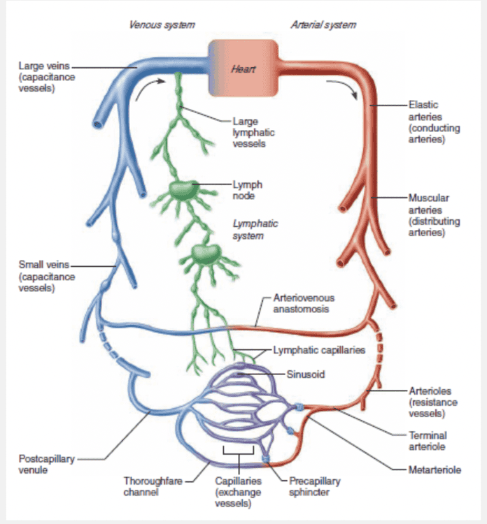
A metarteriole is a vessel that __________.
- is intermediate between the arteriole and the capillary bed
- is intermediate between a capillary and a venule
- drains the capillary bed
- delivers blood to the capillary bed
is intermediate between the arteriole and the capillary bed
Ex.
A metarteriole is a vessel that is intermediate between the arteriole and the capillary bed.
Capillaries do not function independently. Instead they form interweaving networks called capillary beds. The flow of blood from an arteriole to a venule—that is, through a capillary bed—is called the microcirculation. In most body regions, a capillary bed consists of two types of vessels: (1) a vascular shunt (metarteriole– thoroughfare channel), a short vessel that directly connects the arteriole and venule at opposite ends of the bed, and (2) true capillaries, the actual exchange vessels.
The terminal arteriole feeding the bed leads into a metarteriole (a vessel structurally intermediate between an arteriole and a capillary), which is continuous with the thoroughfare channel (intermediate between a capillary and a venule). The thoroughfare channel, in turn, joins the postcapillary venule that drains the bed.
Match the following type of vessel with its structure: Arterioles.
- Smallest of the vessels that lead into capillary beds
- Thick-walled, large vessels near the heart that conduct blood continuously away from the heart
- Contain valves to assist blood flow back toward heart
- Smaller vessels that distribute blood to specific body organs
- Smallest vessels leading away from capillaries
- Smallest blood vessels with thin walls that allow exchange between blood and tissue cells
Smallest of the vessels that lead into capillary beds
Ex.
Arterioles are the smallest of the vessels that lead into capillary beds.
The smallest of the arteries, arterioles have a lumen diameter ranging from 0.3 mm down to 10 μm. Larger arterioles have all three tunics, but their tunica media is chiefly smooth muscle with a few scattered elastic fibers. Smaller arterioles, which lead into the capillary beds, are little more than a single layer of smooth muscle cells spiraling around the endothelial lining.
Minute-to-minute blood flow into the capillary beds is determined by arteriolar diameter, which varies in response to changing neural, hormonal, and local chemical influences. Changing diameter changes resistance to blood flow, and so arterioles are called resistance vessels. When arterioles constrict, the tissues served are largely bypassed. When arterioles dilate, blood flow into the local capillaries increases dramatically.
Which of the following is true when comparing arteries and veins?
- Arteries are less muscular than veins.
- Arteries carry blood away from the heart; veins carry blood to the heart.
- Arteries have valves; veins do not.
- At any given time, there is more blood present in arteries than in veins.
Arteries carry blood away from the heart; veins carry blood to the heart.
The endothelium is composed of __________.
- simple columnar epithelium
- tunica media muscle cells
- simple squamous epithelium
- stratified squamous epithelium
- simple cuboidal epithelium
simple squamous epithelium
Ex.
The endothelium is composed of simple squamous epithelium.
The tunica intima contains the endothelium, the simple squamous epithelium that lines the lumen of all vessels. The endothelium is continuous with the endocardial lining of the heart, and its flat cells fit closely together, forming a slick surface that minimizes friction as blood moves through the lumen. In vessels larger than 1 mm in diameter, a subendothelial layer, consisting of a basement membrane and loose connective tissue, supports the endothelium.
Identify the type of vessel indicated by “D.”
- Postcapillary venule
- Muscular arteries
- Capillaries
- Large veins
- Elastic arteries
- Arterioles
Capillaries
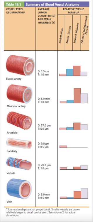
Which of the following types of blood vessels have the proportionally thickest tunica media of all vessels?
- Distributing arteries
- Arterioles
- Vasa vasorum
- Elastic arteries
Distributing arteries
Ex.
Distributing arteries are blood vessels that have the proportionally thickest tunica media of all vessels.
Distally the elastic arteries give way to the muscular arteries, which deliver blood to specific body organs (and so are sometimes called distributing arteries). Muscular arteries account for most of the named arteries studied in the anatomy laboratory. Their internal diameter ranges from that of a little finger to that of a pencil lead.
Proportionately, muscular arteries have the thickest tunica media of all vessels. Their tunica media contains relatively more smooth muscle and less elastic tissue than do elastic arteries. For this reason, they are more active in vasoconstriction and less capable of stretching. In muscular arteries, however, there is an elastic membrane on each face of the tunica media.
The vasa vasorum is a network of tiny blood vessels found in larger blood vessels that nourish the more external tissues of the blood vessel wall.
Which of the layers of an artery wall is regulated by the sympathetic nervous system and many hormones?
- Middle layer
- Inner layer
- Outer layer
- Subendothelial layer
Middle layer
Ex.
The middle layer of an artery wall is regulated by the sympathetic nervous system and many hormones.
The middle tunic, the tunica media (me’de-ah), is mostly circularly arranged smooth muscle cells and sheets of elastin. The activity of the smooth muscle is regulated by sympathetic vasomotor nerve fibers of the autonomic nervous system and a whole battery of chemicals. Depending on the body’s needs at any given moment, regulation causes either vasoconstriction (lumen diameter decreases as the smooth muscle contracts) or vasodilation (lumen diameter increases as the smooth muscle relaxes). The activities of the tunica media are critical in regulating circulatory dynamics because small changes in vessel diameter greatly influence blood flow and blood pressure. Generally, the tunica media is the bulkiest layer in arteries, which bear the chief responsibility for maintaining blood pressure and circulation.
Identify the type of vessel indicated by “A.”
- Capillaries
- Muscular arteries
- Large veins
- Postcapillary venule
- Arterioles
- Elastic arteries
Elastic arteries
Which vessels have a tunica media with relatively more smooth muscle than elastic tissue, and an elastic membrane on each face of the tunica media?
- elastic arteries
- muscular arteries
- conducting arteries
- all arteries
muscular arteries
Ex.
An elastic lamina on both sides of the tunica media is a characteristic of muscular arteries .
Distally the elastic arteries give way to the muscular arteries, which deliver blood to specific body organs (and so are sometimes called distributing arteries ). Muscular arteries have a tunica media with relatively more smooth muscle than elastic tissue, and an elastic membrane on each face of the tunica media. Their internal diameter ranges from that of a little finger to that of a pencil lead.
Proportionately, muscular arteries have the thickest tunica media of all vessels. Their tunica media contains relatively more smooth muscle and less elastic tissue than do elastic arteries. For this reason, they are more active in vasoconstriction and less capable of stretching. In muscular arteries, however, there is an elastic membrane on each face of the tunica media.
Reduction in the lumen diameter of a blood vessel as the smooth muscle contracts is known as __________.
- varicose veins
- vasoconstriction
- vasodilation
- arteriosclerosis
- atherosclerosis
vasoconstriction
Ex.
Reduction in the lumen diameter of a blood vessel as the smooth muscle contracts is known as vasoconstriction.
Depending on the body’s needs at any given moment, regulation causes either vasoconstriction (lumen diameter decreases as the smooth muscle contracts) or vasodilation (lumen diameter increases as the smooth muscle relaxes). The activities of the tunica media are critical in regulating circulatory dynamics because small changes in vessel diameter greatly influence blood flow and blood pressure. Generally, the tunica media is the bulkiest layer in arteries, which bear the chief responsibility for maintaining blood pressure and circulation.
Atherosclerosis and arteriosclerosis are disorders of the arteries usually associated with aging, while varicose veins is a disorder of the veins usually caused by elevated venous pressure.
Which of the following blood vessels is the most susceptible to atherosclerosis?
- The cerebral arteries
- The pulmonary arteries
- The femoral artery
- The aorta
The aorta
Ex.
The aorta is the blood vessel most susceptible to atherosclerosis.
Although all arteries are susceptible to atherosclerosis, those most often affected are the aorta and the coronary and carotid arteries. Atherosclerosis, particularly of the aorta, can cause aneurysms (ballooning of the arterial wall) that may burst.
Identify the type of vessel indicated by “C.”
- Postcapillary venule
- Elastic arteries
- Muscular arteries
- Large veins
- Capillaries
- Arterioles
Arterioles
Identify the artery and vein structure indicated by “C.”
- Internal elastic membrane
- Tunica externa
- Tunica intima
- Tunica media
- External elastic membrane
Tunica media
__________ inhibits fibrinolysis by competing with plasminogen and may contribute to the formation of atherosclerotic plaques.
- Foamy macrophages
- Cholesterol
- Fibrinogen
- Lipoprotein (a)
Lipoprotein (a)
Ex.
Lipoprotein (a) inhibits fibrinolysis by competing with plasminogen and may contribute to the formation of atherosclerotic plaques.
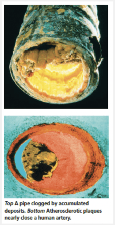
The presence of plaques stiffens artery walls and results in hypertension. The increased pressure stresses the plaques, making them even more unstable. Plaques also constrict the vessel and cause the arterial walls to fray and ulcerate, conditions that encourage blood sludging and backup, platelet adhesion, and thrombus (clot) formation.
Two other factors also promote thrombus formation: (1) Endothelial cells damaged by plaques release less nitric oxide and prostacyclin—chemicals that would otherwise promote vasodilation and inhibit platelet aggregation. (2) Lipoprotein (a), an altered form of LDL found in some individuals, inhibits fibrinolysis.
Although all arteries are susceptible to atherosclerosis, those most often affected are the aorta and the coronary and carotid arteries. Atherosclerosis, particularly of the aorta, can cause aneurysms (ballooning of the arterial wall) that may burst.
Plaque formation also increases the risk of heart attacks and strokes and is responsible for the pain (angina) that occurs when heart muscle is ischemic. We often think of large complicated plaques as being the culprits in heart attacks and strokes, but plaques of any size may rupture and form a clot. At least one-third of all heart attacks are caused by plaques too small to be seen on traditional angiograms. They cause no warning symptoms, and victims appear perfectly healthy until they drop dead! One goal of current research is to find ways to identify these vulnerable plaques.
Match the following type of vessel with its structure: Elastic arteries.
- Contain valves to assist blood flow back toward heart
- Smallest vessels leading away from capillaries
- Smaller vessels that distribute blood to specific body organs
- Smallest blood vessels with thin walls that allow exchange between blood and tissue cells
- Thick-walled, large vessels near the heart that conduct blood continuously away from the heart
- Smallest of the vessels that lead into capillary beds
Thick-walled, large vessels near the heart that conduct blood continuously away from the heart
Ex.
Elastic arteries are the thick-walled arteries near the heart—the aorta and its major branches.
These arteries are the largest in diameter, ranging from 2.5 cm to 1 cm, and the most elastic. Because their large lumens make them low-resistance pathways that conduct blood from the heart to medium-sized arteries, elastic arteries are sometimes called conducting arteries.
Elastic arteries contain more elastin than any other vessel type. It is present in all three tunics, but the tunica media contains the most. There the elastin constructs concentric “holey” sheets of elastic connective tissue that look like slices of Swiss cheese sandwiched between layers of smooth muscle cells. Although elastic arteries also contain substantial amounts of smooth muscle, they are relatively inactive in vasoconstriction. Thus, in terms of function, they can be visualized as simple elastic tubes.
Elastic arteries are pressure reservoirs, expanding and recoiling as the heart ejects blood. Consequently, blood flows fairly continuously rather than starting and stopping with the pulsating rhythm of the heartbeat. If the blood vessels become hard and unyielding, as in atherosclerosis, blood flows more intermittently, similar to the way water flows through a hard rubber garden hose attached to a faucet. When the faucet is on, the high pressure makes the water gush out of the hose. But when the faucet is shut off, the water flow abruptly becomes a trickle and then stops, because the hose walls cannot recoil to keep the water under pressure. Also, without the pressure-smoothing effect of the elastic arteries, the walls of arteries throughout the body experience higher pressures. Battered by high pressures, the arteries eventually weaken and may balloon out (as an aneurysm) or even burst.
Identify the type of vessel indicated by “E.”
- Muscular arteries
- Capillaries
- Arterioles
- Postcapillary venule
- Elastic arteries
- Large veins
Postcapillary venule