Indicate the valve separating the following areas of the heart: Left atrium and left ventricle.
Bicuspid (mitral) valve
Ex.
The valve separating the left atrium and left ventricle is the bicuspid (mitral) valve .
The aortic and pulmonary (semilunar, SL) valves guard the bases of the large arteries issuing from the ventricles (aorta and pulmonary trunk, respectively) and prevent backflow into the associated ventricles. Each SL valve is fashioned from three pocketlike cusps, each shaped roughly like a crescent moon ( semilunar = half-moon). The tricuspid valve separates the right atrium and right ventricle.
A condition in which the valve flaps of the heart become stiff and constrict the opening is called _________.
- a shunt
- stenosis
- angina pectoris
- pericardial friction rub
stenosis
Ex.
A condition in which the valve flaps of the heart become stiff and constricts the opening is called stenosis (“narrowing”).
Heart valves are simple devices, and the heart—like any mechanical pump—can function with “leaky” valves as long as the impairment is not too great. However, severe valve deformities can seriously hamper cardiac function.
An incompetent , or insufficient , valve forces the heart to repump the same blood over and over because the valve does not close properly and blood backflows. In valvular stenosis (“narrowing”), the valve flaps become stiff (typically due to calcium salt deposits or scar tissue that forms following endocarditis) and constrict the opening. This stiffness compels the heart to contract more forcibly than normal. Both conditions increase the heart’s workload and may weaken the heart severely over time.
A shunt is a bypass, or shortcut, e.g., the foramen ovale connecting the two atria in the fetal heart.
Angina pectoris (“choked chest”) is thoracic pain caused by a fleeting deficiency in blood delivery to the myocardium.
Pericardial friction rub is the creaking sound made by an inflamed pericardium (pericarditis).
Match the area of the heart with the “exit” through which the blood leaves: Left ventricle.
- Tricuspid valve
- Aortic semilunar valve
- Bicuspid valve
- Pulmonary semilunar valve
Aortic semilunar valve
Ex.
The exit through which blood leaves the left ventricle is the aortic semilunar valve.
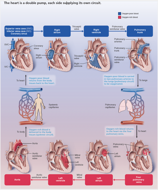
Focus on Blood Flow through the Heart follows a single “spurt” of blood as it passes through all four chambers of the heart and both blood circuits in its ever-repeating journey.
As you work your way through this figure, keep in mind that the left side of the heart is the systemic circuit pump and the right side is the pulmonary circuit pump. Notice how unique the pulmonary circuit is. Elsewhere in the body, veins carry relatively oxygen-poor blood to the heart, and arteries transport oxygen-rich blood from the heart. The opposite oxygenation conditions exist in veins and arteries of the pulmonary circuit.
What makes the heart valves open and close?
- Chordae tendineae
- Papillary muscles
- Blood pressure
- Back-suction
Blood pressure
Failure of which heart valve would allow blood to move from the left ventricle to the left atrium?
- Mitral (bicuspid) valve
- Aortic semilunar valve
- Pulmonary semilunar valve
- Tricuspid valve
Mitral (bicuspid) valve
Ex.
Failure of the m itral (bicuspid) valve would allow blood to move from the left ventricle to the left atrium.
The two atrioventricular (AV) valves, one located at each atrial-ventricular junction, prevent backflow into the atria when the ventricles contract.
- The right AV valve, the tricuspid valve (tri-kus’pid), has three flexible cusps (flaps of endocardium reinforced by connective tissue cores).
- The left AV valve, with two cusps, is called the mitral valve (mi’tral) because it resembles the two-sided bishop’s miter (tall, pointed hat). It is sometimes called the bicuspid valve .
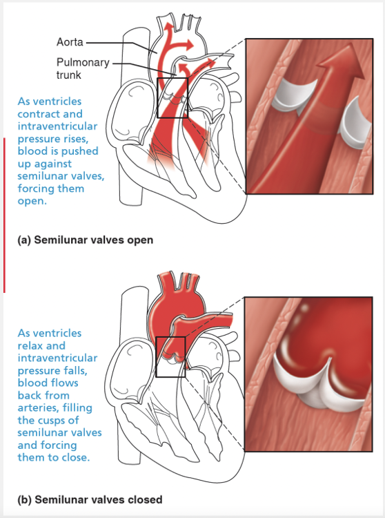
The aortic and pulmonary (semilunar, SL) valves guard the bases of the large arteries issuing from the ventricles (aorta and pulmonary trunk, respectively) and prevent backflow into the associated ventricles.
Match the following term with its correct description: Mitral valve.
- Prevents backflow of blood into the right ventricle
- Valve between the left atrium and left ventricle
- Valve between the right atrium and right ventricle
- Prevents backflow of blood into the left ventricle
Valve between the left atrium and left ventricle
Ex.
The mitral valve is the valve between the left atrium and the left ventricle .
The aortic and pulmonary (semilunar, SL) valves guard the bases of the large arteries issuing from the ventricles (aorta and pulmonary trunk, respectively) and prevent backflow into the associated ventricles. Each SL valve is fashioned from three pocketlike cusps, each shaped roughly like a crescent moon ( semilunar = half-moon). The tricuspid valve is between the right atrium and right ventricle.
Match the following term with its correct description: Tricuspid valve.
- Prevents backflow of blood into the left ventricle
- Valve between the left atrium and left ventricle
- Valve between the right atrium and right ventricle
- Prevents backflow of blood into the right ventricle
Valve between the right atrium and right ventricle
Ex.
The tricuspid valves is the valve between the right atrium and right ventricle .
The two atrioventricular (AV) valves, one located at each atrial-ventricular junction, prevent backflow into the atria when the ventricles contract.
- The right AV valve, the tricuspid valve (tri-kus’pid), has three flexible cusps (flaps of endocardium reinforced by connective tissue cores).
- The left AV valve, with two cusps, is called the mitral valve (mi’tral) because it resembles the two-sided bishop’s miter (tall, pointed hat). It is sometimes called the bicuspid valve.
When the mitral valve closes, it prevents the backflow of blood from the __________.
- right ventricle into the pulmonary trunk
- left ventricle into the left atrium
- left atrium into the left ventricle
- left ventricle into the aorta
- right atrium into the right ventricl
left ventricle into the left atrium
Ex.
When the mitral valve closes, it prevents the backflow of blood from the left ventricle into the left atrium .
The aortic and pulmonary (semilunar, SL) valves guard the bases of the large arteries issuing from the ventricles (aorta and pulmonary trunk, respectively) and prevent backflow into the associated ventricles. Each SL valve is fashioned from three pocketlike cusps, each shaped roughly like a crescent moon ( semilunar = half-moon). The tricuscpid valve prevents backflow of blood from the right ventricle into the right atrium.
Match the area of the heart with the “exit” through which the blood leaves: Right ventricle.
- Pulmonary semilunar valve
- Aortic semilunar valve
- Tricuspid valve
- Bicuspid valve
Pulmonary semilunar valve
Ex.
Blood exits the right ventricle through the pulmonary semilunar valve.
Focus on Blood Flow through the Heart (follows a single “spurt” of blood as it passes through all four chambers of the heart and both blood circuits in its ever-repeating journey.
As you work your way through this figure, keep in mind that the left side of the heart is the systemic circuit pump and the right side is the pulmonary circuit pump. Notice how unique the pulmonary circuit is. Elsewhere in the body, veins carry relatively oxygen-poor blood to the heart, and arteries transport oxygen-rich blood from the heart. The opposite oxygenation conditions exist in veins and arteries of the pulmonary circuit.
Which of the following valves is most often faulty in the heart?
- None of the choices is most often faulty in the heart.
- The mitral, or bicuspid, valve
- The tricuspid valve
- The pulmonary semilunar valve
The mitral, or bicuspid, valve
Ex.
The mitral, or bicuspid, valve is the valve that is most often faulty in the heart.
Heart valves are simple devices, and the heart—like any mechanical pump—can function with “leaky” valves as long as the impairment is not too great. However, severe valve deformities can seriously hamper cardiac function. An incompetent, or insufficient, valve forces the heart to repump the same blood over and over because the valve does not close properly and blood backflows. In valvular stenosis (“narrowing”), the valve flaps become stiff and constrict the opening. [Stenosis is typically due to calcium salt deposits or scar tissue that forms following endocarditis (inflammation of the endocardium, which most often results from infection by bacteria that have entered the bloodstream and usually affects the heart valves).] The constricted opening compels the heart to contract more forcibly than normal. Both types of valve problems increase the heart’s workload and may weaken the heart severely over time. The mitral and aortic valves are most oftenaffected.The faulty valve can be replaced with a mechanical valve, a pig or cow heart valve chemically treated to prevent rejection,or cryopreserved valves from human cadavers. Heart valves tissue-engineered from a patient’s own cells grown on a biodegradable scaffold are being developed.
Indicate the valve separating the following areas of the heart: Left ventricle and aorta.
- Bicuspid (mitral) valve
- Aortic semilunar valve
- Tricuspid valve
- Pulmonary semilunar valve
Aortic semilunar valve
Ex.
The valve separating the left ventricle and aorta is the aortic semilunar valve .
The aortic and pulmonary (semilunar, SL) valves guard the bases of the large arteries issuing from the ventricles (aorta and pulmonary trunk, respectively) and prevent backflow into the associated ventricles. Each SL valve is fashioned from three pocketlike cusps, each shaped roughly like a crescent moon ( semilunar = half-moon). The tricuspid valve separates the right atrium and right ventricle. The bicuspid (mitral) valve separates the left atrium and left ventricle.
The __________ are attached to the AV valve flaps.
- pectinate muscles
- chordae tendineae
- papillary muscles
- trabeculae carneae
chordae tendineae
Ex.
The chordae tendineae are attached to the AV valve flaps.
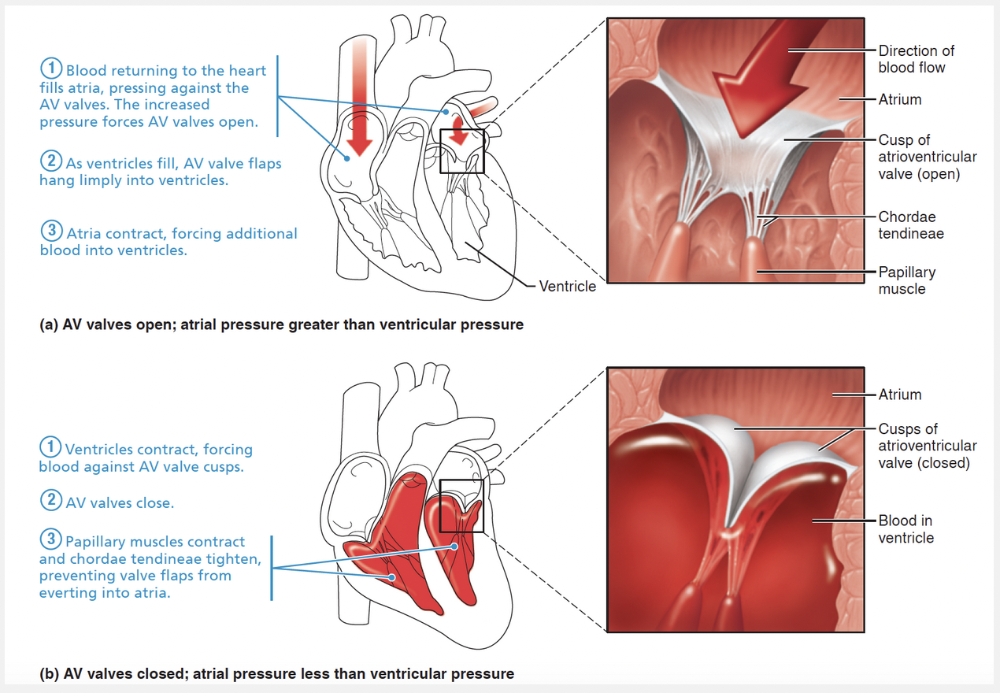
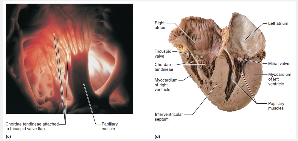
Attached to each AV valve flap are tiny white collagen cords called chordae tendineae (kor′de ten″dĭ′ne-e; “tendinous cords”), “heart strings” which anchor the cusps to the papillary muscles protruding from the ventricular walls.
What muscles prevent the atrioventricular valves from everting during ventricular contraction?
- Interatrial muscles
- The myocardium
- Pectinate muscles
- Papillary muscles
Papillary muscles
Ex.
The papillary muscles prevent the atrioventricular valves from everting during ventricular contraction.
The chordae tendineae and the papillary muscles act as tethers that anchor the valve flaps in their closed position. If the cusps were not anchored, they would be blown upward (everted) into the atria, in the same way an umbrella is blown inside out by a gusty wind. The papillary muscles contract with the other ventricular musculature so that they take up the slack on the chordae tendineae as the full force of ventricular contraction hurls the blood against the AV valve flaps.
Pectinate muscles are are muscle bundles found in the anterior portion of the right atrium.
The myocardium is the muscle in the middle layer of the heart and is composed mainly of cardiac muscle. It forms the bulk of the heart.
Indicate the valve separating the following areas of the heart: Right ventricle and pulmonary trunk.
- Tricuspid valve
- Bicuspid (mitral) valve
- Pulmonary semilunar valve
- Aortic semilunar valve
Pulmonary semilunar valve
Ex.
The valve separating the right ventricle and pulmonary trunk is the pulmonary semilunar valve.
Focus on Blood Flow through the Heart follows a single “spurt” of blood as it passes through all four chambers of the heart and both blood circuits in its ever-repeating journey.
As you work your way through this figure, keep in mind that the left side of the heart is the systemic circuit pump and the right side is the pulmonary circuit pump. Notice how unique the pulmonary circuit is. Elsewhere in the body, veins carry relatively oxygen-poor blood to the heart, and arteries transport oxygen-rich blood from the heart. The opposite oxygenation conditions exist in veins and arteries of the pulmonary circuit.
Indicate the valve separating the following areas of the heart: Right atrium and right ventricle.
- Pulmonary semilunar valve
- Tricuspid valve
- Bicuspid (mitral) valve
- Aortic semilunar valve
Tricuspid valve
Match the following term with its correct description: Tricuspid valve.
- Valve between the left atrium and left ventricle
- Prevents backflow of blood into the left ventricle
- Valve between the right atrium and right ventricle
- Prevents backflow of blood into the right ventricle
Valve between the right atrium and right ventricle
Match the area of the heart with the "exit" through which the blood leaves: Left atrium.
- Tricuspid valve
- Pulmonary semilunar valve
- Aortic semilunar valve
- Mitral (bicuspid) valve
Mitral (bicuspid) valve
Ex.
Blood exits the left atrium through the bicuspid valve .
Focus on Blood Flow through the Heart follows a single “spurt” of blood as it passes through all four chambers of the heart and both blood circuits in its ever-repeating journey.
As you work your way through this figure, keep in mind that the left side of the heart is the systemic circuit pump and the right side is the pulmonary circuit pump . Notice how unique the pulmonary circuit is. Elsewhere in the body, veins carry relatively oxygen-poor blood to the heart, and arteries transport oxygen-rich blood from the heart. The opposite oxygenation conditions exist in veins and arteries of the pulmonary circuit.
Match the following term with its correct description: Pulmonary semilunar valve.
- Prevents backflow of blood into the right ventricle
- Prevents backflow of blood into the left ventricle
- Valve between the right atrium and right ventricle
- Valve between the left atrium and left ventricle
Prevents backflow of blood into the right ventricle
Ex.
The pulmonary semilunar valve prevents backflow of blood into the right ventricle .
The aortic and pulmonary (semilunar, SL) valves guard the bases of the large arteries issuing from the ventricles (aorta and pulmonary trunk, respectively) and prevent backflow into the associated ventricles. Each SL valve is fashioned from three pocketlike cusps, each shaped roughly like a crescent moon ( semilunar = half-moon). The mitral valve is between the left atrium and left ventricle. The tricuspid valve is between the right atrium and right ventricle.
Match the area of the heart with the “exit” through which the blood leaves: Right atrium.
- Pulmonary semilunar valve
- Tricuspid valve
- Aortic semilunar valve
- Bicuspid valve
Tricuspid valve
Ex.
Blood exits the right atrium through the tricuspid valve.
Focus on Blood Flow through the Heart follows a single “spurt” of blood as it passes through all four chambers of the heart and both blood circuits in its ever-repeating journey.
As you work your way through this figure, keep in mind that the left side of the heart is the systemic circuit pump and the right side is the pulmonary circuit pump. Notice how unique the pulmonary circuit is. Elsewhere in the body, veins carry relatively oxygen-poor blood to the heart, and arteries transport oxygen-rich blood from the heart. The opposite oxygenation conditions exist in veins and arteries of the pulmonary circuit.
What is the purpose of the chordae tendineae?
- To open the SL valves
- To open the AV valves
- Anchor the SL valves in the closed position
- Anchor the AV valves in the closed position
Anchor the AV valves in the closed position
Ex.
The chordae tendineae, attached only to the AV valves, act as tethers that anchor the valve flaps in their closed position .
However, neither the chordae tendineae nor the papillary muscles attached to them, are responsible for opening and closing the AV valves. Heart valves open and close in response to differences in blood pressure on their two sides.
Match the area of the heart with the “exit” through which the blood leaves: Left ventricle.
- Pulmonary semilunar valve
- Aortic semilunar valve
- Tricuspid valve
- Bicuspid valve
Aortic semilunar valve
Ex.
The exit through which blood leaves the left ventricle is the aortic semilunar valve.
Focus on Blood Flow through the Heart follows a single “spurt” of blood as it passes through all four chambers of the heart and both blood circuits in its ever-repeating journey.
As you work your way through this figure, keep in mind that the left side of the heart is the systemic circuit pump and the right side is the pulmonary circuit pump. Notice how unique the pulmonary circuit is. Elsewhere in the body, veins carry relatively oxygen-poor blood to the heart, and arteries transport oxygen-rich blood from the heart. The opposite oxygenation conditions exist in veins and arteries of the pulmonary circuit
Match the following term with its correct description: Aortic semilunar valve.
- Valve between the right atrium and right ventricle
- Prevents backflow of blood into the right ventricle
- Prevents backflow of blood into the left ventricle
- Valve between the left atrium and left ventricle
Prevents backflow of blood into the left ventricle
Ex.
The aortic semilunar valve prevents backflow of blood into the left ventricle .
The aortic and pulmonary (semilunar, SL) valves guard the bases of the large arteries issuing from the ventricles (aorta and pulmonary trunk, respectively) and prevent backflow into the associated ventricles. Each SL valve is fashioned from three pocketlike cusps, each shaped roughly like a crescent moon ( semilunar = half-moon).