Match the following term to its correct description: Preload.
- The degree to which cardiac muscle cells are stretched just before they contract
- Back pressure exerted by arterial blood
- The amount of blood pumped by each ventricle in one minute
- The events associated with blood flow through the heart during one complete heartbeat
- The volume of blood pumped by one ventricle with each heartbeat
The degree to which cardiac muscle cells are stretched just before they contract
Explanation:
Preload is the degree to which cardiac muscle cells are stretched just before they contract.
Preload controls stroke volume. In a normal heart, the higher the preload, the higher the stroke volume. This relationship between preload and stroke volume is called the Frank-Starling law of the heart.
The events associated with blood flow through the heart during one complete heartbeat are called the cardiac cycle.
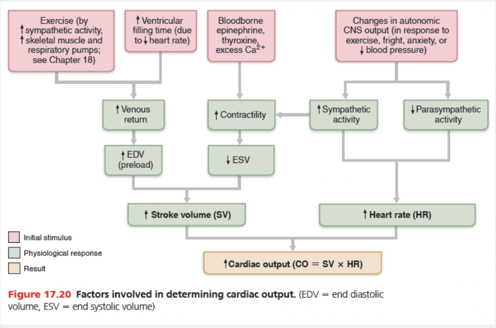
Cardiac output (CO) is the amount of blood pumped out by each ventricle in 1 minute.
It is the product of heart rate (HR) in beats per minute and stroke volume (SV) in ml per beat. Stroke volume is defined as the volume of blood pumped out by one ventricle with each beat. In general, stroke volume correlates with the force of ventricular contraction.
Using normal resting values for heart rate (75 beats/min) and stroke volume (70 ml/beat), the average adult cardiac output can be computed as follows:
CO = HR × SV = 75 beats/min × 70 ml//beat
= 5250 ml/min = 5.25 L min/min
The normal adult blood volume is about 5 L (a little more than 1 gallon). As you can see, the entire blood supply passes through each side of the heart once each minute.
Back pressure exerted by arterial blood is called afterload. It’s the pressure that the ventricles must overcome to eject blood.
The second heart sound (the 'dup' of 'lub-dup') is caused by the __________.

closure of the semilunar valves
Ex. The second heart sound (the 'dup' of 'lub-dup') is caused by the closure of the semilunar valves.
Auscultating (listening to) the thorax with a stethoscope will reveal two sounds during each heartbeat. These heart sounds, often described as lub-dup, are associated with the heart valves closing.
Match the following abbreviation with its definition: EDV.
- Amount of blood ejected by one contraction of the heart
- Amount of blood in the ventricle at the end of contraction
- The frequency of the heart beats
- Amount of blood in the ventricle at the end of relaxation
- The amount of blood ejected from the heart in one minute
Amount of blood in the ventricle at the end of relaxation
Ex.
EDV is the amount of blood in the ventricle at the end of relaxation.
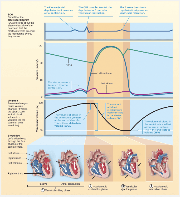
Mathematically, stroke volume (SV) represents the difference between end diastolic volume (EDV), the amount of blood that collects in a ventricle during diastole, and end systolic volume (ESV), the volume of blood remaining in a ventricle after it has contracted. The EDV, determined by how long ventricular diastole lasts and by venous pressure, is normally about 120 ml. (An increase in either factor raises EDV.) The ESV, determined by arterial blood pressure and the force of ventricular contraction, is approximately 50 ml. (The higher the arterial blood pressure, the higher the ESV.) To figure normal stroke volume, simply plug these values into this equation:
SV = EDV − ESV = 120 ml/beat − 50 ml/beat = 70 ml/beat
As you can see, each ventricle pumps out about 70 ml of blood with each beat, which is about 60% of the blood in its chambers. So what is important here—how do we make sense out of this alphabet soup (SV, ESV, EDV)? Although many factors affect SV by altering EDV or ESV, the three most important are preload, contractility, and afterload. Preload affects EDV, whereas contractility and afterload affect the ESV.
Match the following term to its correct description: Cardiac cycle.
- The volume of blood pumped by one ventricle with each heartbeat
- The events associated with blood flow through the heart during one complete heartbeat
- Relaxation
- Contraction
- The amount of blood pumped by each ventricle in one minute
The events associated with blood flow through the heart during one complete heartbeat
Ex.
The cardiac cycle refers to the events associated with blood flow through the heart during one complete heartbeat.
The heart undergoes some dramatic writhing movements as it alternately contracts, forcing blood out of its chambers, and then relaxes, allowing its chambers to refill with blood. The term systole (sis′to-le) refers to these periods of contraction, and diastole (di-as′to-le) refers to those of relaxation. The cardiac cycle includes all events associated with the blood flow through the heart during one complete heartbeat—atrial systole and diastole followed by ventricular systole and diastole. These mechanical events always follow the electrical events seen in the ECG.
Which of the following is the best description of the events in the cardiac cycle?
- Events that cause blood filling of the ventricles
- Events that cause blood ejection from the ventricles
- Events that cause depolarization of cardiac muscle
- All of the events during one heart beat
All of the events during one heart beat
Ex.
The cardiac cycle refers to all of the events during one heart beat .
The heart undergoes some dramatic writhing movements as it alternately contracts, forcing blood out of its chambers, and then relaxes, allowing its chambers to refill with blood. The term systole (sis′to-le) refers to these periods of contraction, and diastole (di-as′to-le) refers to those of relaxation. The cardiac cycle includes all events associated with the blood flow through the heart during one complete heartbeat—atrial systole and diastole followed by ventricular systole and diastole. These mechanical events always follow the electrical events seen in the ECG.
Match the following term to its correct description: Diastole.
- The amount of blood pumped by each ventricle in one minute
- The volume of blood pumped by one ventricle with each heartbeat
- Relaxation
- Contraction
- The events associated with blood flow through the heart during one complete heartbeat
Relaxation
Ex.
Diastole refers to relaxation.
The heart undergoes some dramatic writhing movements as it alternately contracts, forcing blood out of its chambers, and then relaxes, allowing its chambers to refill with blood. The term systole (sis′to-le) refers to these periods of contraction, and diastole (di-as′to-le) refers to those of relaxation. The cardiac cycle includes all events associated with the blood flow through the heart during one complete heartbeat—atrial systole and diastole followed by ventricular systole and diastole. These mechanical events always follow the electrical events seen in the ECG.
A doctor puts his stethoscope on a patient's chest over the location of the heart and hears a swishing sound. Which of the following conditions is the best diagnosis for the patient's condition?
- Incompetent cardiac valve
- Myocardial infarction
- Cardiac tamponade
- Angina pectoris
Incompetent cardiac valve
Ex.
When a doctor puts his stethoscope on a patient's chest over the location of the heart and hears a swishing sound, the best diagnosis for the patient's condition is probably an incompetent cardiac valve.
Blood flows silently as long as its flow is smooth and uninterrupted. If blood strikes obstructions, however, its flow becomes turbulent and generates abnormal heart sounds, called heart murmurs, that can be heard with a stethoscope. Heart murmurs are fairly common in young children (and some elderly people) with perfectly healthy hearts, probably because their heart walls are relatively thin and vibrate with rushing blood. Most often, however, murmurs indicate valve problems. An insufficient or incompetent valve fails to close completely. There is a swishing sound as blood backflows or regurgitates through the partially open valve after the valve has (supposedly) closed. A stenotic valve fails to open completely and its narrow opening restricts blood flow through the valve. In a stenotic aortic valve, for instance, a high-pitched sound or click can be detected when the valve should be wide open during ventricular contraction, but is not.
Match the following abbreviation with its definition: HR
- Amount of blood in the ventricle at the end of contraction
- Amount of blood in the ventricle at the end of relaxation
- Amount of blood ejected by one contraction of the heart
- The frequency of heart beats
- The amount of blood ejected from the heart in one minute
The frequency of heart beats
Ex. HR is the t he frequency of heart beats.
Cardiac output (CO) is the amount of blood pumped out by each ventricle in 1 minute. It is the product of heart rate (HR) in beats per minute and stroke volume (SV) in ml per beat. Stroke volume is defined as the volume of blood pumped out by one ventricle with each beat. In general, stroke volume correlates with the force of ventricular contraction.
Using normal resting values for heart rate (75 beats/min) and stroke volume (70 ml/beat), the average adult cardiac output can be computed as follows:
CO = HR × SV = 75 beats/min × 70 ml//beat
= 5250 ml/min = 5.25 L min/min
The normal adult blood volume is about 5 L (a little more than 1 gallon). As you can see, the entire blood supply passes through each side of the heart once each minute.
Match the following abbreviation with its definition: CO
- Amount of blood in the ventricle at the end of relaxation
- The frequency of heart beats
- Amount of blood pumped out by each ventricle in 1 minute
- Amount of blood in the ventricle at the end of contraction
- Amount of blood ejected by one contraction of the heart
Amount of blood pumped out by each ventricle in 1 minute
Ex.
CO is the amount of blood pumped out by each ventricle in 1 minute.
Cardiac output (CO) is the amount of blood pumped out by each ventricle in 1 minute. It is the product of heart rate (HR) in beats per minute and stroke volume (SV) in ml per beat. Stroke volume is defined as the volume of blood pumped out by one ventricle with each beat. In general, stroke volume correlates with the force of ventricular contraction.
Using normal resting values for heart rate (75 beats/min) and stroke volume (70 ml/beat), the average adult cardiac output can be computed as follows:
CO = HR × SV = 75 beats/min × 70 ml//beat
= 5250 ml/min = 5.25 L min/min
The normal adult blood volume is about 5 L (a little more than 1 gallon). As you can see, the entire blood supply passes through each side of the heart once each minute.
Match the following abbreviation with its definition: ESV.
- Amount of blood ejected by one contraction of the heart
- The amount of blood ejected from the heart in one minute
- The frequency at which the heart beats
- Amount of blood in the ventricle at the end of relaxation
- Amount of blood in the ventricle at the end of contraction
Amount of blood in the ventricle at the end of contraction
Ex.
ESV is the amount of blood in the ventricle at the end of contraction.
Mathematically, stroke volume (SV) represents the difference between end diastolic volume (EDV), the amount of blood that collects in a ventricle during diastole, and end systolic volume (ESV), the volume of blood remaining in a ventricle after it has contracted. The EDV, determined by how long ventricular diastole lasts and by venous pressure, is normally about 120 ml. (An increase in either factor raises EDV.) The ESV, determined by arterial blood pressure and the force of ventricular contraction, is approximately 50 ml. (The higher the arterial blood pressure, the higher the ESV.) To figure normal stroke volume, simply plug these values into this equation:
SV = EDV − ESV = 120 ml/beat − 50 ml/beat = 70 ml/beat
As you can see, each ventricle pumps out about 70 ml of blood with each beat, which is about 60% of the blood in its chambers. So what is important here—how do we make sense out of this alphabet soup (SV, ESV, EDV)? Although many factors affect SV by altering EDV or ESV, the three most important are preload, contractility, and afterload. Preload affects EDV, whereas contractility and afterload affect the ESV.
Cardiac output is __________.
- the amount of blood filling each ventricle at the end of diastole
- the amount of blood pumped out of each ventricle in one minute
- the amount of blood pumped out of the heart during every ventricular contraction
- the number of times the heart beats in one minute
- the number of impulses fired by the SA node in one minute
the amount of blood pumped out of each ventricle in one minute
Ex.
Cardiac output is the amount of blood pumped out of each ventricle in one minute.
It is the product of heart rate (HR) in beats per minute and stroke volume (SV) in ml per beat. Stroke volume is defined as the volume of blood pumped out by one ventricle with each beat. In general, stroke volume correlates with the force of ventricular contraction.
Using normal resting values for heart rate (75 beats/min) and stroke volume (70 ml/beat), the average adult cardiac output can be computed as follows:
CO = HR × SV = 75 beats/min × 70 ml//beat
= 5250 ml/min = 5.25 L min/min
The normal adult blood volume is about 5 L (a little more than 1 gallon). As you can see, the entire blood supply passes through each side of the heart once each minute.
Match the following term to its correct description: Stroke volume.
- The volume of blood pumped by one ventricle with each heartbeat
- The events associated with blood flow through the heart during one complete heartbeat
- Circumflex artery
- The amount of blood pumped by each ventricle in one minute
- Relaxation
- Contraction
The volume of blood pumped by one ventricle with each heartbeat
Ex.
Stroke volume is the volume of blood pumped by one ventricle with each heartbeat.
Mathematically, stroke volume (SV) represents the difference between end diastolic volume (EDV), the amount of blood that collects in a ventricle during diastole, and end systolic volume (ESV), the volume of blood remaining in a ventricle after it has contracted. The EDV, determined by how long ventricular diastole lasts and by venous pressure, is normally about 120 ml. (An increase in either factor raises EDV.) The ESV, determined by arterial blood pressure and the force of ventricular contraction, is approximately 50 ml. (The higher the arterial blood pressure, the higher the ESV.)
To figure normal stroke volume, simply plug these values into this equation:
SV = EDV − ESV = 120 ml/beat − 50 ml/beat = 70 ml/beat
As you can see, each ventricle pumps out about 70 ml of blood with each beat, which is about 60% of the blood in its chambers. So what is important here—how do we make sense out of this alphabet soup (SV, ESV, EDV)? Although many factors affect SV by altering EDV or ESV, the three most important are preload, contractility, and afterload. Preload affects EDV, whereas contractility and afterload affect the ESV.
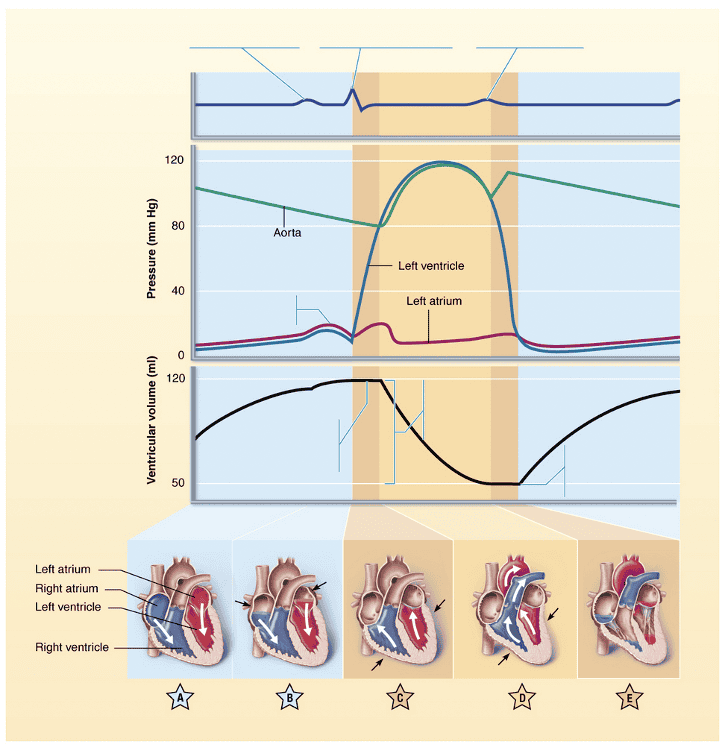
Identify the stage of the cardiac cycle indicated by "D."
- Atrial contraction
- Ventricular filling after completion of the cycle
- Ventricular ejection phase
- Isovolumetric contraction phase
- Passive ventricular filling
- Isovolumetric relaxation
Ventricular ejection phase
Using the graphic, select the letter that identifies the stage(s) of the cardiac cycle during which ventricular filling is occurring.
- A and B
- E
- A
- A and E
- B
A and B
Identify the stage of the cardiac cycle indicated by "A."
- Isovolumetric relaxation
- Isovolumetric contraction phase
- Ventricular filling after completion of the cycle
- Passive ventricular filling
- Atrial contraction
- Ventricular ejection phase
Passive ventricular filling
Identify the stage of the cardiac cycle indicated by "B."
- Atrial contraction
- Ventricular ejection phase
- Passive ventricular filling after completion of the cycle
- Isovolumetric contraction phase
- Isovolumetric relaxation
- Passive ventricular filling
- Atrial contraction
Identify the stage of the cardiac cycle indicated by "C."
- Atrial contraction
- Ventricular filling after completion of the cycle
- Passive ventricular filling
- Isovolumetric contraction phase
- Isovolumetric relaxation
- Ventricular ejection phase
Isovolumetric contraction phase
Identify the stage of the cardiac cycle indicated by "E."
- Ventricular filling after completion of the cycle
- Isovolumetric relaxation
- Atrial contraction
- Passive ventricular filling
- Isovolumetric contraction phase
- Ventricular ejection phase
Isovolumetric relaxation
Which of the following would lead to a decrease in heart rate?
- Sharply decreased blood volume
- Parasympathetic stimulation
- Norepinephrine
- Exercise
Parasympathetic stimulation
Ex.
Parasympathetic stimulation would decrease heart rate.
The parasympathetic division opposes sympathetic effects and effectively reduces heart rate when a stressful situation has passed. Parasympathetic-initiated cardiac responses are mediated by acetylcholine, which hyperpolarizes the membranes of its effector cells by opening K + channels. Because vagal innervation of the ventricles is sparse, parasympathetic activity has little effect on cardiac contractility.
Exercise and norepinephrine would directly increase heart rate.
Sharply decreased blood volume would increase heart rate in order to compensate for the reduced stroke volume caused by the decreased blood volume (and decreased EDV).
Match the following abbreviation with its definition: SV.
- The amount of blood ejected from the heart in one minute
- Amount of blood in the ventricle at the end of relaxation
- Amount of blood in the ventricle at the end of contraction
- The frequency at which the heart beats
- Amount of blood ejected by one contraction of the heart
Amount of blood ejected by one contraction of the heart
Ex.
SV is the amount of blood ejected by one contraction of the heart.
Cardiac output (CO) is the amount of blood pumped out by each ventricle in 1 minute. It is the product of heart rate (HR) in beats per minute and stroke volume (SV) in ml per beat. Stroke volume is defined as the volume of blood pumped out by one ventricle with each beat. In general, stroke volume correlates with the force of ventricular contraction.
Using normal resting values for heart rate (75 beats/min) and stroke volume (70 ml/beat), the average adult cardiac output can be computed as follows:
CO = HR × SV = 75 beats/min × 70 ml//beat
= 5250 ml/min = 5.25 L min/min
The normal adult blood volume is about 5 L (a little more than 1 gallon). As you can see, the entire blood supply passes through each side of the heart once each minute.