The four basic tissue types in the human body.
- epithelial
- connective
- muscular
- nervous
The part of an organ composed of the cells responsible for its specialized functions.
parenchyma
The part of an organ composed of the cells which have a supporting role.
stroma
The tissue that lines all external and internal surfaces of the body.
epithelium
The three principal functions of epithelial tissues.
- protection (e.g. epidermis)
- absorption (e.g. intestine)
- secretion (e.g. glands)
The three basic epithelial cell shapes.
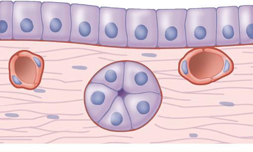
- columnar (tall with elongated nuclei)
- cuboidal (cube-like with spherical nuclei)
- squamous (short with flattened nuclei)
The connective tissue that underlies the epithelia lining the organs of the digestive, respiratory, and urinary systems.
lamina propria
Small evaginations projecting from the connective tissue into the epithelium that increase the surface area between the two tissues.
papillae
The characteristic of epithelial cells of having uneven distribution of organelles and membrane proteins within the cell.
polarity
The region of the epithelial cell contacting the ECM and connective tissue.
basal pole
The region of the epithelial cell opposite the basal pole, usually facing a space.
apical pole
The regions of cuboidal or columnar epithelial cells that adjoin neighboring cells.
lateral surfaces
A thin extracellular, felt-like sheet of macromolecules underlying the basal surface of all epithelial cells which acts as a semipermeable filter.
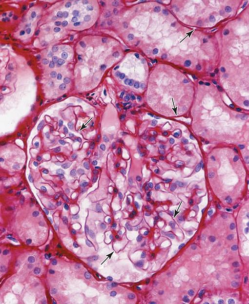
basement membrane
The part of the basement membrane nearest to the epithelial cells, composed of a thin, electron-dense, sheet-like layer of fine fibrils.
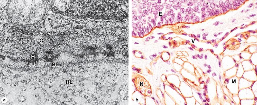
basal lamina
The part of the basement membrane furthest from the epithelial cells, composed of a diffuse and fibrous layer containing collagen III fibers.
reticular lamina
A type of collagen in the basal lamina that self-assembles into a two-dimensional network of evenly spaced subunits resembling the mesh of a window screen.
type IV collagen
Large glycoproteins that attach to transmembrane integrin proteins in the basal cell membrane and project through the mesh formed by the type IV collagen.
laminin
A protein and a proteoglycan which cross-link laminins to the type IV collagen network, helping to provide the basal lamina's three-dimensional structure.
nidogen and perlecan
A type of collagen found in the reticular lamina that binds to the basal lamina by anchoring fibrils of type VI collagen.
type III collagen
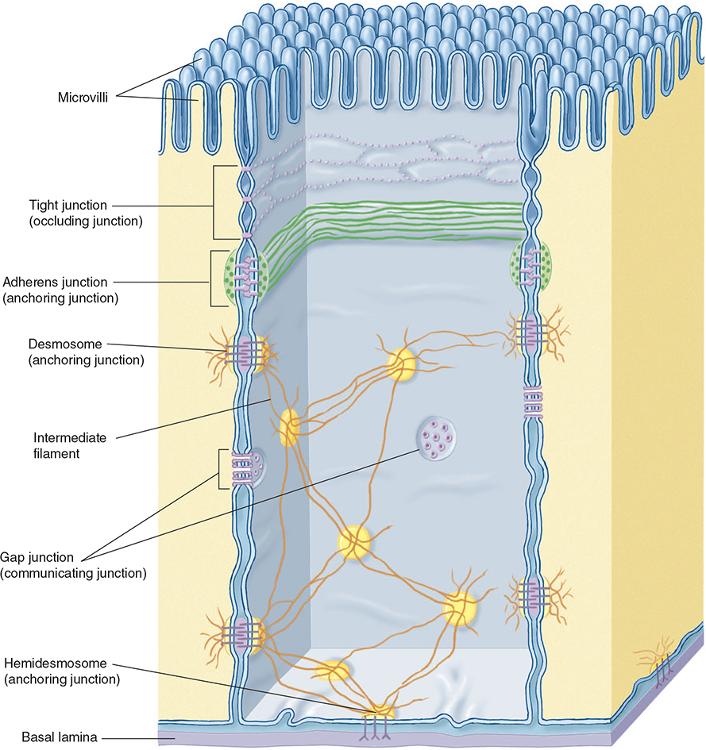
The five types of epithelium cell junctions.
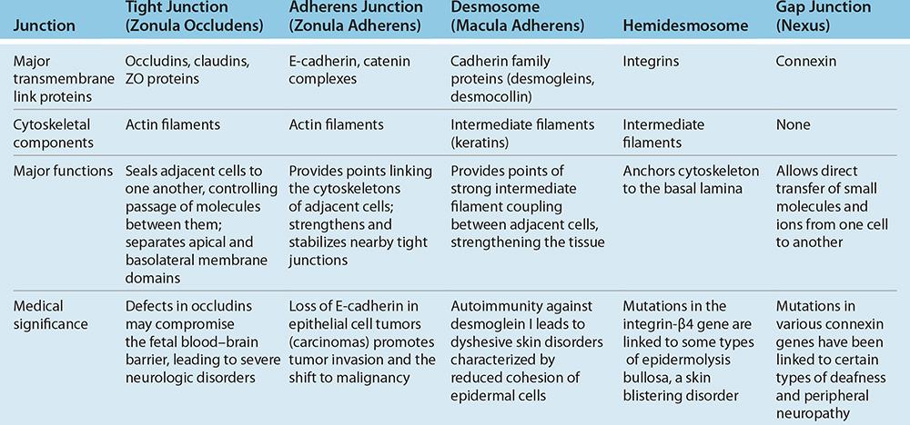
- tight junctions
- adherens junctions
- desmosomes
- hemidesmosomes
- gap junctions
The most apical of the epithelial junctions, which form a seal between adjacent cells.
tight junctions
The two transmembrane proteins responsible for the interactions at tight junctions.
claudin and occludin
A bacterium that secretes an enterotoxin that binds claudin molecules of intestinal cells, preventing insertion of these proteins during maintenance of tight junctions.
Clostridium perfringens
A bacterium that binds the extracellular domains of tight-junction proteins in cells of the stomach and inserts a protein that and disrupts signaling from the junction.
Helicobacter pylori
Epithelial junctions that encircle the epithelial cell, usually immediately below the tight junction, firmly anchoring a cell to its neighbors.
adherens junction
The transmembrane glycoproteins that meditate adherens junctions between cells by binding each other at in the presence of Ca2+
cadherins
Epithelial junctions comprised of disc-shaped structures at the surface of one cell that are matched with identical structures at an adjacent cell surface.
desmosome
Diseases that involve the epidermis or stratified squamous epithelia of the oral mucosa, caused by abnormal desmosome function due to autoimmune reactions.
bullous diseases (e.g. pemphigus vulgaris)
Epithelial junctions that mediate intercellular communication between cells, allowing intercellular exchange of molecules with small diameters.

gap junctions
The transmembrane gap junction proteins that form hexameric complexes, each of which has a central hydrophilic pore about 1.5 nm in diameter.
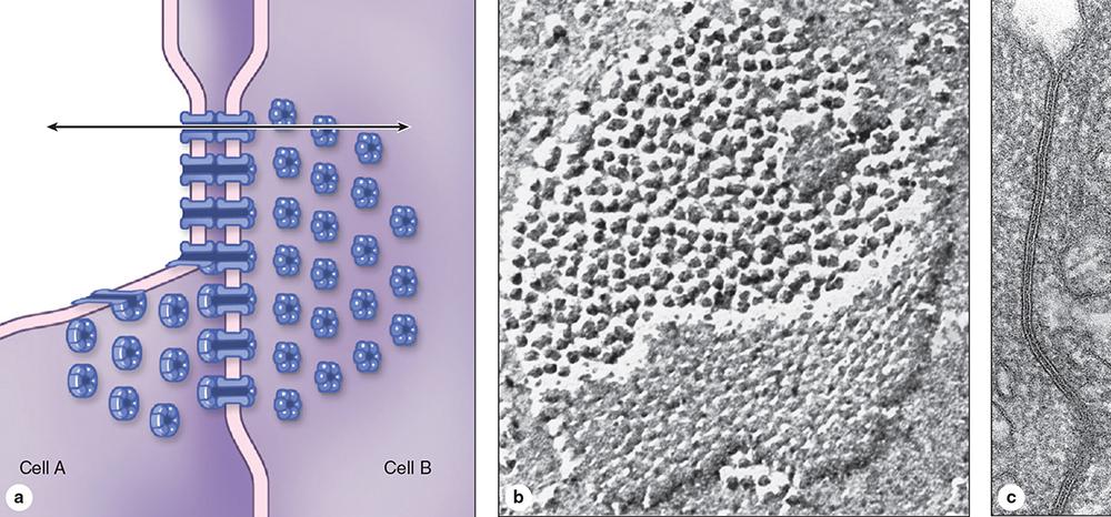
connexins
On the basal epithelial surface, cells attach to the basal lamina by anchoring junctions called (...), which can be seen by TEM.
hemidesmosomes
Another basal anchoring junction found in cells that are moving during epithelial repair or reorganization is the (...).
focal adhesion (or focal contact)
Integrins of focal adhesions are linked via paxillin to (...), a signaling protein which initiates a cascade of intracellular protein phosphorylation affecting cell adhesion.
focal adhesion kinase
In epithelia specialized for absorption the apical cell surfaces are often filled with an array of projecting (...), usually of uniform length.
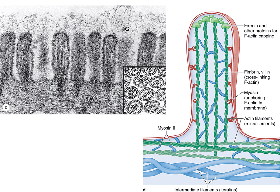
microvilli
In cells such as those lining the small intestine, densely packed microvilli are visible as a (...) projecting into the lumen.
brush or striated border
Each microvillus contains bundled actin filaments, which insert into the (...) of cortical microfilaments at the base of the microvilli.
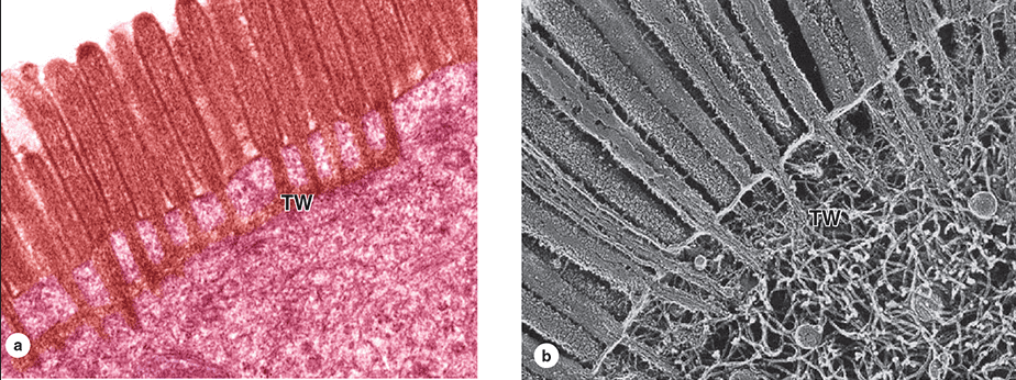
terminal web
A disorder of the small intestine in which there is loss of the microvilli brush border of the absorptive cells caused by an immune reaction against gluten.
Celiac disease (or "sprue")
A much less common type of apical process, best seen on the absorptive epithelial cells lining the male reproductive system.
stereocilia
Long, highly motile apical structures, larger than microvilli, and containing internal arrays of microtubules not microfilaments.
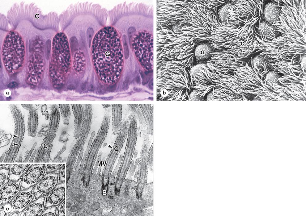
cilia
Each cilium has a core structure consisting of peripheral microtubule doublets arrayed around central microtubules, in a 9 + 2 assembly called an (...).
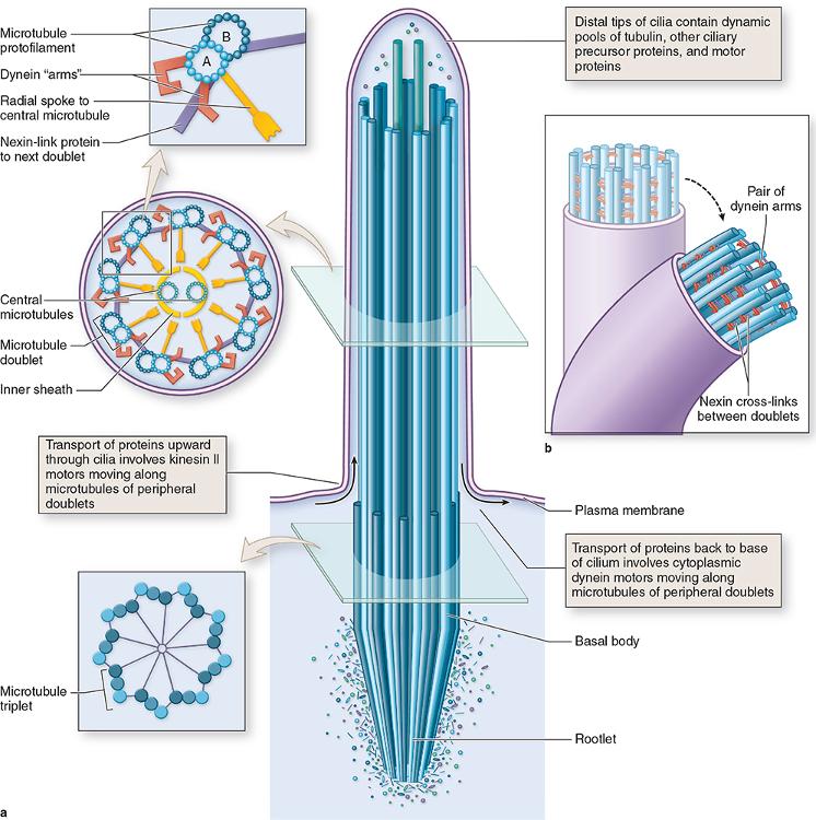
axoneme
Microtubules of axonemes are continuous with those in (...), which are apical cytoplasmic structures just below the cell membrane.
basal bodies
Complexes with (...) bound to one microtubule in each doublet extend as “arms” toward a microtubule of the next doublet. With energy from ATP, sliding of adjacent doublets relative to each other bends the axoneme and a rapid series of these sliding movements produces the beating motion of the cilium.
axonemal dynein
Syndrome of chronic respiratory infections, caused by the lack of ciliary action in the respiratory tract, and male infertility, produced by immotile spermatozoa.
immotile cilia syndrome (Kartagener syndrome)
The two main groups of epithelia.
covering (or lining) and secretory (or glandular)
Epithelium that contain one cell layer.
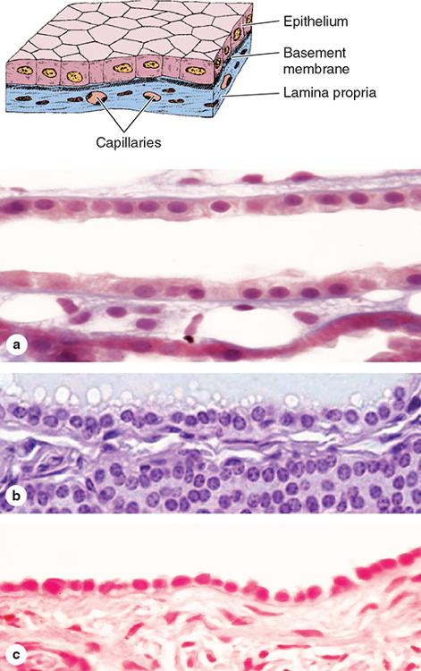
simple epithelium
Epithelium typically specialized as lining of vessels and cavities, where it regulates passage of substances into the underlying tissue.
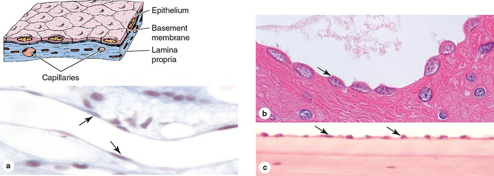
simple squamous
Epithelium found in the renal collecting tubule, rich in mitochondria and other organelles for a active transport across the epithelium.
simple cuboidal
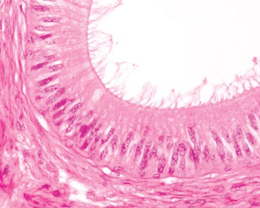
Epithelium with tall cells having apical cilia or microvilli, often specialized for absorption, found in the oviduct lining.
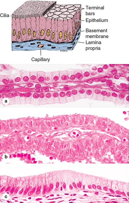
simple columnar
Epithelium contisting of more than one cell layer.
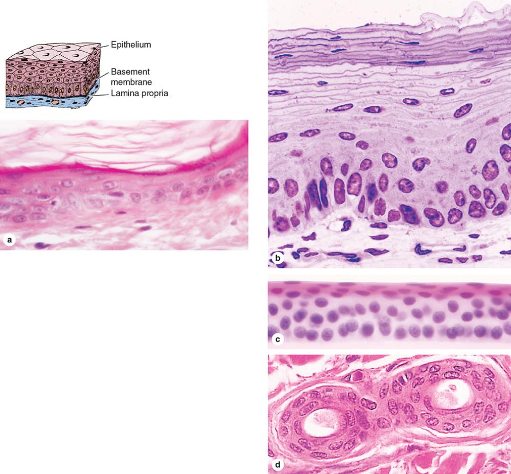
stratified epithelium
The surface cells of stratified squamous epithelia can be “(...)," meaning packed with keratin filaments, or “(...)," meaning they have relatively sparse keratin.
keratinized; nonkeratinized
Epithelium that is found mainly in the epidermis of skin, where it helps prevent dehydration from the tissue.
keratinized stratified squamous epithelium
Epithelium that lines moist internal cavities, such as the mouth, esophagus, and vagina, where water loss is not a problem.
nonkeratinized stratified squamous epithelium
Epithelium found in the excretory ducts of salivary and sweat glands.
stratified cuboidal epithelium
Epithelium found in the conjunctiva lining the eyelids, where it is both protective and mucus secreting.
stratified columnar epithelium
Epithelium found in the urinary tract, characterized by a superficial layer of large, dome-like cells sometimes called umbrella cells, which allow for distension.
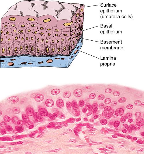
transitional epithelium (or urothelium)
Epithelium found in the upper respiratory tract, characterized by tall, irregular cells attached to the basement membrane but their nuclei are at different levels.
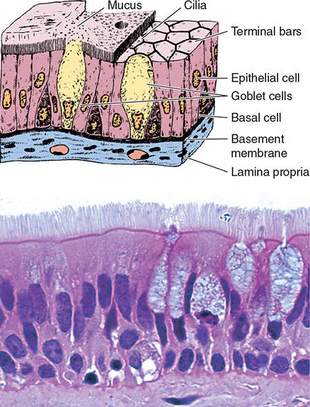
pseudostratified columnar epithelium
Organs comprised of specialized epithelial cells that function mainly to produce and secrete various macromolecules.
glands
Unicellular glands abundant in the lining of the small intestine which secrete lubricating mucus that aids the function of these organs.
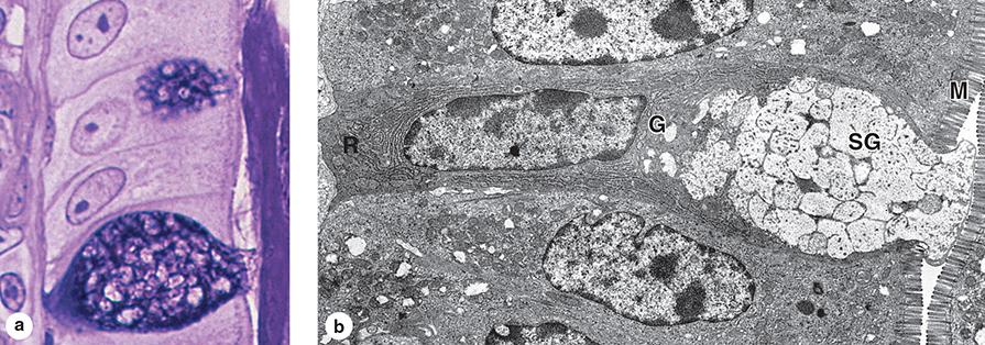
goblet cells
Glands that remain connected with the surface epithelium through tubular ducts lined with epithelium that deliver the secreted material.
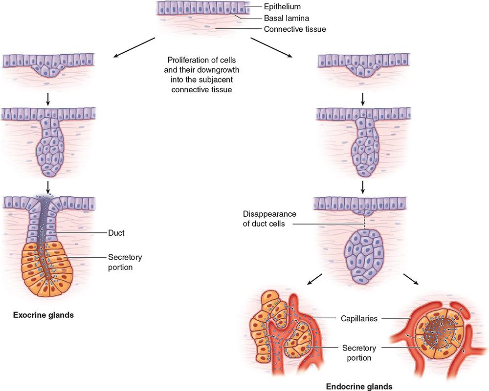
exocrine glands
Glands that lose their connection to epithelium and therefore lack ducts, secreting their hormone products for transport in blood.
endocrine glands
Epithelia of exocrine glands are organized as a continuous system of many small (...) that transport the secretion out of the gland.
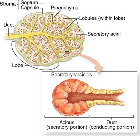
ducts
Glands with ducts that are not branched are called (...), where glands with ducts having two or more branches are termed (...).
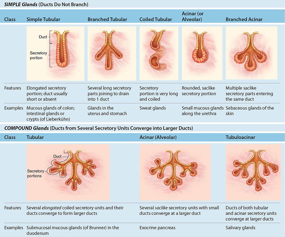
simple; compound
Glands with short or long and coiled secretory portions are called (...), where lands with rounded and saclike secretory portions are termed (...).
tubular; acinar
Glands that have branching ducts and can have multiple tubular, acinar, or tubuloacinar secretory portions.
compound glands
This is the most common method of protein or glycoprotein secretion and involves typical exocytosis from membrane-bound vesicles or secretory granules.
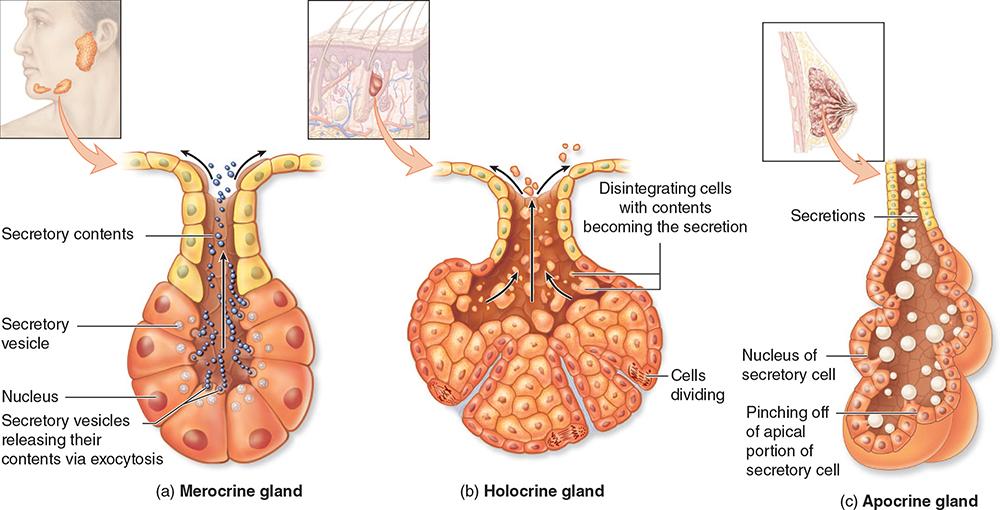
merocrine secretion
Here cells accumulate product continuously as they enlarge and undergo terminal differentiation, culminating in complete cell disruption that releases the product and cell debris into the gland’s lumen (e.g. sebaceous glands).
holocrine secretion
Here product accumulates at the cells’ apical ends, portions of which are then extruded to release the product together with small amounts of cytoplasm and cell membrane (e.g. mammary glands).
apocrine secretion
Mucous cells have RER and Golgi complexes and are filled apically with secretory granules that contain heavily glycosylated proteins called (...), which when released from the cell become hydrated and form a layer of (...).
mucins; mucus
The hydrophilic mucins are usually washed from mucous cells, causing the secretory granules to stain poorly with eosin. However, sufficient oligosaccharides remain to allow mucous cells to be stained by (...)
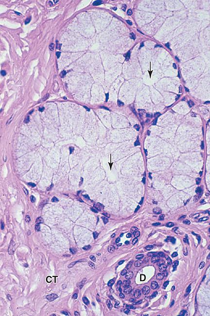
PAS method
Some salivary glands are mixed (...) glands, having both serous acini and mucous tubules with clustered serous cells. The product of such glands is a mixture of digestive enzymes and watery mucus.
seromucous
Many exocrine glands contain contractile (...) at the basal ends of the secretory cells which help propel secretory products from acini into the duct system.
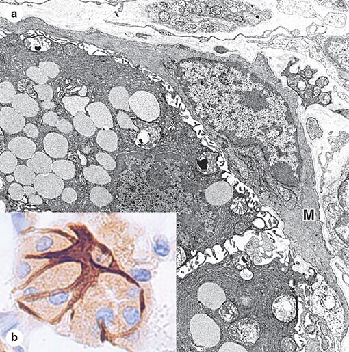
myoepithelial cells
Endocrine glands lack myoepithelial cells and are specialized for (...) or (...) synthesis, with cytoplasmic staining characteristic of RER or SER, respectively.
protein hormone; steroid hormone
Some epithelial cells specialize in the transfer of ions and water in either direction across the epithelium, the process is known as (...).
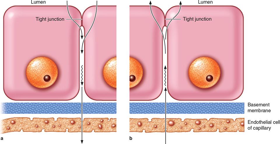
transcellular transport
The process of transport from the apical to the basolateral epithelial cell membrane domains.
absorption
The process of transport from the basolateral to the apical epithelial cell membrane domains.
secretion
Epithelia of kidney tubules are key sites for ion and water transport; the basal membrane of these cells is elaborately folded, with mitochondria located between the folds to supply ATP for (...).
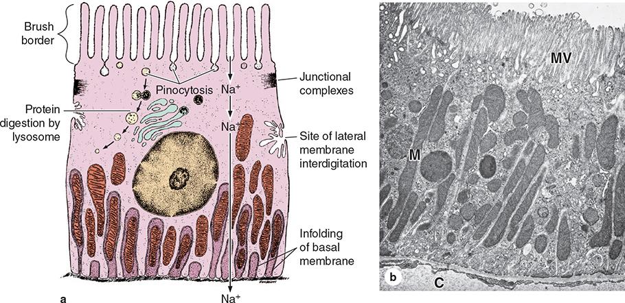
Na+/K+ pumps
Malignant tumors of epithelial origin.
carcinomas
Malignant tumors derived from glandular epithelial tissue.
adenocarcinomas
Some epithelial cells are prone to abnormal growth or dysplasia, which can progress to precancerous growth called (...).
neoplasia
Under certain abnormal conditions, one type of epithelial tissue may undergo transformation into another type in another reversible process called (...). In heavy cigarette smokers, the (...) epithelium lining the bronchi can be transformed into (...) epithelium.
metaplasia; ciliated pseudostratified; stratified squamous