Extremities
Which of the following is recommended to better demonstrate the tarsometatarsal joints in a dorsoplantar projection of the foot?
A Invert the foot.
B Evert the foot.
C Angle the CR 10 degrees posteriorly.
D Angle the CR 10 degrees anteriorly.
Angle the CR 10 degrees posteriorly.
To better demonstrate the interphalangeal joints of the toes, which of the following procedures may be employed?
- Angle the CR 15 degrees caudad.
- Angle the CR 15 degrees cephalad.
- Place a sponge wedge under the foot with the toes elevated 15 degree
2 and 3 only
The scapula shown in Figure 2–29 demonstrates
- its posterior aspect
- its costal surface
- its sternal articular surface
1 only
Which of the following projections of the ankle would best demonstrate the distal tibiofibular joint?
A Medial oblique 15° to 20°
B Lateral oblique 15° to 20°
C Medial oblique 45°
D Lateral oblique 45°
Medial oblique 45°
In the lateral projection of the scapula, the
- vertebral and axillary borders are superimposed.
- acromion and coracoid processes are superimposed.
- inferior angle is superimposed on the ribs.
1 only
The medical term for congenital clubfoot is
A coxa plana. B osteochondritis. C talipes. D muscular dystrophy
talipes.
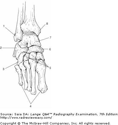
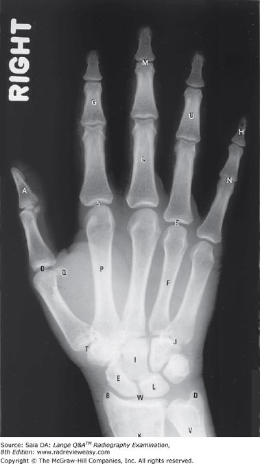
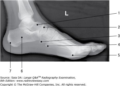
Which of the labeled bones in Figure A identifies the tarsal navicular?
Number 6
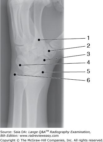
What is the structure labeled number 5 in Figure 2–37?
A Trapezium B Scaphoid C Ulnar styloid D Radial styloid
Radial styloid
Which of the following projections of the elbow should demonstrate the radial head free of ulnar superimposition?
A AP B Lateral C Medial oblique D Lateral oblique
Lateral oblique
Which of the following is proximal to the carpal bones?
A Distal interphalangeal joints B Proximal interphalangeal joints C Metacarpals D Radial styloid
Radial styloid process
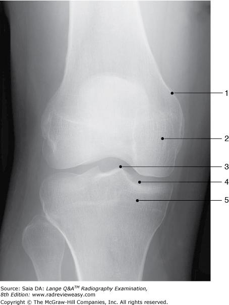
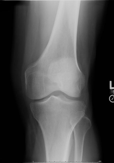
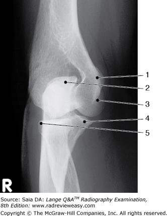
Identify the structure labeled 1 in the AP projection of the knee shown in Figure 2–16.
A Lateral condyle B Lateral epicondyle C Medial condyle D Medial epicondyle
Medial epicondyle
Impingement on the wrist's median nerve causing pain and disability of the affected hand and wrist is known as
A carpal boss syndrome B carpal tunnel syndrome C carpopedal syndrome D radioulnar syndrome
carpal tunnel syndrome
Which of the following is (are) distal to the tibial plateau?
- Intercondyloid fossa
- Tibial condyles
- Tibial tuberosity
2 and 3 only
In the lateral projection of the foot, the
- plantar surface should be perpendicular to the IR.
- metatarsals are superimposed.
- talofibular joint should be visualized.
1 and 2 only

The structure labeled number 4 in Figure 2–41 is the
coracoid process
What is the name of the structure indicated as number 5 in Figure 7–7?
Olecranon fossa
With the patient positioned as shown in Figure 6–13, how should the CR be directed to best demonstrate the intercondyloid fossa?
A Perpendicular to the popliteal depression
B 40 degrees caudad to the popliteal depression
C Perpendicular to the long axis of the femur
D 40 degrees cephalad to the popliteal depression
40 degrees caudad to the popliteal depression
In the lateral projection of the ankle, the
- talotibial joint is visualized.
- talofibular joint is visualized.
- tibia and fibula are superimposed
1 and 3 only
Demonstration of the posterior fat pad on the lateral projection of the adult elbow can be caused by
- trauma or other pathology
- greater than 90-degree flexion
- less than 90-degree flexion
1 and 3 only

Which of the labeled bones in Figure 6–14 identifies the tarsal navicular?
Number 6
Which of the following are components of a trimalleolar fracture?
- Fractured lateral malleolus
- Fractured medial malleolus
- Fractured posterior tibia
1, 2, and 3
Which of the following bones participate(s) in the formation of the knee joint?
- Femur
- Tibia
- Patella
1 and 2 only
Adult orthoroentgenography, or radiographic measurement of long bones of an upper or lower extremity, requires which of the following accessories?
- Bell-Thompson scale
- Bucky tray
- Cannula
1 and 2 only
Which of the following projections of the elbow should demonstrate the coronoid process free of superimposition and the olecranon process within the olecranon fossa?
A AP B Lateral C Medial oblique D Lateral oblique
Medial oblique
With the patient seated at the end of the x-ray table, elbow flexed 80 degrees, and the CR directed 45 degrees laterally from the shoulder to the elbow joint , which of the following structures will be demonstrated best?
Coronoid process
Which of the following correctly identifies the letter T in the radiograph shown in Figure 7–13?
Diarthrotic joint
Which of the following articulations may be described as diarthrotic?
- Knee
- Intervertebral joints
- Temporomandibular joint (TMJ)
1 and 3 only

In which of the following projections was the image in Figure 2–7 made?
medial oblique
All the following structures are associated with the posterior femur except
A popliteal surface B intercondyloid fossa C intertrochanteric line D linea aspera
intertrochanteric line
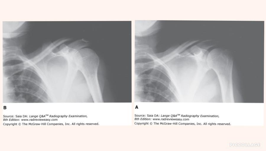
Which of the following statements is (are) true regarding the images shown in Figure 2–33?
- Image A is positioned in internal rotation.
- Image B is positioned in internal rotation.
- The greater tubercle is better demonstrated in image A.
2 and 3 only
All the following can be associated with the elbow joint except
A the capitulum. B the trochlea. C the tubercles. D the epicondyles
the tubercles.

What is the anatomic structure indicated by the number 7 in Figure 6–22?
note: number 6 - coronoid process
Radial notch of the ulna
Which of the following positions would best demonstrate the proximal tibiofibular articulation?
A. AP
B. 90 degrees mediolateral
C. 45-degree internal rotation
D. 45-degree external rotation
45-degree internal rotation
The relationship between the ends of fractured long bones is referred to as
apposition
The bone labeled number 3 in Figure 7–15 is the
A talus B cuboid C navicular D lateral cuneiform
cuboid
Skeletal conditions characterized by faulty bone
calcification include
1. osteoarthritis.
2. osteomalacia.
3. rickets.
2 and 3 only
The term varus refers to
the term valgus refers to
turned inward
turned outward
The AP oblique projection (medial rotation) of the elbow demonstrates
which of the following?
1. Radial head free of superimposition
2. Olecranon process within the olecranon fossa
3. Coronoid process free of superimposition
2 and 3 only
An AP oblique (lateral rotation) of the elbow demonstrates which of the following?
- Radial head free of superimposition
- Capitulum of the humerus
- Olecranon process within the olecranon fossa
1 and 2 only
In the 15° medial oblique projection of the
ankle, demonstrates the entire
1. talofibular joint.
2. tibiotalar joint.
3. ankle mortise.
1, 2, and 3
When examining a patient whose elbow is in partial flexion,
A the AP projection requires two separate positions and exposures.
B the AP projection is made through the partially flexed elbow, resting on the olecranon process, CR perpendicular to IR.
C the AP projection is made through the partially flexed elbow, resting on the olecranon process, CR parallel to the humerus.
D the AP projection is eliminated from the routine.
the AP projection requires two separate positions and exposures.

In Figure 2–29, which of the following is represented by the number 3?
Acromion process
What does the number 8 in Figure 6–14 identify?
Medial malleolus
With the patient positioned as illustrated in Figure 2–20, which of the following structures is best demonstrated?
A Patella B Patellofemoral articulation C Intercondyloid fossa D Tibial tuberosit
Intercondyloid fossa
The primary center of ossification in long bones is the
diaphysis
The secondary center of ossification in long bones is the
epiphysis

Figure A was made in which of the following positions?
Lateral oblique
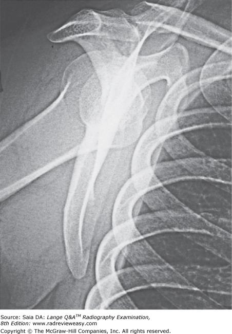
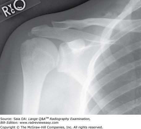
Which of the following statements is (are) true with respect to the radiograph shown in the Figure below?
- The acromion process is seen partially superimposed on the third rib.
- This projection is performed to evaluate the scapula.
- This projection is performed to evaluate the acromioclavicular articulation.
2 only
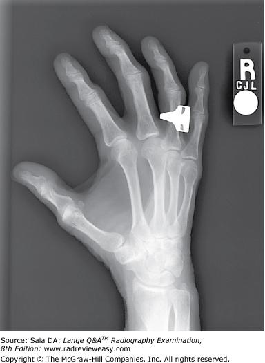
Which of the following fracture classifications describes a small bony fragment pulled from a bony process?
A Avulsion fracture B Torus fracture C Comminuted fracture D Compound fracture
Avulsion fracture
Which of the following statements regarding Figure 2–10 is (are) true?
- Correct degree of rotation is present.
- Midphalanges are foreshortened.
- Fingers are parallel to the IR
1 and 2 only
The carpal scaphoid can be demonstrated in which of the following projection(s) of the wrist?
- PA oblique
- PA with radial flexion
- PA with elbow elevated 20 degrees
1 only
Which position of the shoulder demonstrates the lesser tubercle in profile medially?
Internal rotation

What projection was used to obtain the image seen in Figure 2–41?
AP, external rotation
Which of the following shoulder projections can be used to evaluate the lesser tubercle in profile?
Internal rotation position
Which of the following is most likely to be the correct routine for a radiographic examination of the forearm?
AP and lateral
Which of the following articulations participate(s) in formation of the ankle mortise?
- Talotibial
- Talocalcaneal
- Talofibular
1 and 3 only
What is the most superior structure of the scapula?
A Apex
B Acromion process
C Coracoid process
D Superior angle
Acromion process
All of the following statements regarding the inferosuperior axial (nontrauma, Lawrence method) projection of the shoulder are true, except:
A the coracoid process and lesser tubercle are seen in profile.
B the arm is abducted about 90° from the body.
C the arm should be in internal rotation.
D the CR is directed medially 25° to 30° through the axilla.
the arm should be in internal rotation.
All the following can be associated with the distal ulna except
A head. B radioulnar joint. C styloid process. D trochlear notch.
trochlear notch.
Medial displacement of a tibial fracture would be best demonstrated in the
A AP projection
B lateral projection
C medial oblique projection
D lateral oblique projection
AP projection
Cells concerned with the formation and repair of bone are
A osteoblasts. B osteoclasts. C osteomas. D osteons
osteoblasts.
In which projection of the foot are the interspaces between the first and second cuneiforms best demonstrated?
A Lateral oblique foot B Medial oblique foot C Lateral foot D Weight-bearing foot
Lateral oblique foot
Which of the following is (are) located on the proximal aspect of the humerus?
- Intertubercular groove
- Capitulum
- Coronoid fossa
1 only
Which of the following articulations participate in the formation of the elbow joint?
1. Between the humeral trochlea and the semilunar/trochlear notch
2. Between the capitulum and the radial head
3. The proximal radioulnar joint
1 2 and 3
The term that refers to parts away from the source or beginning is
distal
The radiograph shown in Figure 7–12 can be produced with the
- long axis of the plantar surface perpendicular to the IR
- CR 40 degrees cephalad to the base of the third metatarsal
- CR 20 degrees cephalad to the talotibial joint
1 and 2 only
Which of the following projections require(s) that the humeral epicondyles be perpendicular to the IR?
- AP humerus
- Lateral forearm
- Internal rotation shoulder
2 and 3 only
To better visualize the knee-joint space in the radiograph in Figure 2–31, the radiographer should
A flex the knee more acutely
B flex the knee less acutely
C angle the CR 5 to 7 degrees cephalad
D angle the CR 5 to 7 degrees caudad
angle the CR 5 to 7 degrees cephalad
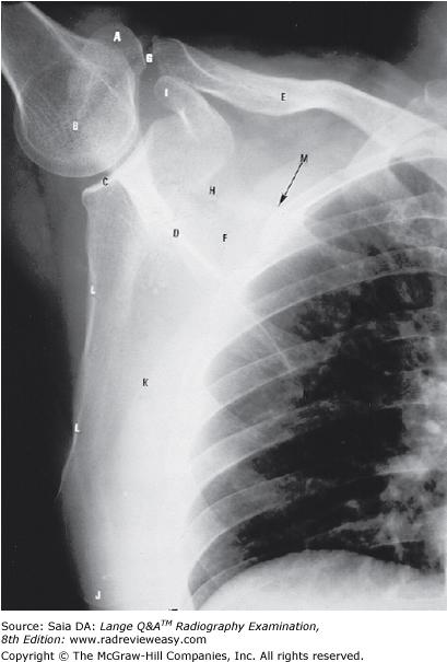
Which of the following indicates the scapular costal surface seen in the figure below?
K
Examples of synovial pivot articulations include the
- atlantoaxial joint
- radioulnar joint
- temporomandibular joint
1 and 2 only
A modified axiolateral inferosuperior projection of the
femoral neck is particularly useful
1. when the
"cross-table" axiolateral is contraindicated.
2. for patients with bilateral hip fractures.
3. for patients with limited movement of the unaffected leg.
1, 2, and 3
In which of the following projections is the talofibular joint best demonstrated?
A AP
B Lateral oblique
C Medial oblique
D Lateral
Medial oblique
Which of the following projections will best demonstrate the carpal scaphoid?
A Lateral wrist B Ulnar deviation C Radial deviation D Carpal tunnel
Ulnar deviation
All the following are posterior structures except
A the linea aspera. B the intertrochanteric line. C the popliteal surface. D the intercondyloid fossa.
the intertrochanteric line.
Which of the following correctly identifies the head of the ulna in the illustration in Figure 6–22?
Number 9
For the AP projection of the scapula, the
- patient's arm is abducted at right angles to the body.
- patient's elbow is flexed.
- exposure is made during quiet breathing.
1, 2, and 3
In the 45-degree medial oblique projection of the ankle, the
- talotibial joint is visualized
- tibiofibular joint is visualized
- plantar surface should be vertical
2 and 3 only
Which of the following projections will best demonstrate acromioclavicular separation?
A AP recumbent, affected shoulder
B AP recumbent, both shoulders
C AP erect, affected shoulder
D AP erect, both shoulders
AP erect, both shoulders
For an AP projection of the knee on a patient whose measurement from ASIS to tabletop is 21 cm, which CR direction will best demonstrate the knee joint?
SN: 19 cm (thin pelvis), the CR should be directed 3 to 5 degrees caudad; when the distance is between 19 to 24 cm, the CR is directed vertically/perpendicular (0 degrees); when the distance is greater than 24 cm (thick pelvis), the CR is directed 3 to 5 degrees cephalad.
0 degrees (perpendicular)
Which of the following articulates with the base of the fifth metatarsal?
A First cuneiform B Third cuneiform C Navicular D Cuboid
Cuboid
Which of the following projections of the ankle would best demonstrate the mortise?
A Medial oblique 15 to 20 degrees
B Lateral oblique 15 to 20 degrees
C Medial oblique 45 degrees
D Lateral oblique 45 degrees
Medial oblique 15 to 20 degrees
How can OID be reduced for a PA projection of the wrist?
A Extend the fingers.
B Flex the metacarpophalangeal joints.
C Extend the forearm.
D Oblique the metacarpals 45 degrees
Flex the metacarpophalangeal joints.
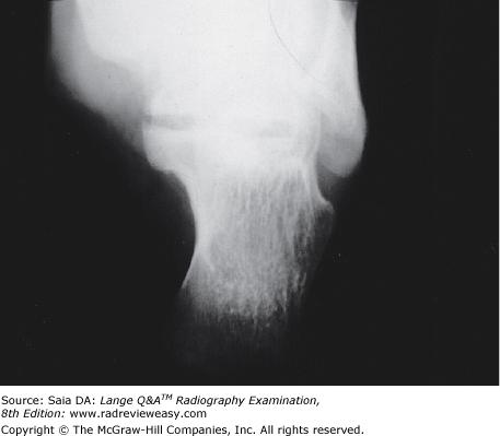
Which of the following conditions is limited specifically to the tibial tuberosity?
A Ewing sarcoma
B Osgood–Schlatter disease
C Gout D
Exostosis
Osgood–Schlatter disease
In the lateral projection of the knee, the central ray is angled 5° cephalad to prevent superimposition of which of the following structures on the joint space?
Medial femoral condyle
Which of the following is (are) valid criteria for a
lateral projection of the forearm?
1. The radius and
ulna should be superimposed proximally and distally.
2. The coronoid process and radial head should be superimposed.
3. The radial tuberosity should face anteriorly.
2 and 3 only
The instrument that is used frequently in quality-control programs to measure varying degrees of x-ray exposure is the
A aluminum step wedge. B spinning top. C densitometer. D sensitometer
densitometer.
Which of the following articulate(s) with the bases of the metatarsals?
- The heads of the first row of phalanges
- The cuboid
- The cuneiforms
2 and 3 only
Which of the following is (are) valid evaluation criteria for a lateral projection of the forearm?
- The radius and the ulna should be superimposed distally.
- The coronoid process and the radial head should be partially superimposed.
- The humeral epicondyles should be superimposed.
1, 2, and 3
When examining a patient whose elbow is in partial flexion, how should an AP projection be obtained?
- With humerus parallel to IR, CR perpendicular
- With forearm parallel to IR, CR perpendicular
- Through the partially flexed elbow, resting on the olecranon process, CR perpendicular
1 and 2 only
The greater tubercle should be visualized in profile in which of the following?
A AP shoulder, external rotation
B AP shoulder, internal rotation
C AP elbow
D Lateral elbow
AP shoulder, external rotation
Which of the following statements regarding the radiograph in Figure A is (are) true?
1. The tibial eminences are well visualized.
2. The intercondyloid fossa is demonstrated between the femoral condyles.
3. The femorotibial articulation is well demonstrated.
1 and 3 only
Which type of articulation is evaluated in arthrography?
A Synarthrodial B Diarthrodial C Amphiarthrodial D Cartilaginous
Diarthrodial
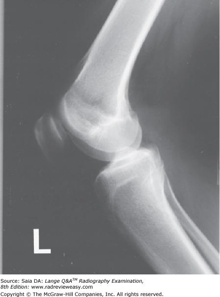
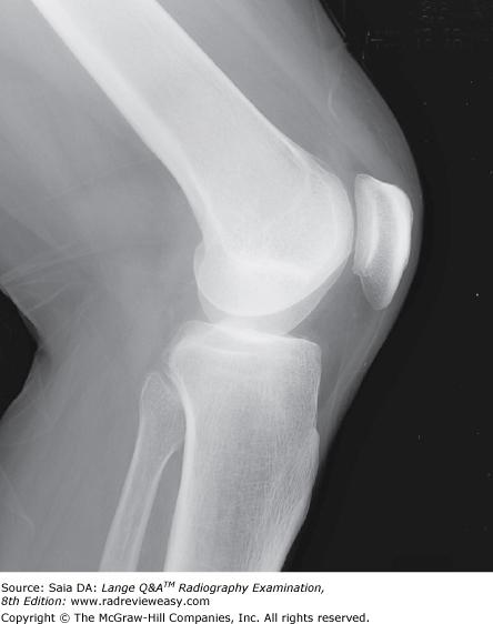
In a lateral projection of the normal knee, the
- fibular head should be somewhat superimposed on the proximal tibia.
- patellofemoral joint should be visualized.
- femoral condyles should be superimposed.
1, 2, and 3

Which of the following correctly identifies the letter L in the radiograph shown in Figure 7–13?
Lunate
Which of the following positions would be the best choice for a right shoulder examination to rule out fracture?
AP and scapular Y

What is the structure labeled number 2 in Figure 2–37?
Trapezium
Which of the following is (are) true regarding radiographic examination of the acromioclavicular joints?
- The procedure is performed in the erect position.
- Use of weights can improve demonstration of the joints.
- The procedure should be avoided if dislocation or separation is suspected.
1 and 2 only
To demonstrate a profile view of the glenoid fossa, the patient is AP recumbent and obliqued 45 degrees
A toward the affected side
B away from the affected side
C with the arm at the side in the anatomic position
D with the arm in external rotation
toward the affected side
The mediolateral projection of the knee shown in Figure 6–1 could best be improved by
angling the CR about 5 degrees cephalad
Which of the following may be used to evaluate the glenohumeral joint?
- Scapular Y projection
- Inferosuperior axial
- Transthoracic lateral
1, 2, and 3
In which of the following tangential axial projections of the patella is complete relaxation of the quadriceps femoris required for an accurate diagnosis?
- Supine flexion 45 degrees (Merchant)
- Prone flexion 90 degrees (Settegast)
- Prone flexion 55 degrees (Hughston)
1 only
Synovial fluid is associated with the
A brain. B spinal canal. C peritoneal cavity. D bony articulations
bony articulations.
Which of the following is an important consideration to avoid excessive metacarpal joint overlap in the oblique projection of the hand?
A Oblique the hand no more than 45 degrees.
B Use a support sponge for the phalanges.
C Clench the fist to bring the carpals closer to the IR.
D Use ulnar flexion.
Oblique the hand no more than 45 degrees.
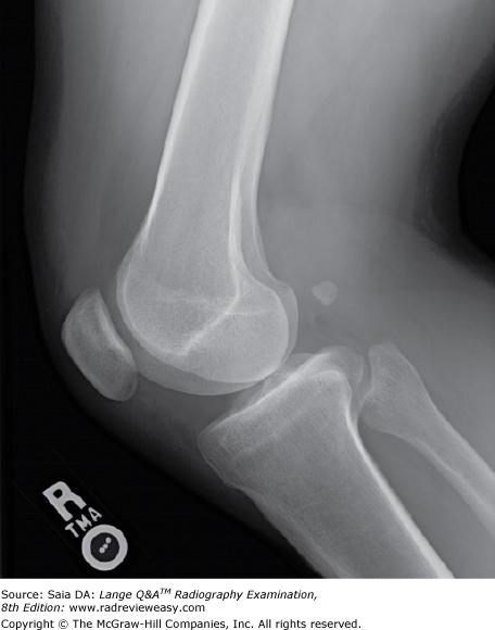
What could be done to improve the mediolateral projection of the knee seen in Figure 2–3?
A Rotate the pelvis slightly forward/anteriorly.
B Rotate the pelvis slightly backward/posteriorly.
C Angle the x-ray tube 5 degrees cephalad.
D Angle the x-ray tube 5 degrees caudad.
Rotate the pelvis slightly backward/posteriorly.
The lesser tubercle of the humerus will be visualized in profile in the
AP shoulder internal rotation radiograph.
Ulnar deviation will best demonstrate which carpal(s)?
- Medial carpals
- Lateral carpals
- Scaphoid
2 and 3 only
A compression fracture of the posterolateral humeral head and associated with an anterior dislocation of the glenohumeral joint is called a(an)
A Hill-Sachs defect.
B Bankart lesion.
C rotator cuff tear.
D adhesive capsulitis.
Hill-Sachs defect.
Which of the following articulates with the base of the first metatarsal?
A First cuneiform B Third cuneiform C Navicular D Cuboid
First cuneiform
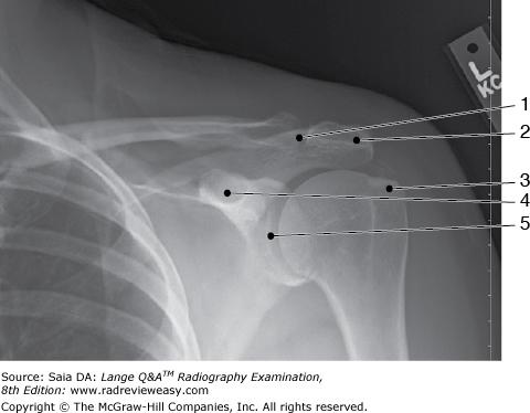
The structure labeled number 5 in Figure 2–41 is the
glenohumeral joint
In which projection of the foot are the sinus tarsi, cuboid, and tuberosity of the fifth metatarsal best demonstrated?
Medial oblique foot
In which of the following positions can the sesamoid bones of the foot be demonstrated to be free of superimposition with the metatarsals or phalanges?
Tangential metatarsals/toes
The first carpometacarpal joint is formed by the articulation of the base of the first metacarpal and the
trapezium.
Which of the following projection(s) require(s) that
the shoulder be placed in internal rotation?
1. AP humerus
2. AP thumb
3. Lateral humerus
2 and 3 only
With which of the following does the trapezium articulate?
A Fifth metacarpal B First metacarpal C Distal radius D Distal ulna
First metacarpal
Which of the following projections will best demonstrate the tarsal navicular free of superimposition?
A AP oblique, medial rotation
B AP oblique, lateral rotation
C Mediolateral
D Lateral weight-bearing
AP oblique, medial rotation
What should be done to better demonstrate the coracoid process shown in Figure 2–22?
A Use a perpendicular CR.
B Angle the CR about 30 degrees cephalad.
C Angle the CR about 30 degrees caudad.
D Angle the MSP 15 degrees toward the affected side.
Angle the CR about 30 degrees cephalad
The fifth metacarpal is located on which aspect of the hand?
Medial
Muscles that contribute to the formation of the rotator cuff include the
1. subscapularis.
2. infraspinatus.
3. teres minor.
1, 2, and 3
The tarsals and metatarsals are arranged to form the
1. transverse arch.
2. longitudinal arch.
3. oblique arch.
1 and 2 only

Which of the following projections/positions would best demonstrate structure number 6 seen in Figure 7–7?
Lateral projection
Important considerations for radiographic examinations of traumatic injuries to the upper extremity include
- only the joint closest to the injured site must be supported during movement.
- both joints must be included in long bone studies.
- two views, at 90 degrees to each other, are required.
2 and 3 only
Which of the following projections require(s) that the shoulder be placed in external rotation?
- AP humerus
- Lateral forearm
- Lateral humerus
1 only
The functions of which body system include mineral homeostasis, protection, and triglyceride storage?
A Endocrine B Integumentary C Skeletal D Muscular
Skeletal

The radiograph shown in Figure 2–15 demonstrates the articulation between the
- talus and the calcaneus
- calcaneus and the cuboid
- talus and the navicular
2 and 3 only
What process is best seen using a perpendicular CR with the elbow in acute flexion and with the posterior aspect of the humerus adjacent to the image receptor?
A Coracoid B Coronoid C Olecranon D Glenoid
Olecranon
Valid evaluation criteria for a lateral projection of the forearm requires that
- the epicondyles be parallel to the IR.
- the radius and ulna be superimposed distally.
- the radial tuberosity should face anteriorly.
2 and 3 only
Conditions in which there is a lack of normal bone calcification include
- rickets.
- osteomalacia.
- osteoarthritis.
1 and 2 only
Which of the following projections or positions will best demonstrate subacromial or subcoracoid dislocation?
A Tangential
B AP axial
C Transthoracic lateral
D PA oblique scapular
PA oblique scapular Y
Tangential axial projections of the patella
can be obtained in which of the following positions?
1. supine
flexion 45° (Merchant)
2. prone flexion 90° (Settegast)
3. prone flexion 55° (Hughston)
1, 2, and 3 only
What projection of the calcaneus is obtained with the leg extended, the plantar surface of the foot vertical and perpendicular to the IR, and the CR directed 40 degrees cephalad?
A Axial plantodorsal projection
B Axial dorsoplantar projection
C Lateral projection
D Weight-bearing lateral projection
Axial plantodorsal projection
Which of the following views would best demonstrate arthritic changes in the knees?
A AP recumbent
B Lateral recumbent
C AP erect
D Medial oblique
AP erect
To evaluate the interphalangeal joints in the oblique and lateral positions, the fingers
A rest on the cassette for immobilization
B must be supported parallel to the IR
C are radiographed in natural flexion
D are radiographed in palmar flexion
must be supported parallel to the IR
A lateral projection of the hand in extension is often recommended to evaluate
- a fracture
- a foreign body
- soft tissue
2 and 3 only
Which of the following can be used to demonstrate the intercondyloid fossa?
- Prone, knee flexed 40 degrees, CR directed caudad 40 degrees to the popliteal fossa
- Supine, IR under flexed knee, CR directed cephalad to knee, perpendicular to tibia
- Prone, patella parallel to IR, heel rotated 5 to 10 degrees lateral, CR perpendicular to knee joint
1 and 2 only
Shoulder arthrography is performed to
- evaluate humeral luxation
- demonstrate complete or partial rotator cuff tear
- evaluate the glenoid labrum
2 and 3 only
Which of the following positions will separate the radial head, neck, and tuberosity from superimposition on the ulna?
Lateral oblique
In the AP knee projection of an asthenic patient who measures less than 19 cm from the anterior superior iliac spine (ASIS) to tabletop, the CR should be directed
5 degrees caudad
Which of the following is (are) accurate positioning or evaluation criteria for an AP projection of the normal knee?
- Femorotibial interspaces equal bilaterally.
- Patella superimposed on distal tibia.
- CR enters ½ in. distal to base of patella.
1 only
Which of the following is most useful for bone age evaluation?
PA hand
Which of the following anatomic structures is indicated by the number 2 in Figure 2–7?
olecranon process
Which of the following should be demonstrated in a true AP projection of the clavicle?
- Clavicular body
- Acromioclavicular joint
- Sternocostal joint
1 and 2 only
A spontaneous fracture most likely would be associated with
A pathology. B crepitus. C trauma. D metabolism
pathology.
With the patient and the x-ray tube positioned as illustrated in Figure 2–2, which of the following will be visualized?
- Intercondyloid fossa
- Patellofemoral articulation
- Tangential patella
2 and 3 only
All the following can be associated with the distal radius except
A head.
B styloid process.
C ulnar notch.
D radioulnar joint.
head
Which of the following is (are) located on the distal
aspect of the humerus?
1. Capitulum
2. Intertubercular groove
3. Coronoid fossa
1 and 3 only
In which of the following positions/projections will the talocalcaneal joint be visualized?
A Dorsoplantar projection of the foot
B Plantodorsal projection of the calcaneus
C Medial oblique position of the foot
D Lateral foot
Plantodorsal projection of the calcaneus

What is the structure indicated by the letter A in Figure 7–3?
acromion process
Which of the following is (are) located on the anterior aspect of the femur?
- Patellar surface
- Intertrochanteric crest
- Linea aspera
1 only
Which of the following statements regarding the Norgaard method, “Ball-Catcher's position,” is (are) correct?
- Bilateral AP oblique hands are obtained.
- It is used for early detection of rheumatoid arthritis.
- The hands are obliqued about 45 degrees, palm up.
1, 2, and 3
What portion of the humerus articulates with the ulna to help form the elbow joint?
A Semilunar/trochlear notch B Radial head C Capitulum D Trochlea
Trochlea
The following procedure can be employed to better
demonstrate the carpal scaphoid:
1. elevate hand and
wrist 20°.
2. place wrist in ulnar deviation.
3. angle CR 20° distally (toward fingers).
1 and 2 only
Which of the following statements regarding knee x-ray arthrography is (are) true?
- Ligament tears can be demonstrated.
- Sterile technique is observed.
- MRI can follow x-ray.
1, 2, and 3
All of the following bones are associated with condyles except the
A femur. B tibia. C fibula. D mandible
fibula.
A patient unable to extend his or her arm is seated at the end of the x-ray table, elbow flexed 90 degrees, with epicondyles perpendicular to IR. The CR is directed 45 degrees medially. Which of the following structures will be demonstrated best ?
- Radial head
- Capitulum
- Coronoid process
1 and 2 only
Which of the following may be used to evaluate the
glenohumeral joint?
1. Scapular Y projection
2. Inferosuperior axial
3. Transthoracic lateral
1, 2, and 3
Which of the following positions is used to demonstrate vertical patellar fractures and the patellofemoral articulation?
A AP knee B Lateral knee C Tangential patella D Tunnel view
Tangential patella
Knee arthrography may be performed to
demonstrate a
1. torn meniscus.
2. Baker's cyst.
3. torn rotator cuff.
1 and 2 only
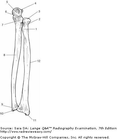
Which of the following correctly identifies the radial styloid process in the illustration in Figure A?
Number 11
In the AP projection of the ankle, the
- plantar surface of the foot is vertical.
- fibula projects more distally than the tibia.
- calcaneus is well visualized.
1 and 2 only
Which projection of the foot will best demonstrate the longitudinal arch?
A Mediolateral B Lateromedial C Lateral weight-bearing D 30-degree medial oblique
Lateral weight-bearing
Which of the following criteria is (are) required for
visualization of the greater tubercle in profile?
1.
Epicondyles parallel to the IR
2. Arm in external rotation
3. Humerus in AP position
1, 2, and 3
Which of the following is used to obtain a lateral projection of the upper humerus on patients who are unable to abduct their arm?
A Bicipital groove projection B Superoinferior lateral C Inferosuperior axial D Transthoracic lateral
Transthoracic lateral
Which of the following projections is most likely to demonstrate the carpal pisiform free of superimposition?
A Radial flexion/deviation B Ulnar flexion/deviation C AP (medial) oblique D AP (lateral) oblique
AP (medial) oblique
Which of the following projections is most likely to demonstrate the carpal pisiform free of superimposition?
AP (medial) oblique
With which of the following does the lateral extremity of the clavicle articulate?
Acromion process
Evaluation criteria for a lateral projection of the humerus include
- epicondyles parallel to the IR
- lesser tubercle in profile
- superimposed epicondyles
2 and 3 only

In Figure 2–29, which of the following is represented by the number 7?
A Medial border B Lateral border C Inferior angle D Superior angle
Lateral border
The secondary center of ossification in long bones is the
epiphysis.
AP stress studies of the ankle may be performed
- to demonstrate fractures of the distal tibia and fibula
- following inversion or eversion injuries
- to demonstrate a ligament tear
2 and 3 only
The best projection to demonstrate the articular surfaces of the femoropatellar articulation is the
tangential (“sunrise”) projection.
To demonstrate the glenoid fossa in profile, the patient is positioned
A 45 degrees oblique, affected side away from IR.
B 45 degrees oblique, affected side adjacent to IR.
C 25 degrees oblique, affected side away from IR.
D 25 degrees oblique, affected side adjacent to IR.
45 degrees oblique, affected side adjacent to IR.
Posterior displacement of a tibial fracture would be best demonstrated in the
A AP projection. B lateral projection. C medial oblique projection. D lateral oblique projection
lateral projection.