Exercise 2-2 Micro Lab: Colony Morphology
How does a colony form?
When a single bacterial cell is deposited on a solid nutrient medium, it begins to divide. One cell makes two, two makes four, four make eight. Eventually a visible mass of cells, a colony, appears.
In what 4 ways are color, size, shape, and texture of microbial growth determined?
1. genetic makeup of the organism
2. nutrient availability
3. temperature
4. incubation
The 5 basic categories of colony morphology
1. colony shape
2. margin (edge)
3. elevation
4. texture
5. pigment production (color)
3 types of colony shape
1. round
2. irregular
3. punctiform (tiny, pinpoint)
5 types of margin
1. entire (smooth with no irregularities)
2. undulate (wavy)
3. lobate (lobed)
4. filamentous
5. rhizoid (branched like roots)
entire
smooth with no irregularities
undulate
wavy
lobate
lobed
rhizoid
branched like roots
5 elevations of colonies
1. flat
2. raised
3. convex
4. pulvinate (very convex)
5. umbonate (raised in the center)
pulvinate
very convex
umbonate
raised in the center
3 types of texture
1. moist
2. mucoid
3. dry
4 types of pigment production (color)
1. opaque
2. translucent
3. shiny
4. dull
colony counter
used to view subtle differences in colony shape and size
what 2 things allow greater observation of detail in a colony counter?
1. transmitted light
2. magnifying glass
colony counter is best determined with ______.
reflected light
The grid in the colony counter background is a ______.
counting aid
6 colonies studied in this lab
1. micrococcus luteus
2. corynebacterium xerosis
3. lactobacillus plantarum
4. mycobacterium smegmatis
5. bacillus subtilis
6. proteus miabilis

Name these elevations, left to right
1. raised
2. raised, spreading edge
3. flat, raised margin
4. growth into medium

Name these elevations, left to right
1. convex
2. umbonate
3. plateau
4. flat
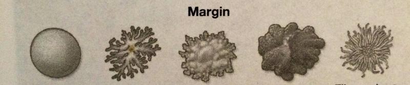
Name these margins, left to right
1. smooth, entire
2. rhizoid
3. irregular (erose)
4. lobate
5. filamentous
3 types of descriptions used to describe colonial morphology:
1. color
2. surface characteristics
3. consistency
4. optical properties
2 types of surface characteristics
1. dull
2. shiny
3 types of consistency
1. dry
2. butyrous-buttery
3. moist
2 types of optical properties
1. opaque
2. translucent
What bacteria is this?
What is the color, shape, elevation and margin?
Staphylococcus epidermidis
Color - white
Shape - circular
Elevation - raised
Margin - entire
What bacteria is this?
What 3 features that describe it?
Providencia stuartii
1. shiny
2. buff
3. convex
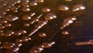
What bacteria is this?
What 3 features that describe it?
Klebsiella pneumoniae
1. mucoid
2. raised
3. shiny
What bacteria is this?
What 3 features that describe it?
Chromobacterium violaceum
1. shiny
2. purple
3. convex
What bacteria is this?
What is the color, shape, elevation and margin of it?
Enterococcus faecium
Color - white
Shape - circular
Elevation - Convex
Margin - Entire
What is this?
5 features of the colonies
A) Bacillus cereus
2) Bacillus anthracis
1. dull
2. dry
3. raised
4. rough-textured
5. gray

What is being shown in these two views? What bacteria is this? What are the 3 features of this bacteria?
A) Alcaligenes Faecalis side view showing a raised center
B) Alcaligenes Faecalis showing spreading edge
1. umbonate
2. opaque center
3. spreading edge
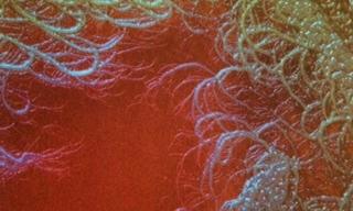
What is being shown here?
Filamentous growth
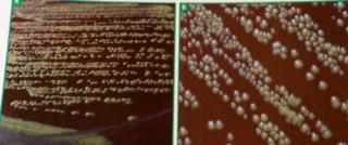
What bacteria is being shown in A and B and what are the differences between them?
What 2 features of the bacteria are being showed here?
Where is this bacteria found?
A) Clostridium sporaogenes grown on sheep blood agar and viewed with reflected light
B) Clostridium sporaogenes grown on nutrient agar and viewed with transmitted light
1. irregular
2. rhizoid
It is found in soil
_____ is an opportunistic pathogen
Staphylococcus epidermidis
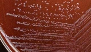
______ is a frequent isolate in urine samples obtained from hospitalized and catheterized patients
Providencia stuartii
______ is found in human and animal feces
Enterococcus faecium
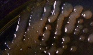
What bacteria is this?
What does this growth demonstrate?
Alcaligenes faecalis
Demonstrates spreading attributable to motility and is translucent
_______ is found in soil and water and rarely produces infections in humans
Chromobacterium violaceum
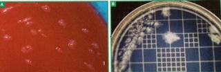
What are these slides showing?
What bacteria is this?
The effect of age on colony morphology after 24 and 48 hours of growth
Bacillus subtilis
A) After 24 hours
B) After 48 hours (note worm like appearance)
What bacteria is this?
This bacteria produces colonies with what 2 features?
Bacillus subtilis
1. raised margin
2. dull surface
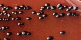
what bacteria is this?
what feature of this colony is being shown here?
Mycobacterium smegmatis
punctiform
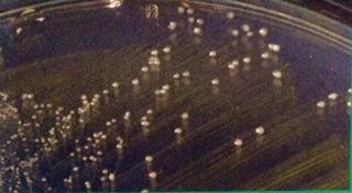
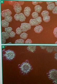
What bacteria is this?
In A, what 4 features of the bacteria are exhibited?
What is being shown here in B?
Coynebacterium xerosis
In A:
1. round
2. dull
3. buff
4. convex
In B: circular
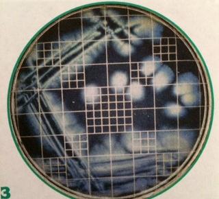
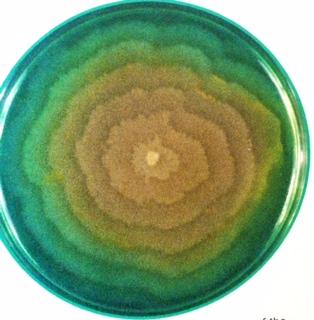
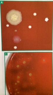
What bacteria is this?
What is being demonstrated in this dish?
Erwinia amylovora
the irregular shape and spreading edges
_________ is a plant pathogen.
Erwinia amylovora
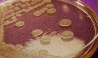
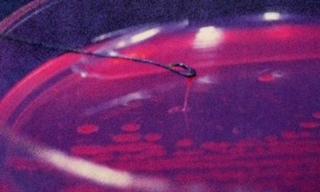
What bacteria is shown here?
What is this slide demonstrating?
Proteus vulgaris
swarming growth pattern due to bacteria motility
What bacteria is shown here?
What is being demonstrated here?
Pseudomonas aeruginosa
Mucoid texture
_____________ is found in soil and water and can cause infections in burn patients
Pseudomonas aeruginosa
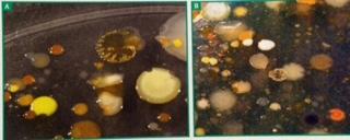
What are these samples showing?
Mixed soil cultures and diversity
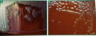
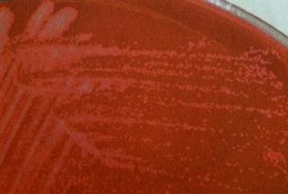
What bacteria is this showing? How do we know?
Staphylococcus aureus
White growth on blood agar plate demonstrating B-hemolysis is characteristic of staph. In A) the agar is darkening from the hemolysis. In B) there is weak hemolysis.
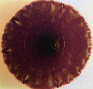
What bacteria is this?
Chromobacterium violaceum
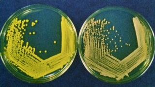
What bacteria is on the left? the right?
What is being demonstrated here?
Left - Micrococcus luteus
Right - Kocuria rosea
Pigment production in similar species of bacteria
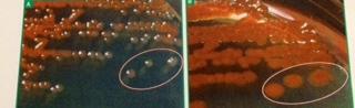
What bacteria is this?
What is being shown here with the encircled sections?
Serratia marcescens
the influence of age on pigment production after 24 and 48 hours
What bacteria is being shown here?
What is being demonstrated here?
Serratia marcescens
influence of temperature on pigment production when grown in 37 degrees C on left and 25 degrees C on right
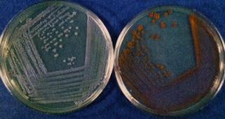
What bacteria is being shown here?
What is being demonstrated here?
Chromobacterium violaceum
The influence of nutrient availability on pigment production. The nutrient agar (right) has less nutrients so less pigment.
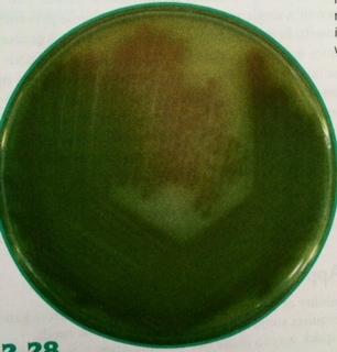
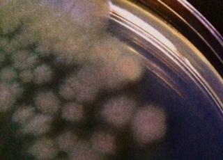
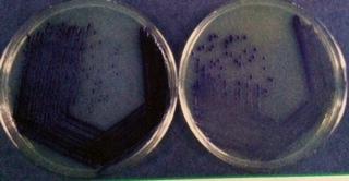
What bacteria is shown here?
What is being demonstrated here?
Pseudomonas aeruginosa
diffusible blue-green pigment