Instructions for Side by Side Printing
- Print the notecards
- Fold each page in half along the solid vertical line
- Cut out the notecards by cutting along each horizontal dotted line
- Optional: Glue, tape or staple the ends of each notecard together
Exercise 2-2 Micro Lab: Colony Morphology
front 1 How does a colony form? | back 1 When a single bacterial cell is deposited on a solid nutrient medium, it begins to divide. One cell makes two, two makes four, four make eight. Eventually a visible mass of cells, a colony, appears. |
front 2 In what 4 ways are color, size, shape, and texture of microbial growth determined? | back 2 1. genetic makeup of the organism
|
front 3 The 5 basic categories of colony morphology | back 3 1. colony shape
|
front 4 3 types of colony shape | back 4 1. round
|
front 5 5 types of margin | back 5 1. entire (smooth with no irregularities)
|
front 6 entire | back 6 smooth with no irregularities |
front 7 undulate | back 7 wavy |
front 8 lobate | back 8 lobed |
front 9 rhizoid | back 9 branched like roots |
front 10 5 elevations of colonies | back 10 1. flat
|
front 11 pulvinate | back 11 very convex |
front 12 umbonate | back 12 raised in the center |
front 13 3 types of texture | back 13 1. moist
|
front 14 4 types of pigment production (color) | back 14 1. opaque
|
front 15 colony counter | back 15 used to view subtle differences in colony shape and size |
front 16 what 2 things allow greater observation of detail in a colony counter? | back 16 1. transmitted light
|
front 17 colony counter is best determined with ______. | back 17 reflected light |
front 18 The grid in the colony counter background is a ______. | back 18 counting aid |
front 19 6 colonies studied in this lab | back 19 1. micrococcus luteus
|
front 20  Name these elevations, left to right | back 20 1. raised
|
front 21  Name these elevations, left to right | back 21 1. convex
|
front 22 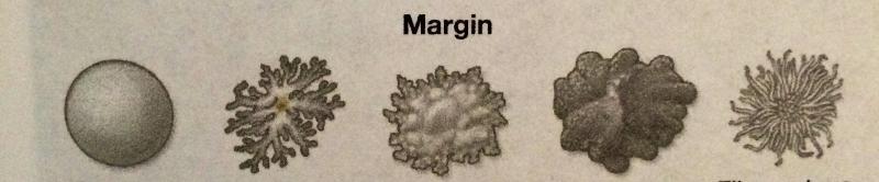 Name these margins, left to right | back 22 1. smooth, entire
|
front 23 3 types of descriptions used to describe colonial morphology: | back 23 1. color
|
front 24 2 types of surface characteristics | back 24 1. dull
|
front 25 3 types of consistency | back 25 1. dry
|
front 26 2 types of optical properties | back 26 1. opaque
|
front 27 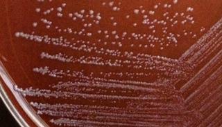 What bacteria is this?
| back 27 Staphylococcus epidermidis
|
front 28 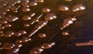 What bacteria is this?
| back 28 Providencia stuartii
|
front 29 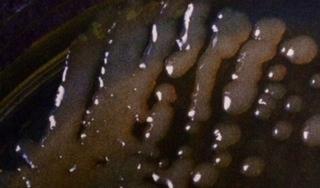 What bacteria is this?
| back 29 Klebsiella pneumoniae
|
front 30 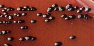 What bacteria is this?
| back 30 Chromobacterium violaceum
|
front 31 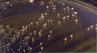 What bacteria is this?
| back 31 Enterococcus faecium
|
front 32 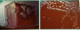 What is this?
| back 32 A) Bacillus cereus
|
front 33  What is being shown in these two views? What bacteria is this? What are the 3 features of this bacteria? | back 33 A) Alcaligenes Faecalis side view showing a raised center
|
front 34 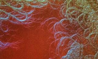 What is being shown here? | back 34 Filamentous growth |
front 35 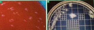 What bacteria is being shown in A and B and what are the differences between them?
| back 35 A) Clostridium sporaogenes grown on sheep blood agar and viewed with reflected light
|
front 36 _____ is an opportunistic pathogen | back 36 Staphylococcus epidermidis |
front 37 ______ is a frequent isolate in urine samples obtained from hospitalized and catheterized patients | back 37 Providencia stuartii |
front 38 ______ is found in human and animal feces | back 38 Enterococcus faecium |
front 39 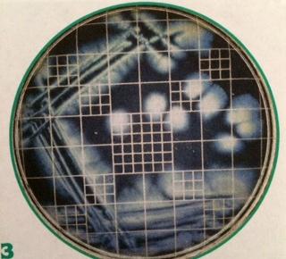 What bacteria is this?
| back 39 Alcaligenes faecalis
|
front 40 _______ is found in soil and water and rarely produces infections in humans | back 40 Chromobacterium violaceum |
front 41 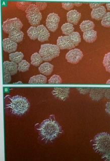 What are these slides showing?
| back 41 The effect of age on colony morphology after 24 and 48 hours of growth
|
front 42 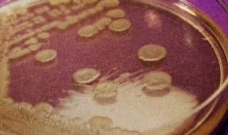 What bacteria is this?
| back 42 Bacillus subtilis
|
front 43 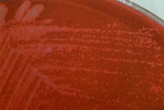 what bacteria is this?
| back 43 Mycobacterium smegmatis
|
front 44 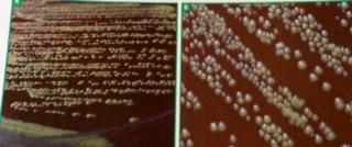 What bacteria is this?
| back 44 Coynebacterium xerosis
|
front 45 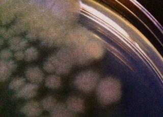 What bacteria is this?
| back 45 Erwinia amylovora
|
front 46 _________ is a plant pathogen. | back 46 Erwinia amylovora |
front 47 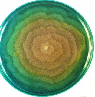 What bacteria is shown here?
| back 47 Proteus vulgaris
|
front 48 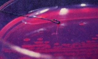 What bacteria is shown here?
| back 48 Pseudomonas aeruginosa
|
front 49 _____________ is found in soil and water and can cause infections in burn patients | back 49 Pseudomonas aeruginosa |
front 50 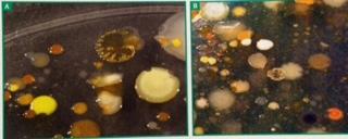 What are these samples showing? | back 50 Mixed soil cultures and diversity |
front 51 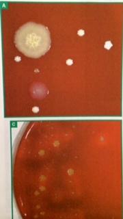 What bacteria is this showing? How do we know? | back 51 Staphylococcus aureus
|
front 52 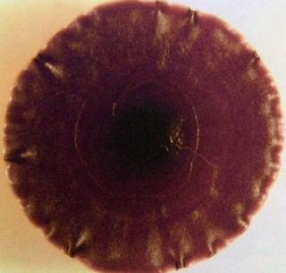 What bacteria is this? | back 52 Chromobacterium violaceum |
front 53 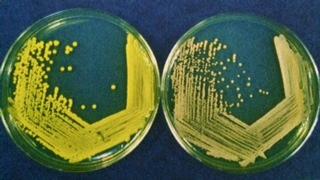 What bacteria is on the left? the right?
| back 53 Left - Micrococcus luteus
|
front 54 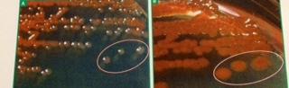 What bacteria is this?
| back 54 Serratia marcescens
|
front 55 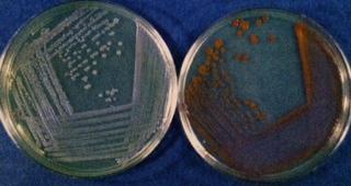 What bacteria is being shown here?
| back 55 Serratia marcescens
|
front 56 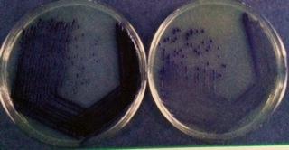 What bacteria is being shown here?
| back 56 Chromobacterium violaceum
|
front 57 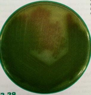 What bacteria is shown here?
| back 57 Pseudomonas aeruginosa
|