Bio 2 Lab Practical 2 - Plant Models and Slides

What is this an image of?
dicot leaf (model)

Name label A
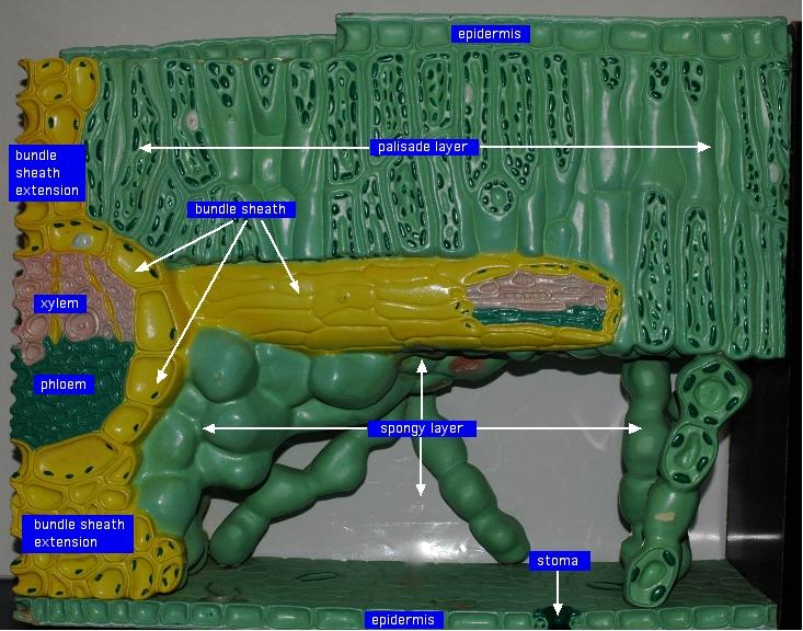
epidermis

name label B
bundle sheath extension

name label c
phloem

name label d
xylem

name label e
bundle sheath extension

name label f
palisade layer

name label g
bundle sheath

name label h
spongy layer

name label i
stoma

What is this an image of?
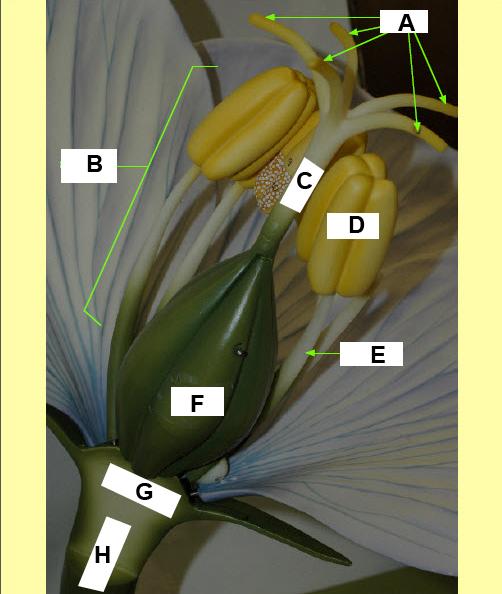
flower (model)

name label a
stigma

name label b
stamen

name label c
style

name label d
anther

name label e
filament

name label f
ovary

name label g
receptacle
name label h
pedicle

What is this an image of?
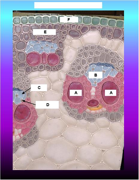
monocot stem (model)

name label a
xylem

name label b
phloem

name label c
sieve cell

name label d
companion cell

name label e
sclerenchyma
name label f
epidermis

What is this an image of?
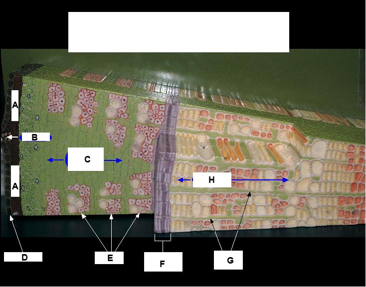
woody dicot leaf (model)

name label a
cork

name label b
lenticel
name label c
phloem ray

name label d
epidermis

name label e
phloem

name label f
vascular cambium

name label g
xylem rays

name label h
xylem

what is this a slide of?
leaf epidermis
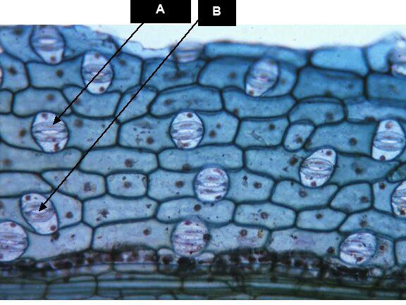
name label a
guard cell

name label b
stoma

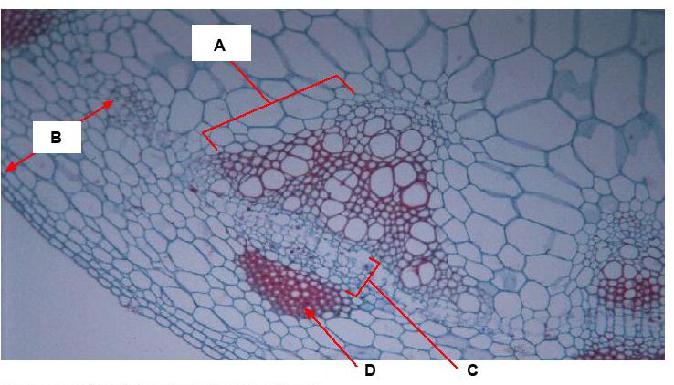
what is this a slide of?
dicot leaf (section cut)

name label a
vascular bundle

name label b
epidermis

name label c
palisade layer

name label d
spongy layer
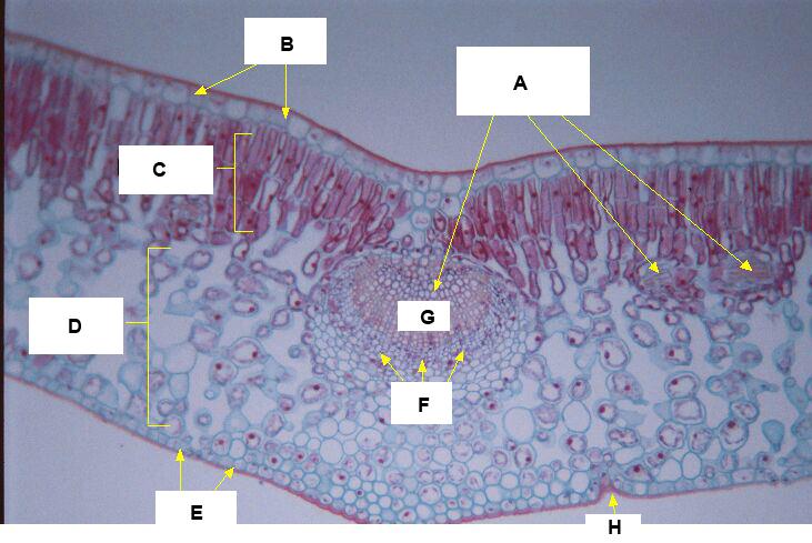
name label e
epidermis

name label f
phloem

name label g
xylem

name label h
stoma
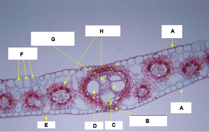
what is this a slide of?
monocot leaf

name label a
epidermis

name label b
sclerenchymal cells

name label c
phloem

name label d
xylem

name label e
stoma

name label f
bulliform cells

name label g
air space (proto xylem)

name label h
vascular bundles

what is this a slide of?
monocot stem

name label a
vascular bundle
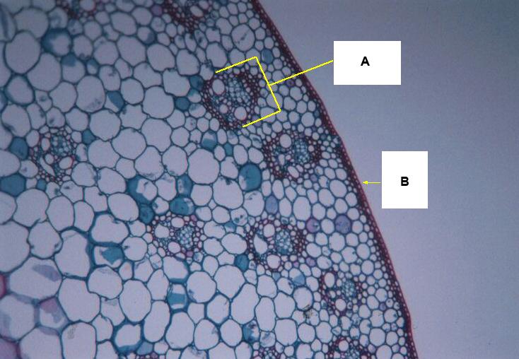
name label b
epidermis

what is this a slide of?
non-woody dicot stem
name label a
xylem

name label b
cortex

name label c
phloem

name label d
schlerenchymal cells

what is this a slide of?
woody dicot stem

name label a
phloem ray
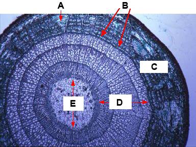
name label b
vascular cambium

name label c
phloem

name label d
xylem

name label e
pith

what is this an image of?
woody dicot stem
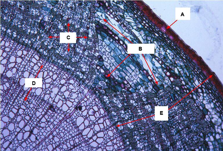
name label a
cork

name label b
phloem ray

name label c
phloem

name label d
xylem

name label e
bark
What is the type of tissue that occupies all the regions between the epidermal and vascular tissues?
ground tissue
What is the type of tissue made up of perpetual embryonic (growth) tissues?
meristem
What is the type of meristem tissue that causes the plant to grow in length?
apical meristems
What is the type of meristem tissue that causes the plant to grow in girth?
lateral meristems
What is the type of lateral meristem tissue that produces new xylem, new phloem, and new rays?
vascular cambium
What is the type of lateral meristem tissue that replaces epidermis with a thick, tough covering (cork)?
cork cambium
Term for all tissues exterior to the vascular cambium
bark
Term for cork cambium plus the cork to its exterior
periderm
Term for openings in the bark that allow for gas transport necessary in local cellular respiration
lenticels
Term for the supporting parenchymal cells that store starches and provide radial transfer of water and minerals
xylem and phloem rays
The mesophyll of the leaf is made up of what two layers?
palisade layer (where most photosynthesis occurs)
spongy layer
bundle of xylem and phloem in a leaf is called
vascular bundle or vein
Name for the yellow ring of collenchymal cells directly surrounding the xylem and phloem. It bunches up the xylem and phloem together
bundle sheath
the yellow column of mainly collenchymal cells above and below the vascular bundle that anchors the vein to the surrounding tissue
bundle sheath extension