Instructions for Side by Side Printing
- Print the notecards
- Fold each page in half along the solid vertical line
- Cut out the notecards by cutting along each horizontal dotted line
- Optional: Glue, tape or staple the ends of each notecard together
Bio 2 Lab Practical 2 - Plant Models and Slides
front 1  What is this an image of? | back 1 dicot leaf (model) |
front 2  Name label A | back 2 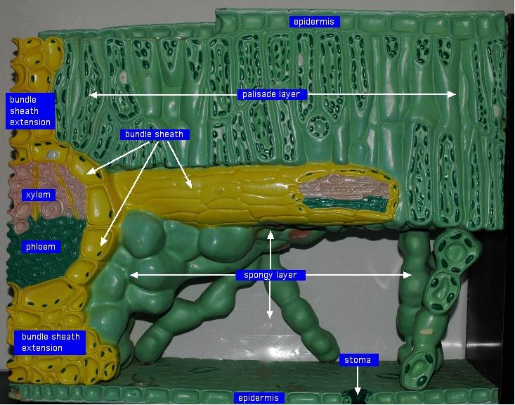 epidermis |
front 3  name label B | back 3 bundle sheath extension |
front 4  name label c | back 4 phloem |
front 5  name label d | back 5 xylem |
front 6  name label e | back 6 bundle sheath extension |
front 7  name label f | back 7 palisade layer |
front 8  name label g | back 8 bundle sheath |
front 9  name label h | back 9 spongy layer |
front 10  name label i | back 10 stoma |
front 11  What is this an image of? | back 11 flower (model) |
front 12  name label a | back 12 stigma |
front 13  name label b | back 13 stamen |
front 14  name label c | back 14 style |
front 15  name label d | back 15 anther |
front 16  name label e | back 16 filament |
front 17  name label f | back 17 ovary |
front 18  name label g | back 18 receptacle |
front 19 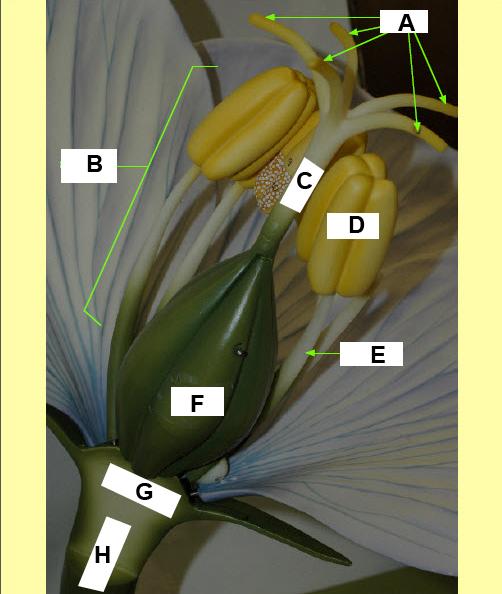 name label h | back 19 pedicle |
front 20  What is this an image of? | back 20 monocot stem (model) |
front 21  name label a | back 21 xylem |
front 22  name label b | back 22 phloem |
front 23  name label c | back 23 sieve cell |
front 24  name label d | back 24 companion cell |
front 25  name label e | back 25 sclerenchyma |
front 26 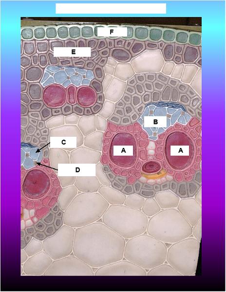 name label f | back 26 epidermis |
front 27  What is this an image of? | back 27 woody dicot leaf (model) |
front 28  name label a | back 28 cork |
front 29  name label b | back 29 lenticel |
front 30 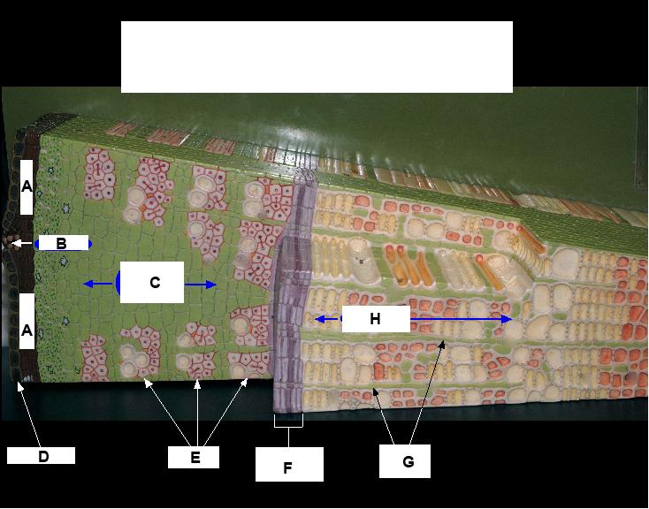 name label c | back 30 phloem ray |
front 31  name label d | back 31 epidermis |
front 32  name label e | back 32 phloem |
front 33  name label f | back 33 vascular cambium |
front 34  name label g | back 34 xylem rays |
front 35  name label h | back 35 xylem |
front 36  what is this a slide of? | back 36 leaf epidermis |
front 37 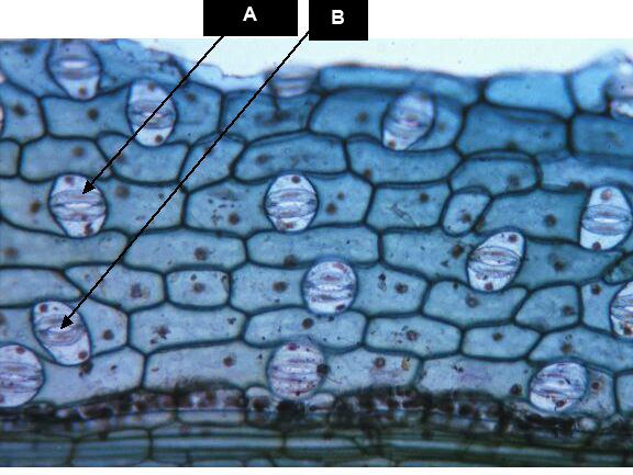 name label a | back 37 guard cell |
front 38  name label b | back 38 stoma |
front 39  what is this a slide of? | back 39 dicot leaf (section cut) |
front 40  name label a | back 40 vascular bundle |
front 41  name label b | back 41 epidermis |
front 42  name label c | back 42 palisade layer |
front 43  name label d | back 43 spongy layer |
front 44 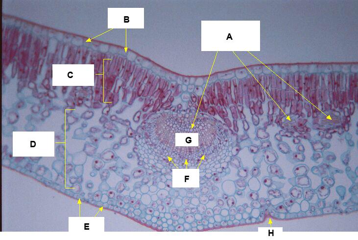 name label e | back 44 epidermis |
front 45  name label f | back 45 phloem |
front 46  name label g | back 46 xylem |
front 47  name label h | back 47 stoma |
front 48 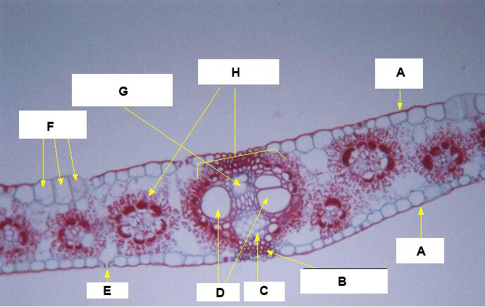 what is this a slide of? | back 48 monocot leaf |
front 49  name label a | back 49 epidermis |
front 50  name label b | back 50 sclerenchymal cells |
front 51  name label c | back 51 phloem |
front 52  name label d | back 52 xylem |
front 53  name label e | back 53 stoma |
front 54  name label f | back 54 bulliform cells |
front 55  name label g | back 55 air space (proto xylem) |
front 56  name label h | back 56 vascular bundles |
front 57  what is this a slide of? | back 57 monocot stem |
front 58  name label a | back 58 vascular bundle |
front 59 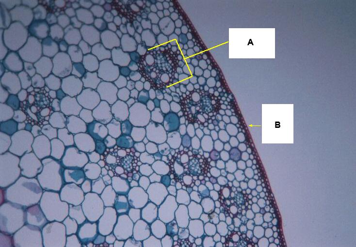 name label b | back 59 epidermis |
front 60  what is this a slide of? | back 60 non-woody dicot stem |
front 61 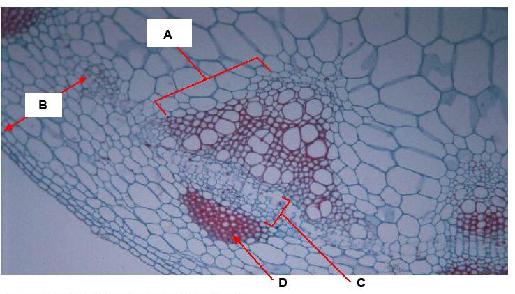 name label a | back 61 xylem |
front 62  name label b | back 62 cortex |
front 63  name label c | back 63 phloem |
front 64  name label d | back 64 schlerenchymal cells |
front 65  what is this a slide of? | back 65 woody dicot stem |
front 66  name label a | back 66 phloem ray |
front 67 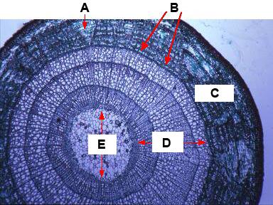 name label b | back 67 vascular cambium |
front 68  name label c | back 68 phloem |
front 69  name label d | back 69 xylem |
front 70  name label e | back 70 pith |
front 71  what is this an image of? | back 71 woody dicot stem |
front 72 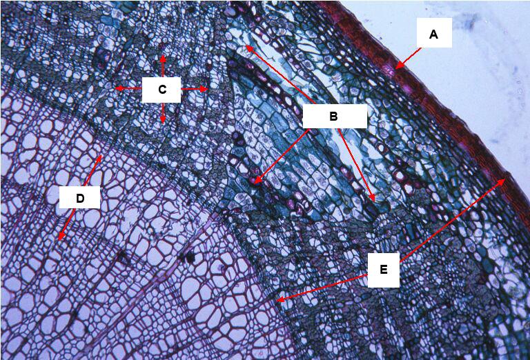 name label a | back 72 cork |
front 73  name label b | back 73 phloem ray |
front 74  name label c | back 74 phloem |
front 75  name label d | back 75 xylem |
front 76  name label e | back 76 bark |
front 77 What is the type of tissue that occupies all the regions between the epidermal and vascular tissues? | back 77 ground tissue |
front 78 What is the type of tissue made up of perpetual embryonic (growth) tissues? | back 78 meristem |
front 79 What is the type of meristem tissue that causes the plant to grow in length? | back 79 apical meristems |
front 80 What is the type of meristem tissue that causes the plant to grow in girth? | back 80 lateral meristems |
front 81 What is the type of lateral meristem tissue that produces new xylem, new phloem, and new rays? | back 81 vascular cambium |
front 82 What is the type of lateral meristem tissue that replaces epidermis with a thick, tough covering (cork)? | back 82 cork cambium |
front 83 Term for all tissues exterior to the vascular cambium | back 83 bark |
front 84 Term for cork cambium plus the cork to its exterior | back 84 periderm |
front 85 Term for openings in the bark that allow for gas transport necessary in local cellular respiration | back 85 lenticels |
front 86 Term for the supporting parenchymal cells that store starches and provide radial transfer of water and minerals | back 86 xylem and phloem rays |
front 87 The mesophyll of the leaf is made up of what two layers? | back 87 palisade layer (where most photosynthesis occurs)
|
front 88 bundle of xylem and phloem in a leaf is called | back 88 vascular bundle or vein |
front 89 Name for the yellow ring of collenchymal cells directly surrounding the xylem and phloem. It bunches up the xylem and phloem together | back 89 bundle sheath |
front 90 the yellow column of mainly collenchymal cells above and below the vascular bundle that anchors the vein to the surrounding tissue | back 90 bundle sheath extension |