Chapter 4 A&P extra stuff
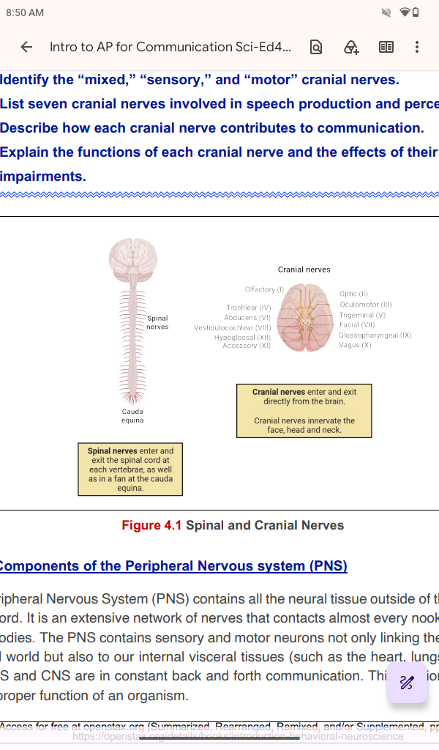
Components of the Peripheral Nervous system (PNS)
The Peripheral Nervous System (PNS) contains all the neural tissue
outside of the brain and spinal cord. It is an extensive network of
nerves that contacts almost every nook and cranny
of our bodies.
The PNS contains sensory and motor neurons not only linking the CNS to
the external world but also to our internal visceral tissues (such as
the heart, lungs, stomach). The PNS and CNS are in constant back and
forth communication. This relationship is vital
for the proper
function of an organism.
The terms used to describe some components in PNS are different from those of CNS. Remember that the collections of neurons are called “nuclei” in CNS and “ganglia” in PNS. The collections of axons are called “tracts” in CNS and “nerves” in PNS
..
collections of neurons are called “nuclei” in CNS a
collections of neurons are called “nuclei” in CNS a
collections of neurons are called ganglia” in PNS
collections of neurons are called ganglia” in PNS
The collections of axons are called “tracts” in CNS and “nerves” in PNS
Tracts CNS
The collections of axons are called “nerves” in PNS
nerves” in PNS
The PNS is not as contained as the CNS because it is defined as
everything that is not the CNS. Some peripheral structures are
incorporated into the other organs of the body. In
describing
the anatomy of the PNS, it is necessary to describe the common
structures, the nerves and the ganglia, as they are found in various
parts of the body. Many of the neural structures that are incorporated
into other organs are features of the digestive system;
these
structures are known as the enteric nervous system and are a special
subset of the PNS.
..
Ganglia
Ganglia (singular is “ganglion”) are neuron cell bodies in the periphery. Ganglia can be categorized, for the most part, as either sensory ganglia or autonomic ganglia, referring to their primary functions.
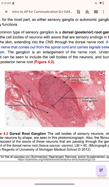
The most common type of sensory ganglion is a dorsal (posterior) root
ganglion. These ganglia are the cell bodies of neurons with axons that
are sensory endings in the periphery, such as in the skin, extending
into the CNS through the dorsal nerve root. A nerve root is
the
part of a nerve that comes out from the spinal cord and carries
signals between the body and the brain. The ganglion is an enlargement
of the nerve root. Under microscopic
inspection, it can be seen
to include the cell bodies of the neurons, and bundles of fibers that
are the posterior nerve root (Figure 4.2).
..
The cell bodies of sensory neurons, which are unipolar neurons by
shape, are seen in this photomicrograph. Also, the fibrous region
is composed of the axons of these neurons that are passing
through the ganglion to be part of the dorsal nerve root (tissue
source: canine).
nerve root is
A nerve root is
the part of a nerve that comes out from the
spinal cord and carries signals between the body and the brain.
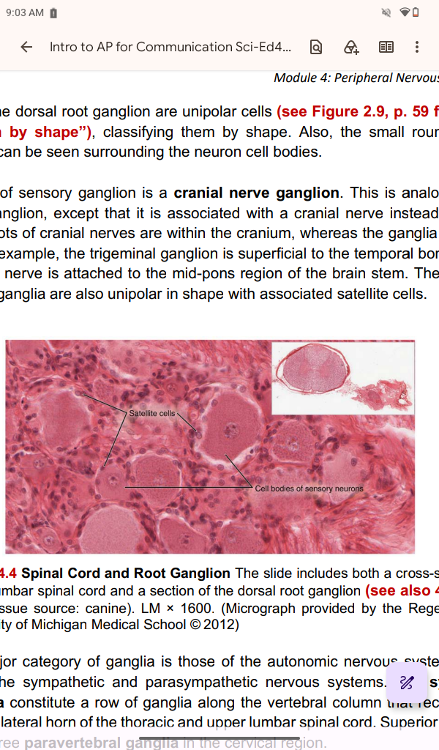
The cells of the dorsal root ganglion are unipolar cells (see Figure 2.9, p. 59 for “neuron classification by shape”), classifying them by shape. Also, the small round nuclei of satellite cells can be seen surrounding the neuron cell bodies.
..
cranial nerve ganglion.
Another type of sensory ganglion is a cranial nerve ganglion. This is analogous to the dorsal root ganglion, except that it is associated with a cranial nerve instead of a spinal nerve. The roots of cranial nerves are within the cranium, whereas the ganglia are outside the skull. For example, the trigeminal ganglion is superficial to the temporal bone, whereas its associated nerve is attached to the mid-pons region of the brain stem. The neurons of cranial nerve ganglia are also unipolar in shape with associated satellite cells.
The other major category of ganglia is those of the autonomic nervous system, which is divided into the sympathetic and parasympathetic nervous systems. The sympathetic chain ganglia constitute a row of ganglia along the vertebral column that receive central input from the lateral horn of the thoracic and upper lumbar spinal cord. Superior to the chain ganglia are three paravertebral ganglia in the cervical region.
..
sympathetic chain ganglia
sympathetic chain ganglia constitute a row of ganglia along the vertebral column that receive central input from the lateral horn of the thoracic and upper lumbar spinal cord.
Superior to the chain
ganglia
Superior to the chain
ganglia are three paravertebral ganglia
in the cervical region
Three other autonomic ganglia related to the sympathetic chain are
the prevertebral ganglia, which are located outside of the chain but
have similar functions. They are referred to as prevertebral because
they are anterior to the vertebral column. The neurons of these
autonomic ganglia are multipolar in shape, with dendrites
radiating around the cell body where synapses from the spinal cord
neurons are made. The neurons of the chain, paravertebral, and
prevertebral ganglia then project to organs in the head and neck,
thoracic, abdominal, and pelvic cavities to regulate the sympathetic
aspect of homeostatic mechanisms.
..
prevertebral
ganglia,
which are located outside of the chain but have similar functions.
They are referred
to as prevertebral because they are anterior
to the vertebral column.
Another group of autonomic ganglia is the terminal ganglia that
receive input from cranial nerves or sacral spinal nerves and are
responsible for regulating the parasympathetic aspect
of
homeostatic mechanisms. These two sets of ganglia, sympathetic and
parasympathetic, often project to the same organs—one input from the
chain ganglia and one input from a terminal ganglion—to regulate the
overall function of an organ. For example, the heart
receives
two inputs: one increases heart rate, and the other decreases it. The
terminal ganglia that receive input from cranial nerves are found in
the head and neck regions, whereas the terminal ganglia that receive
sacral input are located in the lower abdominal
and pelvic
cavities. We will explore the Autonomic Nervous System(ANS) in the
next module
..
autonomic ganglia
autonomic ganglia is the terminal ganglia that receive input from cranial nerves or sacral spinal nerves and are responsible for regulating the parasympathetic aspect of homeostatic mechanisms
These two sets of ganglia, sympathetic and parasympathetic,
often project to the same organs—one input from the chain
ganglia and one input from a terminal ganglion—to regulate the overall
function of an organ.
For example, the heart receives two inputs: one increases heart rate, and the other decreases it.
..
terminal ganglia
The terminal ganglia that receive input from cranial nerves are found in the head and neck region
terminal ganglia that receive sacral
terminal ganglia that receive sacral input are located in the lower abdominal and pelvic cavities
Terminal ganglia below the head and neck are often incorporated into
the wall of the target organ as a plexus. A plexus, in a general
sense, is a network of fibers or vessels. This can apply to nervous
tissue (as in this instance) or structures containing blood vessels
(such as
a choroid plexus). For example, the enteric plexus is
the extensive network of axons and neurons in the wall of the small
and large intestines. The enteric plexus is part of the enteric
nervous system, along with the gastric plexus and the esophageal
plexus. Though the
enteric nervous system receives input
originating from central neurons of the autonomic nervous system, it
does not require CNS input to function. In fact, it operates
independently to regulate the digestive system.
..
plexus
A plexus, in a general sense, is a network of fibers or vessels. This
can
apply to nervous tissue (as in this instance) or structures
containing blood vessels (such as
a choroid plexus).
enteric plexus
For example, the enteric plexus is the extensive network of axons and
neurons in the wall of the small and large intestines. The
enteric plexus is part of the enteric
nervous system, along with
the gastric plexus and the esophageal plexus
Afferent and efferent divisions
Afferent and efferent divisions
The PNS is made up of afferent
(sensory) and efferent (motor) components. The afferent division
brings information from the periphery (receptors found within skin,
joints, muscle,
visceral organs) to the spinal cord and brain
via sensory nerves. The information brought in by afferent neurons is
decoded and processed by the CNS. The efferent component, on the other
hand, utilizes motor neurons that transmit information from the CNS to
the periphery. This includes our muscle tissue (effectors) causing
muscle contraction and thus voluntary body movements. In addition, the
efferent division also controls smooth muscle in visceral organs,
cardiac muscle and glands. It can function with or without
consciousness (voluntary or involuntary).
afferent
The afferent
division brings information from the periphery
(receptors found within skin, joints, muscle, visceral organs) to the
spinal cord and brain via sensory nerves. The information brought in
by afferent neurons is decoded and processed by the CNS
The efferent component
The efferent component, on
the other hand, utilizes motor
neurons that transmit information from the CNS to the periphery. This
includes our muscle tissue (effectors) causing muscle contraction and
thus voluntary body movements. In addition, the efferent division also
controls smooth muscle
in visceral organs, cardiac muscle and
glands. It can function with or without consciousness (voluntary or involuntary).
Nerves
Nerves
Bundles of axons in the PNS are referred to as nerves.
These structures in the periphery are different from the central
counterpart, called a tract. Nerves are composed of more than just
nervous tissue. They have connective tissues invested in their
structure, as well
as blood vessels supplying the tissues with nourishment.
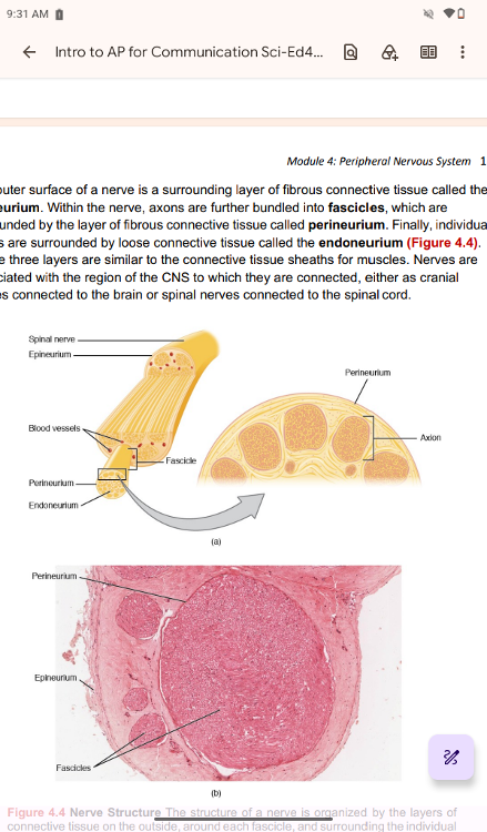
The outer surface of a nerve is a surrounding layer of fibrous connective tissue called the epineurium. Within the nerve, axons are further bundled into fascicles, which are surrounded by the layer of fibrous connective tissue called perineurium. Finally, individual axons are surrounded by loose connective tissue called the endoneurium (Figure 4.4). These three layers are similar to the connective tissue sheaths for muscles. Nerves are associated with the region of the CNS to which they are connected, either as cranial nerves connected to the brain or spinal nerves connected to the spinal cord.
..
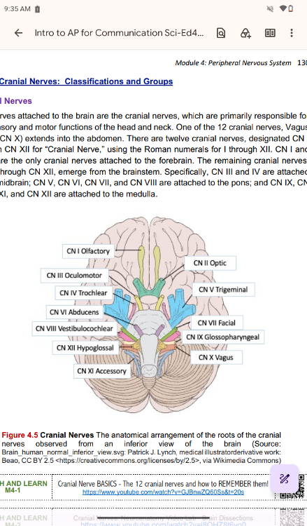
Cranial Nerves: Classifications and Groups
Cranial
Nerves
The nerves attached to the brain are the cranial nerves,
which are primarily responsible for the sensory and motor functions of
the head and neck. One of the 12 cranial nerves, Vagus nerve (CN X)
extends into the abdomen. There are twelve cranial nerves, designated
CN I
through CN XII for “Cranial Nerve,” using the Roman
numerals for I through XII. CN I and CN II are the only cranial nerves
attached to the forebrain. The remaining cranial nerves, CN III
through CN XII, emerge from the brainstem. Specifically, CN III and IV
are attached
to the midbrain; CN V, CN VI, CN VII, and CN VIII
are attached to the pons; and CN IX, CN X, CN XI, and CN XII are
attached to the medulla.
..
Cranial nerves can be classified by their functions as sensory
nerves, motor nerves, or a combination of both (mixed), meaning that
the axons in these nerves originate out of sensory ganglia external to
the cranium or motor nuclei within the brain stem. Sensory axons enter
the brain to synapse in a nucleus. Motor axons connect to
skeletal muscles of the head or neck (most of the cranial nerves).
Three of the nerves are solely composed of sensory fibers, five are
strictly motor, and the remaining four are mixed nerves.
...
Function
General
Target Area
Somatic (S): skin,
skeletal muscle
Direction
Afferent
(A)
Classification
General Somatic
Afferent (GSA)
Description
Carries general
sensory information
(e.g., touch, pain,
proprioception)
Associated CNs
CN V, VII, IX, X
..
Function
General
Target Area
Somatic (S):
Skeletal (striated)
muscle
Direction
Efferent
Classification
General Somatic
Efferent (GSE)
Description
Sends motor
commands to
skeletal muscles
Associated CNs
CN III, IV, VI, XII
..
Function
General
Target Area
Visceral (V):
Internal organs
(viscera
Direction
Afferent
Classification
General Visceral
Afferent (GVA)
Description
Conveys sensory
input from viscera
Associated CNs
CN IX, X
..
Function
General
Target Area
Visceral (V):
Smooth muscle,
glands, cardiac
Direction
Efferent
Classification
General Visceral
Efferent (GVE)
Description
Autonomic motor
control of organs
and glands
Associated CNs
CN III, VII, IX, X
..
Function
Special
Target Area
Somatic (S): Eye,
ear (vision,
hearing, balance)
Direction
Afferent
Classification
Special Visceral
Afferent (SVA)
Description
Special senses:
vision, hearing,
balance
Associated CNs
CN II, VIII
..
Function
Special
Target Area
Visceral (V):
Tongue, nose
(taste, smell)
Direction
Afferent
Classification
Special Visceral
Afferent (SVA)
Description
Special senses
related to internal
structures: taste
and smell
Associated CNs
CN I, VII, IX, X
..
Function
Special
Target Area
Visceral (V):
Muscles of face,
jaw, pharynx,
larynx (branchial
arch)
Direction
Efferent
Classification
Special Visceral
Efferent
(Branchial)
(SVE)
Description
Motor to muscles
from branchial
arches
Associated CNs
CN V, VII, IX, X,
XI
..
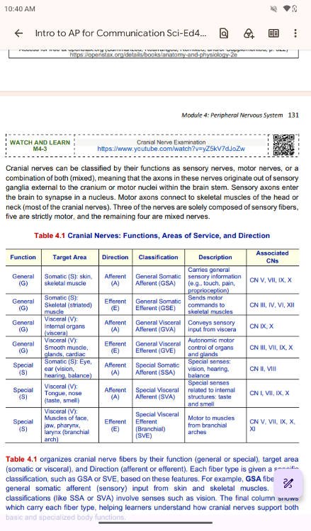
4.1 organizes cranial nerve fibers by their function (general or
special), target area (somatic or visceral), and Direction (afferent
or efferent). Each fiber type is given a specific classification, such
as GSA or SVE, based on these features. For example, GSA fibers carry
general somatic afferent (sensory) input from skin and skeletal
muscles. Special classifications (like SSA or SVA) involve senses such
as vision. The final column shows which carry each fiber type, helping
learners understand how cranial nerves support both
basic and
specialized body functions.
..
GSA
For example, GSA fibers carry
general somatic afferent
(sensory) input from skin and skeletal muscles.
Special
classifications (
Special
classifications (like SSA or SVA) involve senses such
as vision
Learning the cranial nerves is a tradition in anatomy courses, and
students have always used mnemonic devices to remember the nerve
names. A traditional mnemonic is “On Old Olympus’ Towering Tops/A Finn
And German Viewed Some Hops,” in which the initial letter
of
each word corresponds to the initial letter in the name of each nerve.
The names of the nerves have changed over the years to reflect current
usage and more accurate naming. An exercise to help learn this sort of
information is to generate a mnemonic using words that
have
personal significance. The names of the cranial nerves are listed in
..
Another important aspect of the cranial nerves that lends itself to a
mnemonic is each nerve's functional role. The nerves fall into one of
three basic groups. They are sensory (S), motor (M), or both (B). The
sentence, “Some Say Marry Money, But My Brother Says Brains
Beauty
Matter More,” corresponds to the basic function of each
nerve. See the summary below. • The first, second, and eighth nerves
are purely sensory: the olfactory (CN I), optic (CN II), and
vestibulocochlear (CN VIII) nerves.
• The three eye-movement
nerves are all motor: the oculomotor (CN III), trochlear (CN IV), and
abducens (CN VI).
• The spinal accessory (CN XI) and hypoglossal
(CN XII) nerves are also strictly motor.
• The remainder of the
nerves contain both sensory and motor fibers. They are the trigeminal
(CN V), facial (CN VII), glossopharyngeal (CN IX), and vagus (CN X) nerves.
..
The nerves that convey both (mixed cranial nerves) are often related
to each other. Cranial nerve V (CN V: trigeminal nerve) and cranial
nerve VII (CN VII: facial nerve) concern the face; one concerns the
sensations, and the other concerns the muscle movements. Cranial nerve
VII (CN VII: facial nerve) and cranial nerve IX (CN IX:
glossopharyngeal nerve) are both responsible for conveying gustatory
(taste) sensations, as well as controlling salivary glands.
This
is not an exhaustive list of what these combination nerves do, but
there is a thread of relation between them.
..