Instructions for Side by Side Printing
- Print the notecards
- Fold each page in half along the solid vertical line
- Cut out the notecards by cutting along each horizontal dotted line
- Optional: Glue, tape or staple the ends of each notecard together
Chapter 4 A&P extra stuff
front 1 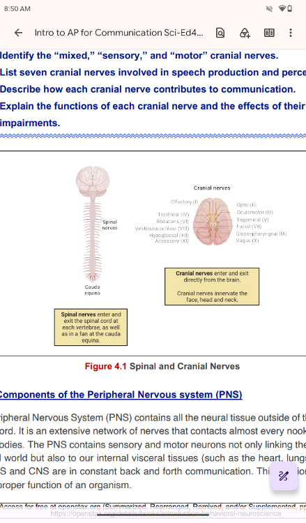 Components of the Peripheral Nervous system (PNS) | back 1 The Peripheral Nervous System (PNS) contains all the neural tissue
outside of the brain and spinal cord. It is an extensive network of
nerves that contacts almost every nook and cranny |
front 2 The terms used to describe some components in PNS are different from those of CNS. Remember that the collections of neurons are called “nuclei” in CNS and “ganglia” in PNS. The collections of axons are called “tracts” in CNS and “nerves” in PNS | back 2 .. |
front 3 collections of neurons are called “nuclei” in CNS a | back 3 collections of neurons are called “nuclei” in CNS a |
front 4 collections of neurons are called ganglia” in PNS | back 4 collections of neurons are called ganglia” in PNS |
front 5 The collections of axons are called “tracts” in CNS and “nerves” in PNS | back 5 Tracts CNS |
front 6 The collections of axons are called “nerves” in PNS | back 6 nerves” in PNS |
front 7 The PNS is not as contained as the CNS because it is defined as
everything that is not the CNS. Some peripheral structures are
incorporated into the other organs of the body. In | back 7 .. |
front 8 Ganglia | back 8 Ganglia (singular is “ganglion”) are neuron cell bodies in the periphery. Ganglia can be categorized, for the most part, as either sensory ganglia or autonomic ganglia, referring to their primary functions. |
front 9 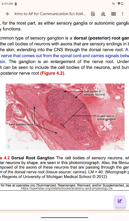 The most common type of sensory ganglion is a dorsal (posterior) root
ganglion. These ganglia are the cell bodies of neurons with axons that
are sensory endings in the periphery, such as in the skin, extending
into the CNS through the dorsal nerve root. A nerve root is | back 9 .. The cell bodies of sensory neurons, which are unipolar neurons by
shape, are seen in this photomicrograph. Also, the fibrous region
|
front 10 nerve root is | back 10 A nerve root is |
front 11 The cells of the dorsal root ganglion are unipolar cells (see Figure 2.9, p. 59 for “neuron classification by shape”), classifying them by shape. Also, the small round nuclei of satellite cells can be seen surrounding the neuron cell bodies. | back 11 .. |
front 12 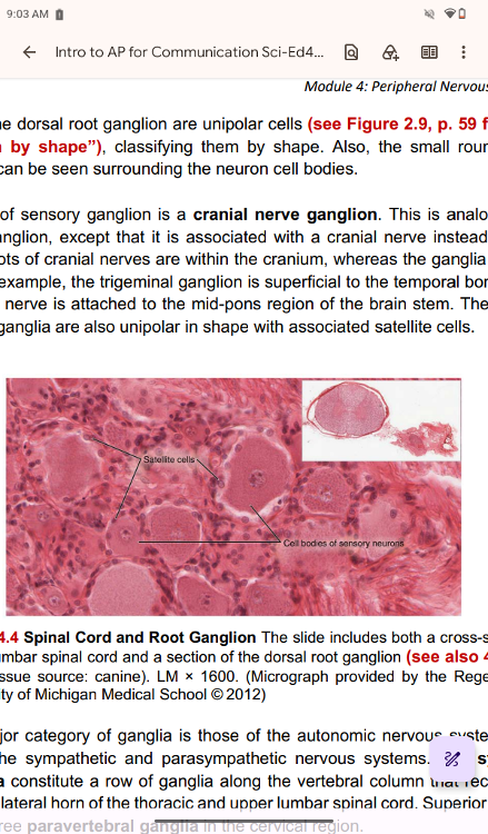 cranial nerve ganglion. | back 12 Another type of sensory ganglion is a cranial nerve ganglion. This is analogous to the dorsal root ganglion, except that it is associated with a cranial nerve instead of a spinal nerve. The roots of cranial nerves are within the cranium, whereas the ganglia are outside the skull. For example, the trigeminal ganglion is superficial to the temporal bone, whereas its associated nerve is attached to the mid-pons region of the brain stem. The neurons of cranial nerve ganglia are also unipolar in shape with associated satellite cells. |
front 13 The other major category of ganglia is those of the autonomic nervous system, which is divided into the sympathetic and parasympathetic nervous systems. The sympathetic chain ganglia constitute a row of ganglia along the vertebral column that receive central input from the lateral horn of the thoracic and upper lumbar spinal cord. Superior to the chain ganglia are three paravertebral ganglia in the cervical region. | back 13 .. |
front 14 sympathetic chain ganglia | back 14 sympathetic chain ganglia constitute a row of ganglia along the vertebral column that receive central input from the lateral horn of the thoracic and upper lumbar spinal cord. |
front 15 Superior to the chain | back 15 Superior to the chain |
front 16 Three other autonomic ganglia related to the sympathetic chain are
the prevertebral ganglia, which are located outside of the chain but
have similar functions. They are referred to as prevertebral because
they are anterior to the vertebral column. The neurons of these
| back 16 .. |
front 17 prevertebral | back 17 which are located outside of the chain but have similar functions.
They are referred |
front 18 Another group of autonomic ganglia is the terminal ganglia that
receive input from cranial nerves or sacral spinal nerves and are
responsible for regulating the parasympathetic aspect | back 18 .. |
front 19 autonomic ganglia | back 19 autonomic ganglia is the terminal ganglia that receive input from cranial nerves or sacral spinal nerves and are responsible for regulating the parasympathetic aspect of homeostatic mechanisms |
front 20 These two sets of ganglia, sympathetic and parasympathetic,
For example, the heart receives two inputs: one increases heart rate, and the other decreases it. | back 20 .. |
front 21 terminal ganglia | back 21 The terminal ganglia that receive input from cranial nerves are found in the head and neck region |
front 22 terminal ganglia that receive sacral | back 22 terminal ganglia that receive sacral input are located in the lower abdominal and pelvic cavities |
front 23 Terminal ganglia below the head and neck are often incorporated into
the wall of the target organ as a plexus. A plexus, in a general
sense, is a network of fibers or vessels. This can apply to nervous
tissue (as in this instance) or structures containing blood vessels
(such as | back 23 .. |
front 24 plexus | back 24 A plexus, in a general sense, is a network of fibers or vessels. This
can |
front 25 enteric plexus | back 25 For example, the enteric plexus is the extensive network of axons and
|
front 26 Afferent and efferent divisions | back 26 Afferent and efferent divisions |
front 27 afferent | back 27 The afferent |
front 28 The efferent component | back 28 The efferent component, on |
front 29 Nerves | back 29 Nerves |
front 30 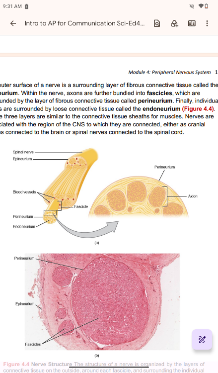 The outer surface of a nerve is a surrounding layer of fibrous connective tissue called the epineurium. Within the nerve, axons are further bundled into fascicles, which are surrounded by the layer of fibrous connective tissue called perineurium. Finally, individual axons are surrounded by loose connective tissue called the endoneurium (Figure 4.4). These three layers are similar to the connective tissue sheaths for muscles. Nerves are associated with the region of the CNS to which they are connected, either as cranial nerves connected to the brain or spinal nerves connected to the spinal cord. | back 30 .. |
front 31 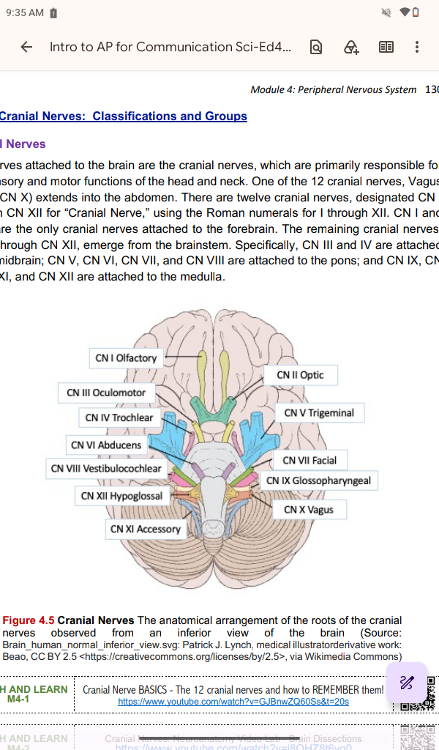 Cranial Nerves: Classifications and Groups | back 31 .. |
front 32 Cranial nerves can be classified by their functions as sensory
nerves, motor nerves, or a combination of both (mixed), meaning that
the axons in these nerves originate out of sensory ganglia external to
the cranium or motor nuclei within the brain stem. Sensory axons enter
| back 32 ... |
front 33 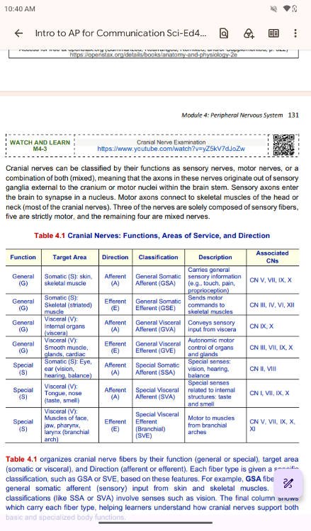 Function General Target Area Somatic (S): skin, Direction Afferent Classification General Somatic Description Carries general Associated CNs CN V, VII, IX, X | back 33 .. |
front 34 Function General Target Area Somatic (S): Direction Efferent Classification General Somatic Description Sends motor Associated CNs CN III, IV, VI, XII | back 34 .. |
front 35 Function General Target Area Visceral (V): Direction Afferent Classification General Visceral Description Conveys sensory Associated CNs CN IX, X | back 35 .. |
front 36 Function General Target Area Visceral (V): Direction Efferent Classification General Visceral Description Autonomic motor Associated CNs CN III, VII, IX, X | back 36 .. |
front 37 Function Special Target Area Somatic (S): Eye, Direction Afferent Classification Special Visceral Description Special senses: Associated CNs CN II, VIII | back 37 .. |
front 38 Function Special Target Area Visceral (V): Direction Afferent Classification Special Visceral Description Special senses Associated CNs CN I, VII, IX, X | back 38 .. |
front 39 Function Special Target Area Visceral (V): Direction Efferent Classification Special Visceral Description Motor to muscles Associated CNs CN V, VII, IX, X, | back 39 .. |
front 40 4.1 organizes cranial nerve fibers by their function (general or
special), target area (somatic or visceral), and Direction (afferent
or efferent). Each fiber type is given a specific classification, such
as GSA or SVE, based on these features. For example, GSA fibers carry
| back 40 .. |
front 41 GSA | back 41 For example, GSA fibers carry |
front 42 Special | back 42 Special |
front 43 Learning the cranial nerves is a tradition in anatomy courses, and
students have always used mnemonic devices to remember the nerve
names. A traditional mnemonic is “On Old Olympus’ Towering Tops/A Finn
And German Viewed Some Hops,” in which the initial letter | back 43 .. |
front 44 Another important aspect of the cranial nerves that lends itself to a
mnemonic is each nerve's functional role. The nerves fall into one of
three basic groups. They are sensory (S), motor (M), or both (B). The
sentence, “Some Say Marry Money, But My Brother Says Brains
Beauty | back 44 .. |
front 45 The nerves that convey both (mixed cranial nerves) are often related
to each other. Cranial nerve V (CN V: trigeminal nerve) and cranial
nerve VII (CN VII: facial nerve) concern the face; one concerns the
sensations, and the other concerns the muscle movements. Cranial nerve
VII (CN VII: facial nerve) and cranial nerve IX (CN IX:
glossopharyngeal nerve) are both responsible for conveying gustatory
(taste) sensations, as well as controlling salivary glands. | back 45 .. |