3063 chapter 3 part 2
Neurons
Neurons
The billions of highly specialized cells that compose
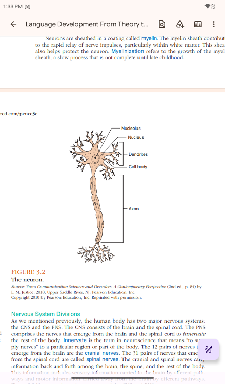
the nervous system are called neurons. A neuron is functionally
divided into four components: cell body, axon, presynaptic terminal,
and dendrites.
cell body
The cell body is the center of the neuron,
containing its
nucleus; the nucleus contains DNA material (genes, chromosomes) and
proteins. The human brain uses an estimated 30,000–40,000 genes, more
than any other organ of the body
The axon and the dendrites are extensions from the cell body, serving
as vehicles for the cell body to receive and transmit information from
other neurons, as shown in Figure 3.2. The information carried by
neurons is in the form of electrochemical nerve impulses; these
impulses transmit information to and away from the cell
body.
Each neuron has a single efferent nerve extension, the axon, which
carries nerve impulses away from the cell body. The axon extends from
the cell body for a distance of 1 mm to 1 m, at which point it
arborizes into a number of terminal branches
..
Axon
Each neuron has a single efferent nerve extension, the axon, which carries nerve impulses away from the cell body. The axon extends from the cell body for a distance of 1 mm to 1 m, at which point it arborizes into a number of terminal branches
The distal end of each terminal branch is a presyn-
aptic
terminal. These terminals are the sites at which the axonal connection
of one neuron corresponds with the dendritic extension of another neuron.
..
Dendrites are
the afferent extensions of a neuron, meaning they
bring nerve impulses into the cell
body from the axonal
projections of other neurons.
..
A single cell body contains a
number of dendritic extensions;
many dendrites are studded with small protuber-
ances (called
spines), which increase the surface area of the afferent connections
of
the neuron (Noback et al., 2005).
..
Neurons communicate by means of electrochemical nerve impulses that
travel along the dendrite of one neuron and into its cell body, then
along the axon to the
dendrite of another neuron
..
synapse
The synapse is the site where two neurons meet. For
the two
neurons to communicate, the nerve impulse must cross the synapse
Neurotransmitters
For the two neurons to communicate, the nerve impulse must cross the
synapse. Neurotransmitters are chemical agents that help transmit
information across the synap-
tic cleft, which is the space
between the axon of the transmitting neuron and the
dendrite of
the receiving neuron.
synaptogenesis
When a synapse is created, that is, when one neu-
ron forges a
connection with another neuron, this is referred to as synaptogenesis
nervous tissue
The tissue formed by the linkages of thousands of neurons is called nervous tissue
The two primary types of nervous tissue are gray matter and white
matter. Gray matter consists of the cell bodies of neurons and the
dendrites. White matter
is the tissue that carries information
among gray matter, consisting primarily of axonal fibers that carry
information among gray matter tissues. Thus, gray matter is where
information is generated and processed, whereas white matter serves as
an
information conduit.
..
Gray matter
Gray matter consists of the cell bodies of neurons and the dendrites.
Thus, gray matter is where information is generated and processed,
White matter
White matter
is the tissue that carries information among gray
matter, consisting primarily of axonal fibers that carry information
among gray matter tissues.
whereas white matter serves as an
information conduit.
myelin.
Neurons are sheathed in a coating called myelin. The myelin sheath
contributes
to the rapid relay of nerve impulses, particularly
within white matter. This sheath
also helps protect the neuron
Myelinization
refers to the growth of the myelin
sheath, a slow process that
is not complete until late childhood.
Nervous System Divisions
Nervous System Divisions
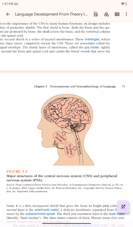
As we mentioned previously, the human
body has two major nervous systems:
the CNS and the PNS. The CNS
consists of the brain and the spinal cord. The PNS
comprises the
nerves that emerge from the brain and the spinal cord to
innervate
the rest of the body.
Innervate
Innervate is the term in neuroscience that means “to sup-
ply
nerves” to a particular region or part of the body
cranial nerves
The 12 pairs of nerves that
emerge from the brain are the
cranial nerves.
spinal nerves
The 31 pairs of nerves that emerge
from the spinal cord are
called spinal nerves.
The cranial and spinal nerves carry
information back and forth
among the brain, the spine, and the rest of the body. This information
includes sensory information carried to the brain by afferent pathways
and motor information carried away from the brain by efferent pathways.
..
Central Nervous System
Central Nervous System. The CNS consists of the brain and the spinal cord
The brain is essentially the chief executive operator of the entire CNS: It initiates and regulates virtually all motor, sensory, and cognitive processes.
..
The spinal cord acts
primarily as a conduit of information,
carrying not only sensory information from the body to the brain
through afferent pathways, but also motor commands from the brain to
the rest of the body through efferent pathways
..
First shield is bone
Given the importance of the CNS to many human functions, its design
includes a number of protective shields. The first shield is bone.
Both the brain and the spinal cord are protected by bone; the skull
covers the brain, and the vertebral column
covers the spinal cord.
The second shield
The second shield is a series of layered membranes
meninges
These meninges, which
comprise three layers, completely encase
the CNS. These are sometimes called the
meningeal envelope
pia mater
The inside layer of membrane, called the pia mater, tightly
wraps around the brain and spinal cord and carries the blood
vessels that serve the brain. It is a thin, transparent shield that
gives the brain its bright pink color.
The second layer is the arachnoid mater, a delicate membrane separated from the pia mater by the subarachnoid space.
..
dura mater
The third and outermost layer is the dura mater
(literally,
“hard mother”). The dura mater consists of thick, fibrous tissue that
com-
pletely encases the brain and the spinal cord.
cerebrospinal fluid (CSF).
The third shield is a layer of fluid called cerebrospinal fluid (CSF).
CSF circu-
lates between the two innermost layers of the
meninges—the pia mater and the
arachnoid mater—within the
subarachnoid space. CSF carries chemicals important
to metabolic
processes, but it is also an important buffer against jolts to the CNS.
spinal tap
Perhaps you have heard of a procedure called a spinal tap (not the
fictional rock band, which is perhaps a more amusing connotation).
Also called a lumbar puncture, a spinal tap involves inserting a
needle between two of the lower (lumbar)
vertebrae and
extracting CSF from the subarachnoid space. It is a procedure often
used to diagnose meningitis, which is an infection or inflammation of
the meninges. [Meningitis is also very serious, so it is important to
know the symptoms. Typically, these include headache, neck stiffness,
high fever, and altered mental state
(Glimåker et al., 2015).]
Meningitis
It is a procedure often
used to diagnose meningitis, which is
an infection or inflammation of the menin-
ges. [Meningitis is
also very serious, so it is important to know the symptoms.
Typ-
ically, these include headache, neck stiffness, high fever,
and altered mental state
(Glimåker et al., 2015).]
Peripheral Nervous System
Peripheral Nervous System.
The PNS is the system of nerves connected to the
brainstem
and the spinal cord. These nerves carry sensory information to the
CNS
and motor commands away from the CNS, thus controlling
nearly all voluntary and
involuntary activity of the human body.
The PNS consists of two sets of nerves: cranial nerves and spinal
nerves. The 12 pairs of cranial nerves run between the brainstem and
the facial and neck regions and are particularly important for speech,
language, and hearing. The cranial nerves transmit information
concerning four of the five senses (vision, hearing, smell, and
taste) to the brain. They also carry motor impulses from the
brain to the face and neck muscles, including those activating the
tongue and the jaw, both of which are involved with speech.
..
cranial nerves
cranial nerves run between the brainstem and the facial and neck
regions and are particularly important for speech, language, and
hearing. The cranial nerves transmit information concerning four of
the five senses (vision, hearing, smell, and taste) to the brain. They
also carry motor impulses from the brain to the face and
neck
muscles, including those activating the tongue and the jaw, both of
which are involved with speech.
The seven cranial nerves most closely involved with speech
and
language production are the following:
• Trigeminal (V): Facial
sensation; jaw movements, including chewing
• Facial (VII): Taste
sensation; facial movements, including smiling
• Acoustic (VIII):
Hearing and balance
• Glossopharyngeal (IX): Tongue sensation;
palatal and pharyngeal movement,
including gagging
• Vagus
(X): Taste sensation; palatal, pharyngeal, and laryngeal movement,
in-
cluding voicing
Accessory (XI): Palatal, pharyngeal,
laryngeal, head, and shoulder movement
• Hypoglossal (XII):
Tongue movement
..
The 31 pairs of spinal nerves run between the spinal cord and all
peripheral
areas of the human body, including the arms and the
legs. These nerves mediate reflexes, sensory activity, and conscious
(volitional) motor activity.
..
contralateral.
An important fea-
ture of the CNS and PNS is that almost
everything is organized to be contralateral.
This means the
right side of the brain processes information from the left side of
the body, and vice versa. In the simplest terms, damage to the
left side of the brain
will affect the functioning of the right
side of the body
What are the Major Structures and
Functions of the Human Brain?
What are the Major Structures and
Functions of the Human
Brain?
The brain is the commander in chief, or mediator, of the
entire human body, and
it is viewed as the most complex and
sophisticated organ of the human body.
The relatively small
volume and murky gray appearance of the brain belie its
significance to the human species’ capacity for thought and
language. Weighing
only about 2 lb (1,100–1,400 g) and
comprising about 2% of the total weight of
the body (Jerison, 2012), the brain is extraordinarily important to the entire functioning of the human body and mind. In fact, the human brain—and its capacity for abstract thought and language—differentiates humans most significantly from other species
..
The growth of the human brain in both size and weight is one of
the most important evolutionary changes in the anatomy of the
human species. Proportionally, the relative size of the human brain
and its sheer demand for energy (consuming one-fifth of the metabolic
resources of the body) far exceed
those of any other mammal
(Jerison, 2012).
..
The most important evolutionary change in the human brain, accounting for these increases in weight and mass, is the enlargement of the outer layers of the brain
..
..
These enlarged regions are called the neocortex, meaning “new cortex”
(or, more literally, “new rind”), which has grown over the original
human brain. The neocortex controls most of the functions that
exemplify human thought and language, including speech, language,
reasoning, planning, and problem solving. Recent research has exposed
deficits in regions of the neocortex of children with
autism
spectrum disorder, which may help us understand why these brain-based
functions are so impaired in this population (Stoner et al., 2014)