Instructions for Side by Side Printing
- Print the notecards
- Fold each page in half along the solid vertical line
- Cut out the notecards by cutting along each horizontal dotted line
- Optional: Glue, tape or staple the ends of each notecard together
3063 chapter 3 part 2
front 1 Neurons | back 1 Neurons |
front 2 cell body | back 2 The cell body is the center of the neuron, |
front 3 The axon and the dendrites are extensions from the cell body, serving
as vehicles for the cell body to receive and transmit information from
other neurons, as shown in Figure 3.2. The information carried by
neurons is in the form of electrochemical nerve impulses; these
impulses transmit information to and away from the cell | back 3 .. |
front 4 Axon | back 4 Each neuron has a single efferent nerve extension, the axon, which carries nerve impulses away from the cell body. The axon extends from the cell body for a distance of 1 mm to 1 m, at which point it arborizes into a number of terminal branches |
front 5 The distal end of each terminal branch is a presyn- | back 5 .. |
front 6 Dendrites are | back 6 .. |
front 7 A single cell body contains a | back 7 .. |
front 8 Neurons communicate by means of electrochemical nerve impulses that
travel along the dendrite of one neuron and into its cell body, then
along the axon to the | back 8 .. |
front 9 synapse | back 9 The synapse is the site where two neurons meet. For |
front 10 Neurotransmitters | back 10 For the two neurons to communicate, the nerve impulse must cross the
synapse. Neurotransmitters are chemical agents that help transmit
information across the synap- |
front 11 synaptogenesis | back 11 When a synapse is created, that is, when one neu- |
front 12 nervous tissue | back 12 The tissue formed by the linkages of thousands of neurons is called nervous tissue |
front 13 The two primary types of nervous tissue are gray matter and white
matter. Gray matter consists of the cell bodies of neurons and the
dendrites. White matter | back 13 .. |
front 14 Gray matter | back 14 Gray matter consists of the cell bodies of neurons and the dendrites. Thus, gray matter is where information is generated and processed, |
front 15 White matter | back 15 White matter whereas white matter serves as an |
front 16 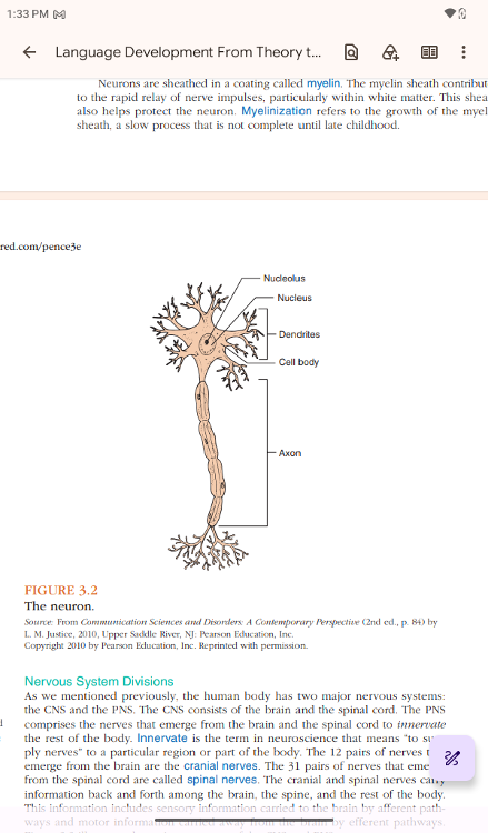 myelin. | back 16 Neurons are sheathed in a coating called myelin. The myelin sheath
contributes |
front 17 Myelinization | back 17 refers to the growth of the myelin |
front 18 Nervous System Divisions | back 18 Nervous System Divisions |
front 19 Innervate | back 19 Innervate is the term in neuroscience that means “to sup- |
front 20 cranial nerves | back 20 The 12 pairs of nerves that |
front 21 spinal nerves | back 21 The 31 pairs of nerves that emerge |
front 22 The cranial and spinal nerves carry | back 22 .. |
front 23 Central Nervous System | back 23 Central Nervous System. The CNS consists of the brain and the spinal cord |
front 24 The brain is essentially the chief executive operator of the entire CNS: It initiates and regulates virtually all motor, sensory, and cognitive processes. | back 24 .. |
front 25 The spinal cord acts | back 25 .. |
front 26 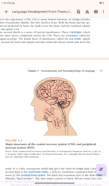 First shield is bone | back 26 Given the importance of the CNS to many human functions, its design
includes a number of protective shields. The first shield is bone.
Both the brain and the spinal cord are protected by bone; the skull
covers the brain, and the vertebral column |
front 27 The second shield | back 27 The second shield is a series of layered membranes |
front 28 meninges | back 28 These meninges, which |
front 29 pia mater | back 29 The inside layer of membrane, called the pia mater, tightly
|
front 30 The second layer is the arachnoid mater, a delicate membrane separated from the pia mater by the subarachnoid space. | back 30 .. |
front 31 dura mater | back 31 The third and outermost layer is the dura mater |
front 32 cerebrospinal fluid (CSF). | back 32 The third shield is a layer of fluid called cerebrospinal fluid (CSF). CSF circu- |
front 33 spinal tap | back 33 Perhaps you have heard of a procedure called a spinal tap (not the
fictional rock band, which is perhaps a more amusing connotation).
Also called a lumbar puncture, a spinal tap involves inserting a
needle between two of the lower (lumbar) |
front 34 Meningitis | back 34 It is a procedure often |
front 35 Peripheral Nervous System | back 35 Peripheral Nervous System. The PNS is the system of nerves connected to the |
front 36 The PNS consists of two sets of nerves: cranial nerves and spinal
nerves. The 12 pairs of cranial nerves run between the brainstem and
the facial and neck regions and are particularly important for speech,
language, and hearing. The cranial nerves transmit information
concerning four of the five senses (vision, hearing, smell, and
| back 36 .. |
front 37 cranial nerves | back 37 cranial nerves run between the brainstem and the facial and neck
regions and are particularly important for speech, language, and
hearing. The cranial nerves transmit information concerning four of
the five senses (vision, hearing, smell, and taste) to the brain. They
also carry motor impulses from the brain to the face and |
front 38 The seven cranial nerves most closely involved with speech | back 38 .. |
front 39 The 31 pairs of spinal nerves run between the spinal cord and all
peripheral | back 39 .. |
front 40 contralateral. | back 40 An important fea- |
front 41 What are the Major Structures and | back 41 What are the Major Structures and |
front 42 the body (Jerison, 2012), the brain is extraordinarily important to the entire functioning of the human body and mind. In fact, the human brain—and its capacity for abstract thought and language—differentiates humans most significantly from other species | back 42 .. |
front 43 The growth of the human brain in both size and weight is one of
| back 43 .. |
front 44 The most important evolutionary change in the human brain, accounting for these increases in weight and mass, is the enlargement of the outer layers of the brain | back 44 .. |
front 45 .. | back 45 These enlarged regions are called the neocortex, meaning “new cortex”
(or, more literally, “new rind”), which has grown over the original
human brain. The neocortex controls most of the functions that
exemplify human thought and language, including speech, language,
reasoning, planning, and problem solving. Recent research has exposed
deficits in regions of the neocortex of children with |