Anatomy JV Exam 3: Eye & Orbit
The eyelids are covered externally by ______ ______ and internally by the ______ ______, which is continuous with the ______ ______.
thin skin, palpebral conjunctivum, bulbar conjunctivum
The muscular portion of the eyelid is formed by the ______ ______, which is innervated by the ______ nerve, also known as cranial nerve ______.
orbicularis oculi, facial, VII
The edges of the eyelids are lubricated by the ______ glands, also known as ______ glands.
tarsal, Meibomian
In the corneal reflex, the afferent limb is carried by the ______ nerve (CN ______), and the efferent limb is carried by the ______ nerve (CN ______), which contracts the ______ ______ muscle.
trigeminal, V, facial, VII, orbicularis oculi
Loss of the corneal reflex due to ______ nerve injury can lead to ______ damage and eventual ______.
facial, corneal, ulceration
The ______ ______ ______ elevates the upper eyelid and is innervated by the ______ nerve (CN ______).
levator palpebrae superioris, oculomotor, III
Müller’s muscle, also called the ______ ______ muscle, is a ______ muscle innervated by ______ ______ axons from the ______ ______ ______.
superior tarsal, smooth, postganglionic sympathetic, superior cervical ganglion
Paralysis of Müller’s muscle can lead to ______, or ______ of the upper eyelid, which is a feature of ______ ______.
ptosis, drooping, Horner's Syndrome
Müller’s muscle, also known as the ______ ______ muscle, is a ______ muscle located in the ______ eyelid.
superior tarsal, smooth, upper
Müller’s muscle is innervated by ______ ______ axons from the ______ ______ ______.
postganglionic sympathetic, superior cervical ganglion
Tear production by the lacrimal gland is primarily controlled by ______ innervation from the ______ nerve (CN ______), with postganglionic fibers originating from the ______ ______.
parasympathetic, facial, VII, pterygopalatine ganglion
In addition to parasympathetic input, the lacrimal gland also receives ______ innervation, which plays a supporting role in ______ control.
sympathetic, tear
Tears contain ______ and ______, which help protect the eye from infection.
lysozyme, IgA
Tears drain into the ______ ______, then through the ______ into the ______ ______, which empties via the ______ ______ into the ______ ______ ______.
lacrimal puncta, canaliculi, lacrimal sac, nasolacrimal duct, inferior nasal meatus
The orbit is a ______-shaped cavity with its apex located at the ______ ______ in the ______ wing of the ______ bone.
pyramidal, optic canal, lesser, sphenoid
The superior wall of the orbit is formed by the ______ portion of the ______ bone, and the medial wall is formed mostly by the ______ bone.
orbital, frontal, ethmoid
The lateral wall of the orbit is formed by the ______ portion of the ______ bone and the ______ wing of the ______ bone.
frontal, zygomatic, greater, sphenoid
The inferior wall of the orbit is formed mostly by the ______ bone, with additional contributions from the ______ and ______ bones.
maxillary, zygomatic, palatine
at what angle do the orbital axes diverage?
______ deg
45 deg
The apex of the orbit is located at the ______ ______ in the ______ wing of the ______ bone.
optic canal, lesser, sphenoid
A blowout fracture occurs when the ______ walls of the orbit fracture, often involving nearby ______.
thin, sinuses
Medial wall ______ fractures involve the ______ bone and can affect the ______ sinuses.
blowout, ethmoid, ethmoid
Inferior wall blowout fractures involve the ______ bone and can affect the ______ sinuses.
maxillary, maxillary
Superior wall blowout fractures can affect the ______ lobe of the ______.
frontal, brain
Bleeding into the orbit posteriorly after a fracture may cause the eye to protrude, a condition called ______.
exophthalmos
Exophthalmos is the ______ of the ______, which can be caused by various conditions such as bleeding into the ______ ______.
protrusion, eyeball, eye socket
The orbit contains the ______ (periosteum), ______ ______ that provide padding and allow ______ movements, ______ and ______, ______ muscles, the ______ (bulbus oculi), and the ______ apparatus.
periorbita, orbital fat, eye, nerves, vessels, extraocular, eye, lacrimal
PONEEL
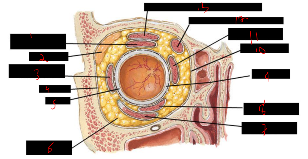
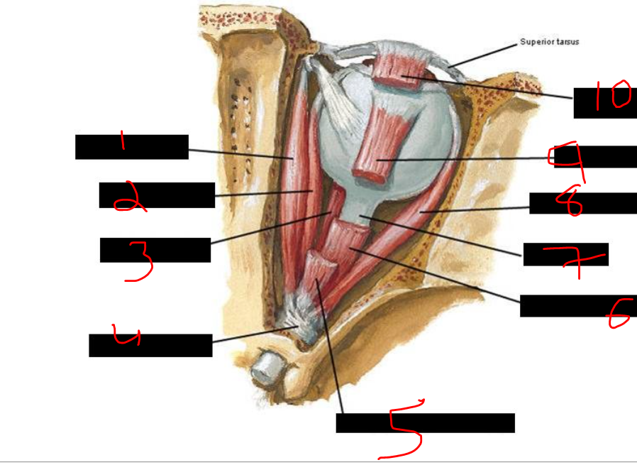
What is 1?
superior rectus muscle and fascial sheath
What is 2?
lacrimal gland
What is 3?
lateral rectus muscle and fascial sheath
What is 4?
periorbita
What is 5?
sclera
What is 6?
retrobulbar fat (orbital fat body)
What is 7?
inferior oblique muscle and fascial sheath
What is 8?
inferior rectus muscle and fascial sheath
What is 9?
sclera
What is 10?
periorbita
What is 11?
medial rectus muscle and fascial sheath
What is 12?
superior oblique muscle and fascial sheath
What is 13?
levator palpebrae superioris muscle and fascial sheath
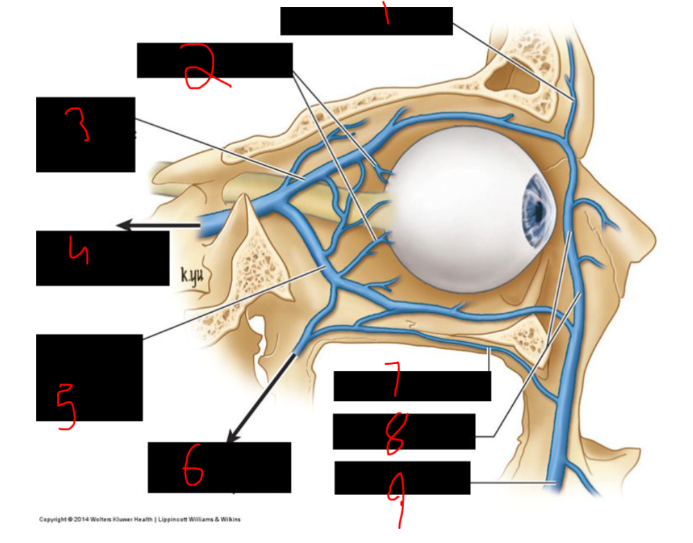
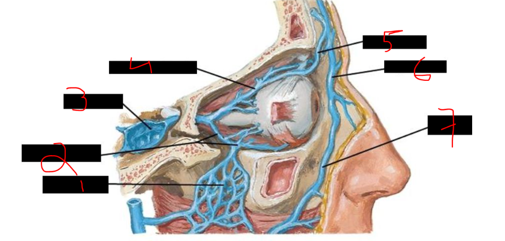
The major blood supply to the orbit is the ______ artery, which is the first branch of the ______ ______ artery.
ophthalmic, internal carotid
The ______ artery of the ______ enters the optic nerve; its occlusion can cause ______ in the eye.
central, retina, blindness
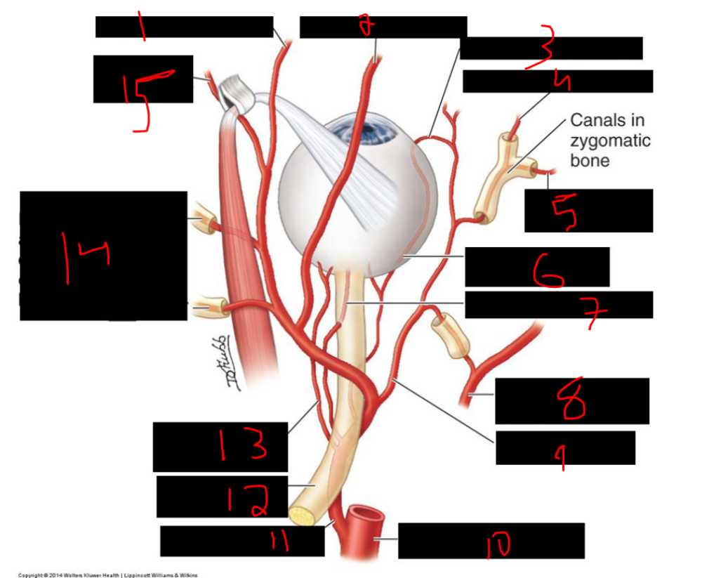
What is 1?
supratrochlear artery

What is 2?
supra-orbital artery
What is 3?
anterior ciliary artery
What is 4?
zygomagitcofacial artery
What is 5?
zygomaticotemporal artery
What is 6?
long posterior ciliary artery
What is 7?
central artery of retina
What is 8?
middle meningeal artery
What is 9?
lacrimal artery
What is 10?
internal carotid artery
What is 11?
opthalmic artery
What is 12?
optic nerve (CN II)
What is 13?
short posterior ciliary artery
What is 14?
anterior and posterior
ethmoidal arteries in canals in ethmoid bone

What is 15?
dorsal nasal artery
The ______ artery of the ______ enters the ______ nerve to supply the ______ retina.
central, retina, optic, inner
The ______ ______ arteries penetrate the ______ to supply the ______ and ______ retina, including the pigment epithelium and rods and cones.
posterior ciliary, sclera, choroid, outer
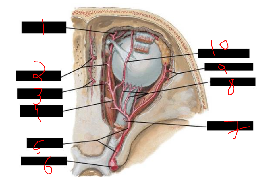
What is 1?
supratrochlear artery
What is 2?
anterior ethmoidal artery
What is 3?
posterior ethmoidal artery
What is 4?
continuation of opthalmic artery
What is 5?
opthalmic artery
What is 6?
internal carotid artery
What is 7?
central retinal artery
What is 8?
posterior ciliary artery
What is 9?
zygomatic branches
What is 10?
supraorbital artery
The ______ artery, a branch of the ______ artery, forms an anastomosis with the ______ artery.
infraorbital, maxillary, ophthalmic
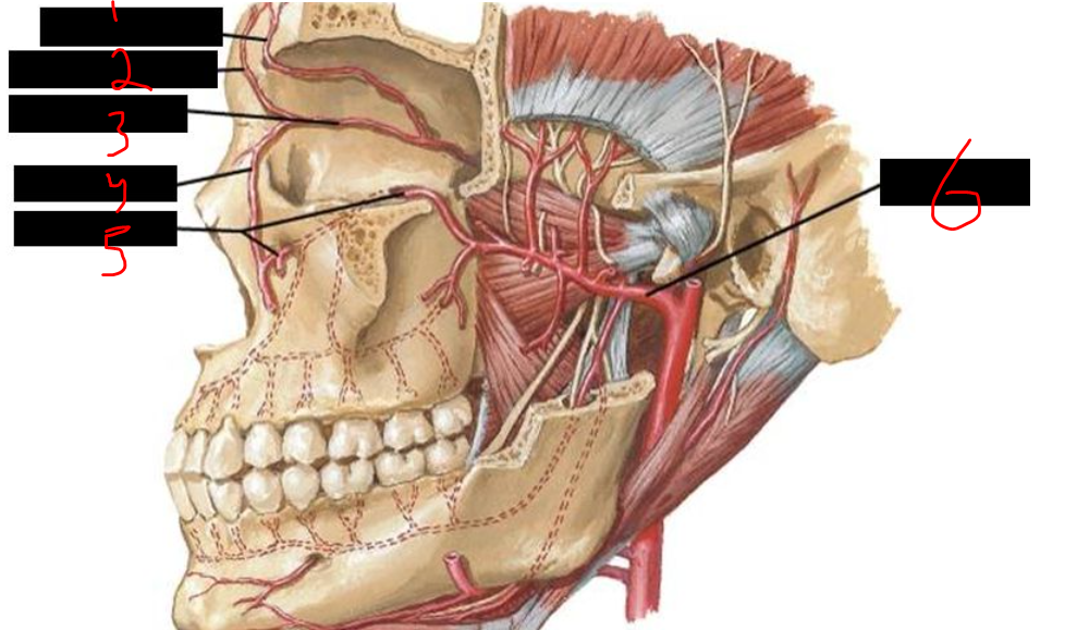
What is 1?
supraorbital artery
What is 2?
supratrochlear artery
What is 3?
opthalmic artery
What is 4?
angular artery
What is 5?
infraorbital artery
What is 6?
maxillarty artery
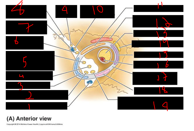
What is 1?
supra-orbital vein
What is 2?
vorticose vein
What is 3?
superior ophthalmic vein
What is 4?
to cavernous sinus
What is 5?
inferior ophthalmic vein
What is 6?
to pterygoid venous plexus
What is 7?
infra-orbital vein
What is 8?
angular vein
What is 9?
facial vein
What is 1?
pterygoid plexus
What is 2?
inferior ophthalmic vein
What is 3?
cavernous sinus
What is 4?
superior ophthalmic vein
What is 5?
nasofrontal vein
What is 6?
angular vein
What is 7?
facial vein
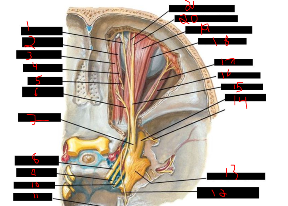
The following structures pass through the tendinous ring: the ______ nerve (both superior and inferior divisions, CN ______), the ______ nerve, the ______ nerve (CN ______), the ______ nerve (CN ______), and the ______ artery (within the ______ nerve).
oculomotor, III, nasociliary, abducent, VI, optic, II, ophthalmic, optic
The extraocular eye muscles that do not attach to the tendinous ring are the ______ ______ and the ______ ______.
superior oblique, inferior oblique
The muscles that attach to the tendinous ring and their functions are:
- ______ ______: abduction
- ______ ______: downward gaze
- ______ ______: adduction
- ______ ______: upward gaze
lateral rectus
inferior rectus
medial rectus
superior rectus
What is 1?
inferior ophthalmic vein

What is 2?
abducens nerve (CN VI)
What is 3?
lateral rectus
What is 4?
nasociliary nerve
What is 5?
oculomotor nerve (CN III), superior division
What is 6?
trochlear nerve (CN IV)
What is 7?
superior ophthalmic vein
What is 8?
superior orbital fissure
What is 9?
frontal nerve
What is 10?
lacrimal nerve (CN V1)
What is 11?
superior rectus
What is 12?
levator palpebral superioris
What is 13?
optic nerve (CN II)
What is 14?
superior oblique
What is 15?
medial rectus
What is 16?
ophthalmic artery
What is 17?
common tendinous ring
What is 18?
inferior rectus
What is 19?
oculomotor nerve (CN III), inferior divison
What is 1?
supratrochlear nerve

What is 2?
medial rectus muscle
What is 3?
superior oblique muscle
What is 4?
infratrochlear nerve
What is 5?
nasociliary nerve
What is 6?
trochlear nerve (IV)
What is 7?
ophthalmic nerve (V1)
What is 8?
oculomotor nerve (III)
What is 9?
trochlear nerve (IV)
What is 10?
abducens nerve (VI)
What is 11?
tentorium cerebelli
What is 12?
tentorial nerve (meningeal) branch of ophthalmic nerve
What is 13?
trigeminal (semilunar) ganglion
What is 14?
maxillary nerve (V2)
What is 15?
frontal nerve
What is 16?
lateral rectus muscle
What is 17?
lacrimal nerve
What is 18?
superior rectus muscle
What is 19?
levator palpebral superioris muscle
What is 20?
lateral branch of supraorbital nerve
What is 21?
medial branch of supraorbital nerve
The extrinsic eye muscles and their innervation are:
- ______ nerve (CN ______): superior rectus, medial rectus, inferior rectus, inferior oblique, and levator palpebrae superioris
- ______ nerve (CN ______): superior oblique
- ______ nerve (CN ______): lateral rectus (abducts eye)
oculomotor, III
trochlear, IV
abducens, VI
What is 1?
superior oblique muscle

What is 2?
medial rectus muscle
What is 3?
inferior rectus muscle
What is 4?
common tendinous ring
What is 5?
leavtor palpebrae superioris muscle
What is 6?
superior rectus muscle
What is 7?
optic nerve
What is 8?
lateral rectus muscle
What is 9?
superior rectus muscle
What is 10?
levator palpebrae superioris muscle
What is 1?
superior oblique
What is 2?
superior rectus
What is 3?
medial rectus
What is 4?
inferior rectus
What is 5?
lateral rectus
What is 6?
inferior oblique
action of medial rectus?
_____ _____
adduct eyeball
action of lateral rectus?
_____ _____
abducts eyeball
The ______ ______ elevates the eyeball and has secondary actions of ______ and ______ rotation.
superior rectus, adduction, medial
The ______ ______ depresses the eyeball and has secondary actions of ______ and ______ rotation.
inferior rectus, adduction, lateral
The ______ ______ depresses the eyeball and also ______ and ______ rotates it. It is the primary ______ rotator.
superior oblique, abducts, medially, medial
The ______ ______ elevates the eyeball and also ______ and ______ rotates it. It is the primary ______ rotator.
inferior oblique, abducts, laterally, lateral
what is the primary medial rotator of the eyeball?
______ ______
superior oblique
what is the primary lateral rotator?
______ ______
inferior oblique
how do you clinically test superior rectus?
have patient look ______ , then ______
have patient look laterally, then up
how do you clinically test inferior rectus?
have patient look ______ , then ______
have patient look laterally, then down
how do you clinically test superior oblique?
have patient look ______ , then ______
have patient look medially, then down
how do you clinically test inferior oblique?
have patient look ______ , then ______
have patient look medially, then up
how do you clinically test medial & lateral recti?
look ______ (medial oblique) & ______ (lateral oblique)
look medially (medial oblique) & laterally (lateral oblique)
What is 1?
superior rectus (III)
What is 2?
lateral rectus (VI)
What is 3?
inferior rectus (III)
What is 4?
inferior oblique (III)
What is 5?
medial rectus (III)
What is 6?
superior oblique (IV)
What is 7?
superior rectus (III)
What is 8?
lateral rectus (VI)
What is 9?
inferior rectus (III)
With abducens nerve palsy, during ______ gaze, the affected eye is pulled ______ due to unopposed action of the ______ ______, because the ______ ______ is denervated.
primary, medially, medial rectus, lateral rectus
what nerve has been damaged/affected?
abducent nerve (CN VI) palsy
In abducent nerve (CN ______) palsy, during ______ gaze, the affected eye is pulled ______ due to unopposed ______ ______.
VI, primary, medially, medial rectus
In trochlear nerve palsy:
- ______ occurs, where the affected eye looks ______ in primary gaze due to denervated ______ ______
- ______ occurs, where the eye is rotated ______
- Patients experience ______
- They tilt their head ______ and ______ from the affected eye to compensate
hypertropia, upward, superior oblique
extorsion, laterally
diplopia
downward, away

what nerve has been damaged/affected?
trochlear nerve (CN IV) palsy
In oculomotor nerve palsy:
- The affected eye is positioned ______ and ______ due to unopposed actions of the ______ ______ and ______ ______ muscles
- The patient cannot ______ the upper eyelid
- The patient cannot ______ the eye to follow an object toward the face (impaired ______)
- ______ is present
- The pupil on the affected side is ______ due to unopposed ______ stimulation of the ______ ______ muscle
down, out, lateral rectus, superior oblique
elevate
adduct, adduction
diplopia
dilated, sympathetic, dilator pupillae
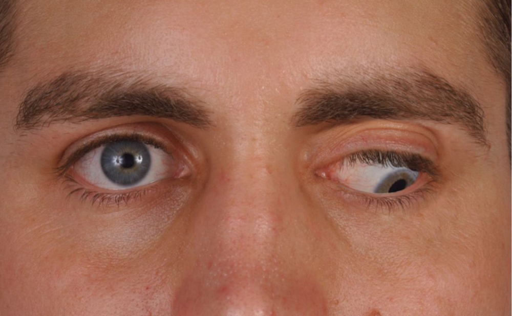
What is this?
oculomotor nerve palsy
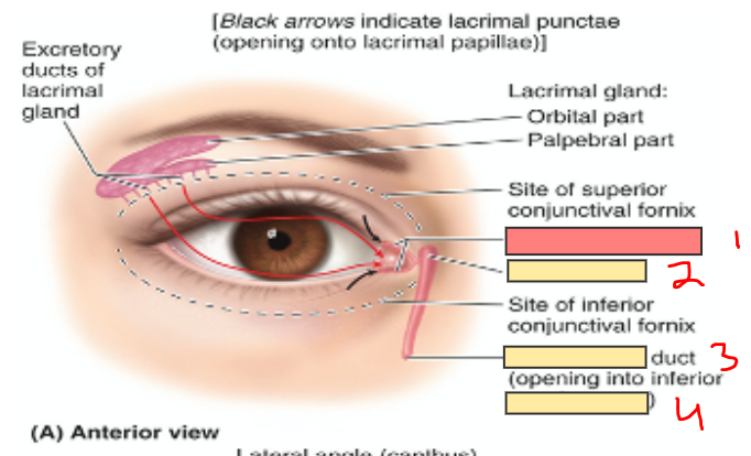
What is 1?
lacrimal canaliculi
What is 2?
lacrimal sac
What is 3?
nasolacrimal
What is 4?
inferior nasal meatus
Axons from the _____ act as the afferent part of pupillary reflexes.
retina
Pupillary reflexes axons terminate in the _____ area and the _____ _____
pretectal
superior colliculus
Some retinal ______ cell axons bypass the ______ ______ nucleus and terminate in the ______ area and the ______ ______. At least some of these axons are involved in ______ reflexes.
ganglion, lateral geniculate, pretectal, superior colliculus, optic
In the pupillary light reflex, shining a light into one eye causes the ______ to ______, testing the integrity of ______ innervation to the pupil.
pupil, constrict, parasympathetic
The consensual reflex refers to constriction of the pupil in the ______ eye when light is shined into the ______ eye.
contralateral, opposite
The consensual reflex is the ______ of the pupil in the ______ eye when light is shined into the ______ eye.
constriction, contralateral, opposite
In the accommodation reflex, when a patient looks at a ______ object, the pupils ______ bilaterally.
near, constrict
During the accommodation reflex, the ______ muscles contract to allow the ______ to thicken for ______ vision.
ciliary, lenses, near
Dilated pupils that do not respond to light suggest unopposed action of the ______ ______ muscle, which is innervated by the ______ nervous system.
dilator pupillae, sympathetic
Non-reactive dilated pupils may indicate brainstem damage involving the ______ -______ nucleus.
Edinger-Westphal
In presbyopia, the ______ loses its ______ and can no longer ______, making it difficult to focus on ______ objects.
lens, flexibility, thicken, near
Under parasympathetic control, the ______ muscles contract, the ______ thickens, and the ______ constrict.
ciliary, lens, pupils
The test used to detect Marcus Gunn pupil (afferent pupillary defect) is the ______ ______ test.
swinging light
When shining a light into a normal eye during the swinging light test, the pupil in that eye ______ and the pupil in the other eye ______ (consensual light reflex).
constricts, constricts
In a normal swinging light test, when the light is swung to the other eye, that pupil should ______ more.
constrict
If during the swinging light test the pupil ______ when the light is shined in that eye, it indicates an ______ defect, often due to an ______ nerve problem.
dilates, afferent, optic
Argyll Robertson pupils are characterized by small, irregular pupils that fail to ______ to ______ but do constrict during ______.
Argyll Robertson pupils are associated with ______
constrict, light, accommodation
neurosyphilis