(OTD 515) Special Senses: Vision, Audition, and Olfaction Overview
Cranial Nerve VIII
Transmits auditory information from cochlea.
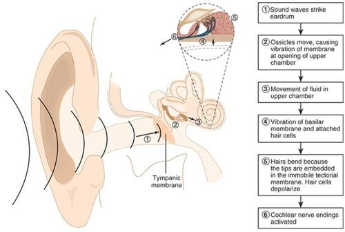
Sound Waves
Vibrations characterized by frequency and amplitude.
Frequency
Pitch measured in Hertz (Hz) of oscillations.
Amplitude
Loudness determined by magnitude of oscillations.
Cochlea
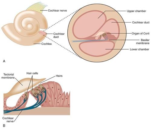
auditory structure
Organ of Corti
coverts mechanical energy from sound waves into neural signals
Basilar Membrane
- Codes sound frequency through its shape.
- narrowest at middle ear = shorter fivers for higher frequencies
- widest at free end = longer hairs for lower frequencies
Hair Cells
Sensory cells that generate neural signals.
Auditory Pathway (from choclear nuceli)
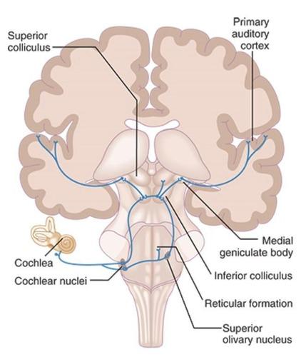
reticular formation --> inferior colliculus --> medial geniculate body
Cochlear Nuclei
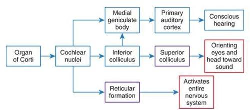
First relay station for auditory signals.
Inferior Colliculus
Integrates auditory information from both ears.
Medial Geniculate Body
Thalamic relay for auditory signals to cortex.
Primary Auditory Cortex
Processes complex sounds, sound localization, selective attention to specific sounds, discrimination of auditory patterns, performance of difficult auditory tasks
Auditory Patterns
Recognition and discrimination of sound sequences.
Round Window
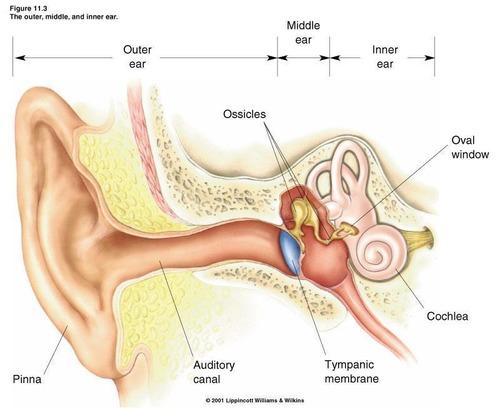
Flexible membrane that relieves cochlear pressure.
Oval Window
Membrane transmitting vibrations from stapes to cochlea.
Reticular Formation
Regulates arousal and attention to auditory stimuli.
Auditory Nerve
Transmits signals from cochlea to brainstem.
Olfactory receptors
Hair cells in nostrils detecting odors.
Olfactory pathway

olfactory receptors --> olfactory bulb --> Primary olfactory cortex/amygdala/parahippocampal gyrus
Amygdala sends olfactory information to (3 places)
- Hypothalamus (hunger)
- Medial parahippocampal gyrus
(quality of aromas and odors)
Sends to secondary olfactory area in orbitofrontal cortex (frontal lobe) for value judgements - Lateral parahippocampal gyrus (declarative memory)
Primary olfactory cortex
Initial processing area for olfactory signals.
Orbitofrontal cortex
Processes value judgments of odors.
Visual system
System responsible for sight and visual processing.
Eye movement control
Regulates movements for visual targeting.
Lens
Refracts light before it enters the pupil.
Pupil
Opening for light entry, controlled by ciliary bodies.
Retina
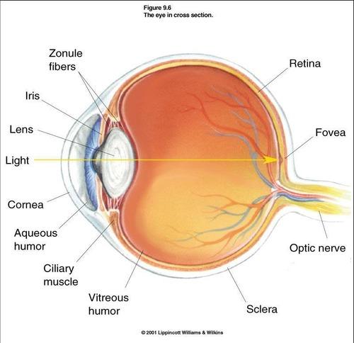
Receptors that convert light into neural signals
Optic nerve (CN II)
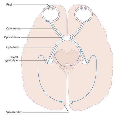
First order neuron in visual pathway.
Visual pathway
Sequence of neural connections for vision.
Optic chiasm
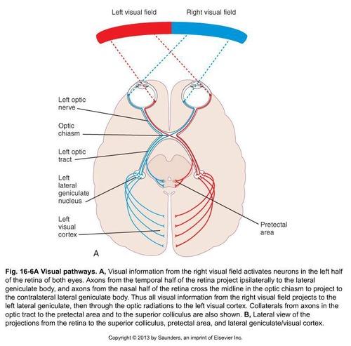
Point where retinal axons cross to opposite cortex.
Lateral geniculate nucleus
Thalamic relay for visual information.
Primary visual cortex
Processes basic visual features like shape.
Dorsal stream
Visual pathway for action and movement adjustments.
Ventral stream
Visual pathway for object recognition.
Tectal system
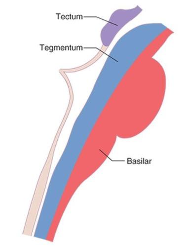
Midbrain structure for auditory and visual reflexes.
Eye movement system
- Normal eye movements require synthesis of information
concerning
- head movements (vestibular), visual objects (visual info)
- eye movement and position (proprioception)
- selection of visual target (brainstem & cortical areas)
Conjugate Movements
Both eyes move in the same direction.
Vergence Movements
Eyes move toward or away from midline.
Gaze Stabilization
Maintains stable vision during head movements.
Nystagmus
Involuntary oscillating movements of the eyes.
Optokinetic Nystagmus
Reflex elicited by moving moving visual stimuli.
Physiologic Nystagmus
Normal response elicited by head rotation or stimulation.
Saccades
Fast eye movements between visual targets.
Smooth Pursuits
Eye movements tracking a moving object.
Convergence
Eyes aim at midline for closer objects.
Optic Nerve Lesion
Causes total vision loss in ipsilateral eye.
Optic Chiasm Lesion
Results in bitemporal hemianopia.
Complete Optic Tract Lesion (before LGN)
Leads to contralateral homonymous hemianopia.
Incomplete Optic Tract Lesion (after LGN)
Causes partial vision loss in contralateral field.
Ciliary Muscles
Contract to increase lens curvature for focus.
Frontal Eye Fields
Involved in voluntary eye movement control.
Superior Colliculus
Processes visual information for saccades.
Eye movement system objectives
- Keeping the position of the eyes stable during head movements to ensure that the environment does not appear to bounce.
- Directing the gaze at visual targets
Eye movements are either
- Conjugate (both eyes move in the same direction)
- Vergence (eyes move toward the midline or away from the midline)