Lab Practical 2 A&P II LAB
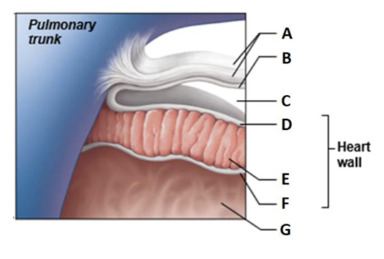
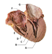
Label A
Fibrous pericardium
Label B
Parietal layer of serous pericardium
Label C
Pericardial cavity
Label D
Epicardium (visceral layer of serous pericardium)
Label E
Myocardium
Label F
Endocardium
Label G
Heart chamber
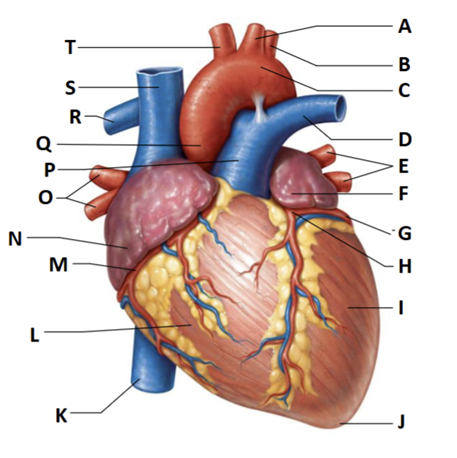
Label A
left common carotid artery
Label B
left subclavian artery
Label C
Aortic arch
Label D
Left pulmonary trunk
Label E
left pulmonary veins
Label F
auricle of left atrium
Label G
circumflex artery
Label H
left coronary artery (in coronary sulcus)
Label I
left ventricle
Label J
apex
Label K
inferior vena cava
Label L
right ventricle
Label M
right coronary artery (in coronary sulcus)
Label N
right atrium
Label O
right pulmonary veins
Label P
pulmonary trunk
Label Q
ascending aorta
Label R
right pulmonary artery
Label S
superior vena cava
Label T
brachiocephalic trunk
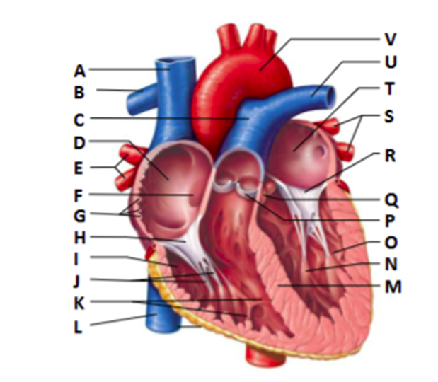
Label A
Superior vena cava
Label B
right pulmonary artery
Label C
pulmonary trunk
Label D
right atrium
Label E
right pulmonary veins
Label F
fossa ovalis
Label G
pectinate muscles
Label H
tricuspid valve
Label I
right ventricle
Label J
chordae tendineae
Label K
trabeculae carneae
Label L
inferior vena cava
Label M
interventricular septum
Label N
papillary muscle
Label O
left ventricle
Label P
pulmonary valve
Label Q
aortic valve
Label R
mitral (bicuspid) valve
Label S
left pulmonary vein
Label T
left atrium
Label U
left pulmonary artery
Label V
aorta
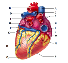
Label A
superior vena cava
Label C
right pulmonary artery
Label E
right pulmonary veins
Label G
right atrium
Label J
inferior vena cava
Label M
right coronary artery (in coronary sulcus)
Label N
posterior interventricular artery (in posterior interventricular sulcus)
Label P
right ventricle
Label L
coronary sinus
Label B
aorta
Label D
left pulmonary artery
Label F
left pulmonary veins
Label H
auricle of left atrium
Label I
left atrium
Label K
great cardiac vein
Label O
left ventricle
Label Q
apex
Label A
myocardium of left ventricle
Label B
papillary muscle
Label C
interventricular septum
Label D
chordae tendineae
Label E
myocardium of right ventricle
Label F
tricuspid valve
Label G
mitral valve
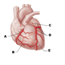
Label A
right coronary artery
Label B
right marginal artery
Label C
left coronary artery
Label D
circumflex artery
Label E
anterior interventricular artery
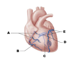
Label A
anterior cardiac veins
Label B
small cardiac vein
Label C
middle cardiac vein
Label D
coronary sinus
Label E
great cardiac vein
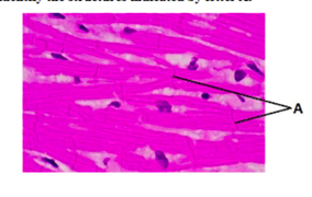
Identify A
Intercalated discs
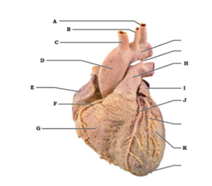
Label A
left subclavian artery
Label B
left common carotid artery
Label C
brachiocephalic trunk
Label D
ascending aorta
Label E
right atrium
Label F
right coronary artery (in coronary sulcus)
Label G
right ventricle
Label H
pulmonary trunk
Label I
auricle of left atrium
Label J
anterior interventricular artery (in anterior interventricular sulcus)
Label K
left ventricle
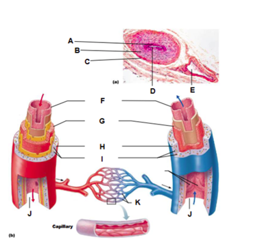
Label A
tunica intima
Label B
tunica media
Label C
tunica externa
Label D
artery
Label E
vein
Label F
tunica intima
Label G
tunica media
Label H
tunica externa
Label I
vasa vasorum
Label K
capillary network
Label J
Lumen
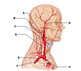
Label A
vertebral artery
Label B
internal carotid artery
Label C
external carotid artery
Label D
common carotid artery
Label E
subclavian artery
Label F
axillary artery
Label G
superficial temporal artery
Label H
brachiocephalic trunk
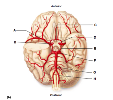
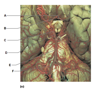
Label A
middle cerebral artery
Label B
internal carotid artery
Label C
anterior communicating artery
Label D
anterior cerebral artery
Label E
posterior communicating artery
Label F
posterior cerebral artery
Label G
basilar artery
Label H
vertebral artery
Label A
anterior communicating artery
Label B
anterior cerebral artery
Label C
posterior communicating artery
Label D
posterior cerebral artery
Label E
basilar artery
Label F
vertebral artery
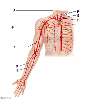
Label A
vertebral artery
Label B
axillary artery
Label C
brachial artery
Label D
radial artery
Label E
ulnar artery
Label F
common carotid arteries
Label G
right subclavian artery
Label H
left subclavian artery
Label I
brachiocephalic trunk
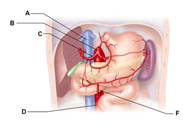
Label A
inferior vena cava
Label B
celiac trunk
Label C
common hepatic artery
Label D
abdominal aorta
Label F
superior mesenteric artery
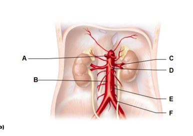
Label A
celiac trunk
Label B
abdominal aorta
Label C
renal artery
Label D
superior mesenteric artery
Label E
inferior mesenteric artery
Label F
common iliac artery
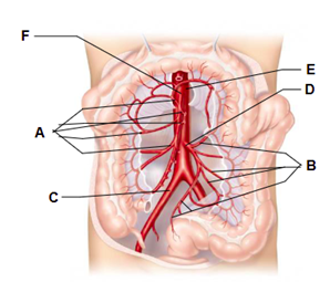
Label A
branches of the superior mesenteric artery
Label B
branches of the inferior mesenteric artery
Label C
right common iliac artery
Label D
inferior mesenteric artery
Label E
abdominal aorta
Label F
superior mesenteric artery
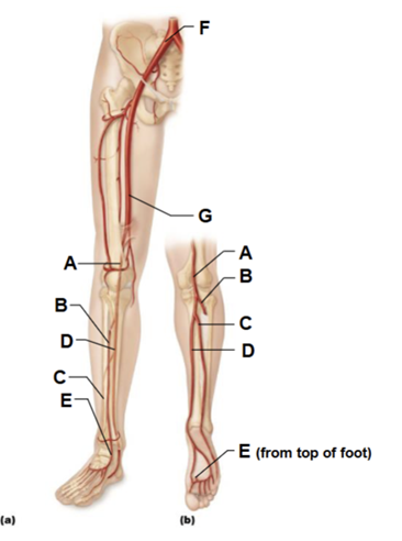
Label A
popliteal artery
Label B
anterior tibial artery
Label C
fibular artery
Label D
posterior tibial artery
Label E
dorsalis pedis artery (from top of foot)
Label F
common iliac artery
Label G
femoral artery
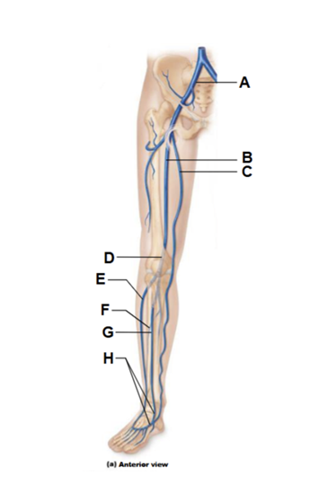
Label A
common iliac vein
Label B
femoral vein
Label C
great saphenous vein (superficial)
Label D
popliteal vein
Label E
small saphenous vein
Label F
fibular vein
Label G
anterior tibial vein
Label H
dorsalis pedis vein
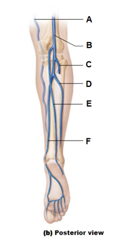
Label A
great saphenous vein
Label B
popliteal vein
Label C
anterior tibial vein
Label D
fibular vein
Label E
small saphenous vein (superficial)
Label F
posterior tibial vein
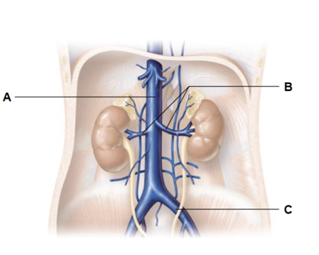
Label A
inferior vena cava
Label B
renal veins
Label C
common iliac vein
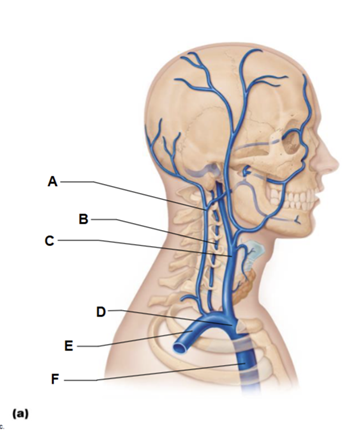
Label A
external jugular vein
Label B
vertebral vein
Label C
internal jugular vein
Label D
brachiocephalic vein
Label E
subclavian vein
Label F
superior vena cava
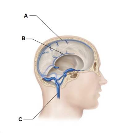
Label A
superior sagittal sinus
Label B
inferior sagittal sinus
Label C
internal jugular vein
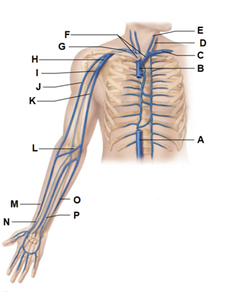
Label A
inferior vena cava
Label B
superior vena cava
Label C
left subclavian vein
Label D
external jugular vein
Label E
Internal jugular vein
Label F
brachiocephalic veins
Label G
right subclavian vein
Label H
axillary vein
Label I
brachial vein
Label J
cephalic vein
Label K
basilic vein
Label L
median cubital vein
Label M
cephalic vein
Label N
radial vein
Label O
basilic vein
Label P
ulnar vein

Label A
left pulmonary artery
Label B
aortic arch
Label C
pulmonary trunk
Label D
right pulmonary artery
Label E
three lobar arteries to right lung
Label F
right pulmonary veins
Label G
right atrium
Label H
right ventricle
Label I
two lobar arteries to left lung
Label J
left pulmonary veins
Label K
left atrium
Label L
left ventricle
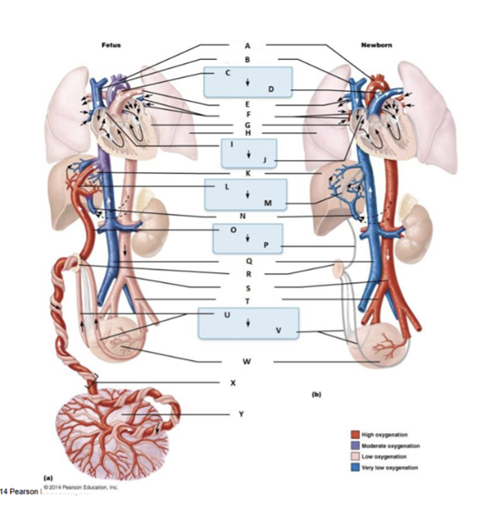
Label A
aortic arch
Label B
Superior vena cava
Label C
ductus arteriosus
Label D
ligamentum arteriosum
Label E
pulmonary artery
Label F
pulmonary veins
Label G
heart
Label H
lung
Label I
foramen ovale
Label J
fossa ovalis
Label K
liver
Labal L
ductus venosus
Label M
ligamentum venosum
Label N
hepatic portal vein
Label O
umbilical vein
Label P
ligamentum teres
Label Q
inferior vena cava
Label R
umbilicus
Label S
abdominal aorta
Label T
common iliac artery
Label U
umbilical arteries
Lable V
medial umbilical ligaments
Label W
urinary bladder
Label X
umbilical cord
Label Y
placenta
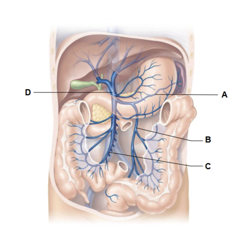
Label A
splenic vein
Label B
inferior mesenteric vein
Label C
superior mesenteric vein
Label D
hepatic portal vein
List the elements of the intrinsic conduction system in
order,
starting from the beginning
SA node → AV node → AV bundle (bundle of His) → left and right bundle
branches
→ subendocardial conducting network (Purkinje fibers)
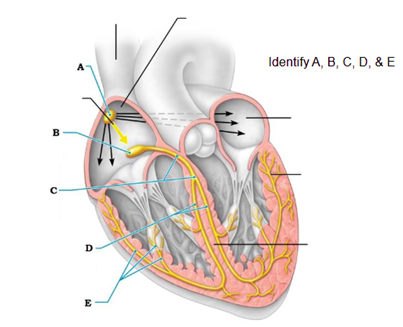
Label A
the sinoatrial (SA) node pacemaker generates impulses
Label B
the impulses pause (0.1 sec) at the atrioventricular (AV) node
Label C
the atrioventricular (AV) bundle conducts the impulses to the bundle branches
Label D
the bundle branches conduct the impulses through the interventricular septum
Label E
the subendocardial conducting network depolarizes the contractile cells of both ventricles
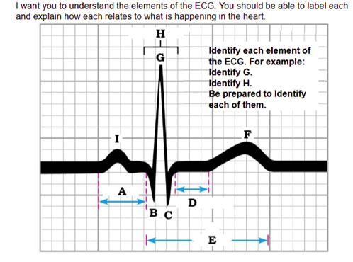
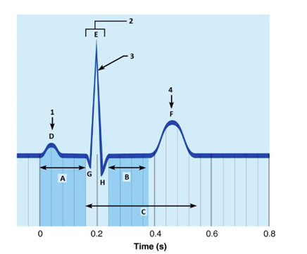
Label A
P-R interval
Label B
Q wave
Label C
S wave
Label D
S-T segment
Label E
Q-T interval
Label F
T wave
Label G
R wave
Label H
QRS complex
Label I
P wave
Label A
P-R interval
Label B
S-T segment
Label C
Q-T interval
Label D
P wave
Label E
R wave
Label F
T wave
Label G
Q wave
Label H
S wave
What is happening at 1
atrial depolarization
What is 2
QRS complex
What is happening at 3
ventricular depolarization
What is happening at 4
ventricular repolarization
immediately before the P wave:
The heart is in diastole
during the P wave:
Depolarization of the atria
immediately after the P wave:
Contraction of the atria.
during the QRS complex:
Depolarization of the ventricles and
repolarization of the atria.
immediately after the QRS complex (S-T segment):
Contraction of the ventricles
during the T wave:
Repolarization of the ventricles.
Enlarged R wave
enlarged ventricles
prolonged P-R interval
First-degree heart block
Prolonged Q-T interval
increased risk of ventricular arrhythmias
S-T segment elevated from baseline
myocardial infarction (heart attack)
WHAT IS 1
Complete the following statements.
The two monosyllables
describing the
heart sounds are 1 . The first heart sound
is
a result of closure of the 2 valves,
whereas the second is a
result of closure
of the 3 valves. The heart chambers
that
have just been filled when you hear the
first heart
sound are the 4 , and the
chambers that have just emptied are
the
5 . Immediately after the second heart
sound, both the 6
and 7 are filling
with blood
lub-dup
WHAT IS 2
Complete the following statements.
The two monosyllables
describing the
heart sounds are 1 . The first heart sound
is
a result of closure of the 2 valves,
whereas the second is a
result of closure
of the 3 valves. The heart chambers
that
have just been filled when you hear the
first heart
sound are the 4 , and the
chambers that have just emptied are
the
5 . Immediately after the second heart
sound, both the 6
and 7 are filling
with blood
atrioventricular
WHAT IS 3
Complete the following statements.
The two monosyllables
describing the
heart sounds are 1 . The first heart sound
is
a result of closure of the 2 valves,
whereas the second is a
result of closure
of the 3 valves. The heart chambers
that
have just been filled when you hear the
first heart
sound are the 4 , and the
chambers that have just emptied are
the
5 . Immediately after the second heart
sound, both the 6
and 7 are filling
with blood
aortic and pulmonary (semilunar)
WHAT IS 4
Complete the following statements.
The two monosyllables
describing the
heart sounds are 1 . The first heart sound
is
a result of closure of the 2 valves,
whereas the second is a
result of closure
of the 3 valves. The heart chambers
that
have just been filled when you hear the
first heart
sound are the 4 , and the
chambers that have just emptied are
the
5 . Immediately after the second heart
sound, both the 6
and 7 are filling
with blood
ventricles
WHAT IS 5
Complete the following statements.
The two monosyllables
describing the
heart sounds are 1 . The first heart sound
is
a result of closure of the 2 valves,
whereas the second is a
result of closure
of the 3 valves. The heart chambers
that
have just been filled when you hear the
first heart
sound are the 4 , and the
chambers that have just emptied are
the
5 . Immediately after the second heart
sound, both the 6
and 7 are filling
with blood
atria
WHAT IS 6
Complete the following statements.
The two monosyllables
describing the
heart sounds are 1 . The first heart sound
is
a result of closure of the 2 valves,
whereas the second is a
result of closure
of the 3 valves. The heart chambers
that
have just been filled when you hear the
first heart
sound are the 4 , and the
chambers that have just emptied are
the
5 . Immediately after the second heart
sound, both the 6
and 7 are filling
with blood
atria
WHAT IS 7
Complete the following statements.
The two monosyllables
describing the
heart sounds are 1 . The first heart sound
is
a result of closure of the 2 valves,
whereas the second is a
result of closure
of the 3 valves. The heart chambers
that
have just been filled when you hear the
first heart
sound are the 4 , and the
chambers that have just emptied are
the
5 . Immediately after the second heart
sound, both the 6
and 7 are filling
with blood
ventricles
Define pulse
Pressure surges in an artery occurring during each
contraction
and relaxation of the left ventricle.
Identify the artery palpated at each of the pressure points
listed.
at the wrist:
radial
Identify the artery palpated at each of the pressure points listed. on the dorsum of the foot:
dorsalis pedis
Identify the artery palpated at each of the pressure points listed. in front of the ear:
temporal
Identify the artery palpated at each of the pressure points listed. at the side of the neck:
carotid
When you were palpating the various pulse or pressure points, which
appeared to
have the greatest amplitude or tension?
_______________ Why do you think this
was so?
carotid artery, The carotid arteries are the major arteries that deliver blood to the brain and they are closest to the heart.
Assume someone has been injured in an auto accident and is
hemorrhaging badly.
What pressure point would you compress to
help stop bleeding from each of the
following areas THE THIGH
femoral artery
Assume someone has been injured in an auto accident and is
hemorrhaging badly.
What pressure point would you compress to
help stop bleeding from each of the
following areas THE CALF
popliteal artery
Assume someone has been injured in an auto accident and is
hemorrhaging badly.
What pressure point would you compress to
help stop bleeding from each of the
following areas THE FOREARM
brachial artery
Assume someone has been injured in an auto accident and is
hemorrhaging badly.
What pressure point would you compress to
help stop bleeding from each of the
following areas THE THUMB
radial artery
How could you tell by simple observation whether bleeding is arterial or venous?
If it spurts, it is arterial. It will flow evenly if it is venous blood
Define blood pressure.
Pressure exerted by blood against the walls of the blood
vessels.
Identify the phase of the cardiac cycle to which each of the following applies: systolic pressure
systole (ventricular contraction)
Identify the phase of the cardiac cycle to which each of the following applies: diastolic pressure
diastole (relaxation)
What are the sounds of Korotkoff?
Sounds that can be auscultated over a partially
occluded artery
What causes the systolic sound?
Sound of turbulent blood flow as it first begins to
move through
the constricted artery
What causes the disappearance of the sound?
Blood is flowing freely; the artery is no longer constricted
Define pulse pressure.
Why is this measurement important?
Systolic pressure minus diastolic pressure
It indicates the actual working pressure (actual amount of blood forced out of the heart during systole)
Explain why pulse pressure is different from pulse rate.
Pulse pressure is what generates the pulse felt calculated as the systolic pressure minus the diastolic pressure; pulse rate is the number of pulsations per minute
How do venous pressures compare to arterial pressures? Why?
Venous pressures are lower.
Veins are far removed from the pumping action of the heart
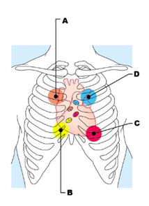
Label A
aortic valve sounds are heard in 2nd intercostal space at right sternal margin
Label B
tricuspid valve sounds are heard in right sternal margin of 5th intercostal space variations include over sternum or over left sternal margin in 5th intercostal space
Label C
mitral valve sounds are heard over heart apex, in 5th intercostal space in line with middle of clavicle
Label D
pulmonary valve sounds are heard in 2nd intercostal space at left sternal margin
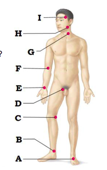
Label A
dorsalis pedis artery
Label B
posterior tibial artery
Label C
popliteal artery
Label D
femoral artery
Label E
radial artery
Label F
brachial artery
Label G
common carotid artery
Label H
facial artery
Label I
superficial temporal artery