Instructions for Side by Side Printing
- Print the notecards
- Fold each page in half along the solid vertical line
- Cut out the notecards by cutting along each horizontal dotted line
- Optional: Glue, tape or staple the ends of each notecard together
Lab Practical 2 A&P II LAB
front 1 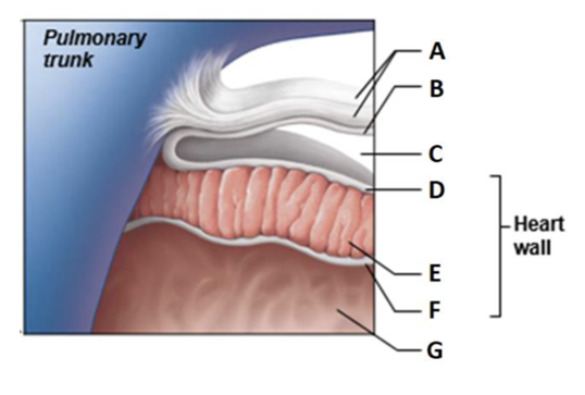 Label A | back 1 Fibrous pericardium |
front 2 Label B | back 2 Parietal layer of serous pericardium |
front 3 Label C | back 3 Pericardial cavity |
front 4 Label D | back 4 Epicardium (visceral layer of serous pericardium) |
front 5 Label E | back 5 Myocardium |
front 6 Label F | back 6 Endocardium |
front 7 Label G | back 7 Heart chamber |
front 8 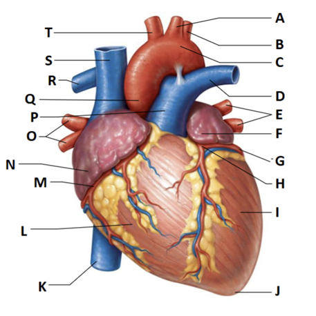 Label A | back 8 left common carotid artery |
front 9 Label B | back 9 left subclavian artery |
front 10 Label C | back 10 Aortic arch |
front 11 Label D | back 11 Left pulmonary trunk |
front 12 Label E | back 12 left pulmonary veins |
front 13 Label F | back 13 auricle of left atrium |
front 14 Label G | back 14 circumflex artery |
front 15 Label H | back 15 left coronary artery (in coronary sulcus) |
front 16 Label I | back 16 left ventricle |
front 17 Label J | back 17 apex |
front 18 Label K | back 18 inferior vena cava |
front 19 Label L | back 19 right ventricle |
front 20 Label M | back 20 right coronary artery (in coronary sulcus) |
front 21 Label N | back 21 right atrium |
front 22 Label O | back 22 right pulmonary veins |
front 23 Label P | back 23 pulmonary trunk |
front 24 Label Q | back 24 ascending aorta |
front 25 Label R | back 25 right pulmonary artery |
front 26 Label S | back 26 superior vena cava |
front 27 Label T | back 27 brachiocephalic trunk |
front 28 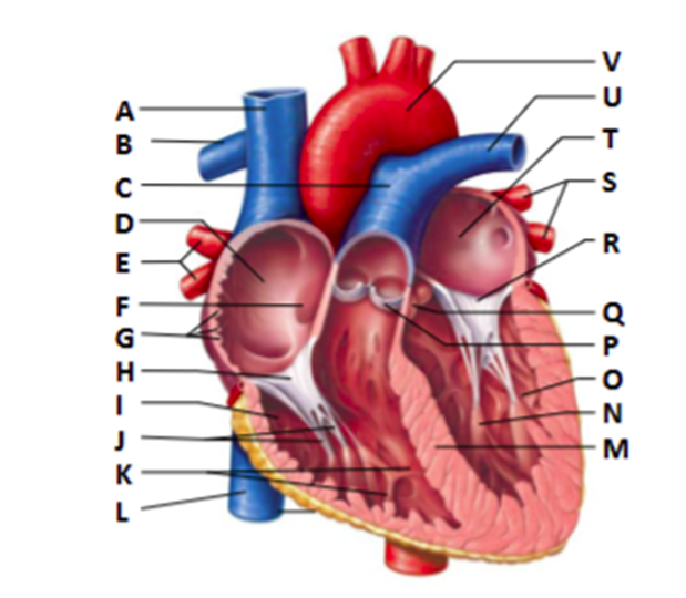 Label A | back 28 Superior vena cava |
front 29 Label B | back 29 right pulmonary artery |
front 30 Label C | back 30 pulmonary trunk |
front 31 Label D | back 31 right atrium |
front 32 Label E | back 32 right pulmonary veins |
front 33 Label F | back 33 fossa ovalis |
front 34 Label G | back 34 pectinate muscles |
front 35 Label H | back 35 tricuspid valve |
front 36 Label I | back 36 right ventricle |
front 37 Label J | back 37 chordae tendineae |
front 38 Label K | back 38 trabeculae carneae |
front 39 Label L | back 39 inferior vena cava |
front 40 Label M | back 40 interventricular septum |
front 41 Label N | back 41 papillary muscle |
front 42 Label O | back 42 left ventricle |
front 43 Label P | back 43 pulmonary valve |
front 44 Label Q | back 44 aortic valve |
front 45 Label R | back 45 mitral (bicuspid) valve |
front 46 Label S | back 46 left pulmonary vein |
front 47 Label T | back 47 left atrium |
front 48 Label U | back 48 left pulmonary artery |
front 49 Label V | back 49 aorta |
front 50 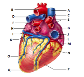 Label A | back 50 superior vena cava |
front 51 Label C | back 51 right pulmonary artery |
front 52 Label E | back 52 right pulmonary veins |
front 53 Label G | back 53 right atrium |
front 54 Label J | back 54 inferior vena cava |
front 55 Label M | back 55 right coronary artery (in coronary sulcus) |
front 56 Label N | back 56 posterior interventricular artery (in posterior interventricular sulcus) |
front 57 Label P | back 57 right ventricle |
front 58 Label L | back 58 coronary sinus |
front 59 Label B | back 59 aorta |
front 60 Label D | back 60 left pulmonary artery |
front 61 Label F | back 61 left pulmonary veins |
front 62 Label H | back 62 auricle of left atrium |
front 63 Label I | back 63 left atrium |
front 64 Label K | back 64 great cardiac vein |
front 65 Label O | back 65 left ventricle |
front 66 Label Q | back 66 apex |
front 67 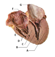 Label A | back 67 myocardium of left ventricle |
front 68 Label B | back 68 papillary muscle |
front 69 Label C | back 69 interventricular septum |
front 70 Label D | back 70 chordae tendineae |
front 71 Label E | back 71 myocardium of right ventricle |
front 72 Label F | back 72 tricuspid valve |
front 73 Label G | back 73 mitral valve |
front 74 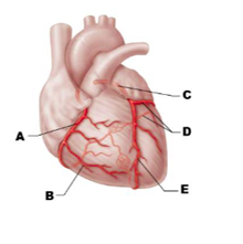 Label A | back 74 right coronary artery |
front 75 Label B | back 75 right marginal artery |
front 76 Label C | back 76 left coronary artery |
front 77 Label D | back 77 circumflex artery |
front 78 Label E | back 78 anterior interventricular artery |
front 79 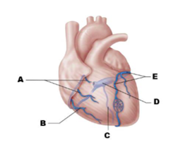 Label A | back 79 anterior cardiac veins |
front 80 Label B | back 80 small cardiac vein |
front 81 Label C | back 81 middle cardiac vein |
front 82 Label D | back 82 coronary sinus |
front 83 Label E | back 83 great cardiac vein |
front 84 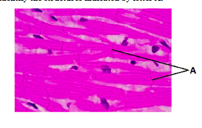 Identify A | back 84 Intercalated discs |
front 85 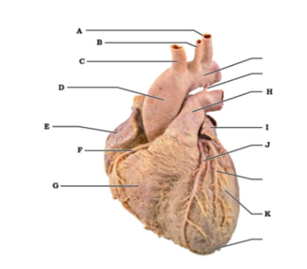 Label A | back 85 left subclavian artery |
front 86 Label B | back 86 left common carotid artery |
front 87 Label C | back 87 brachiocephalic trunk |
front 88 Label D | back 88 ascending aorta |
front 89 Label E | back 89 right atrium |
front 90 Label F | back 90 right coronary artery (in coronary sulcus) |
front 91 Label G | back 91 right ventricle |
front 92 Label H | back 92 pulmonary trunk |
front 93 Label I | back 93 auricle of left atrium |
front 94 Label J | back 94 anterior interventricular artery (in anterior interventricular sulcus) |
front 95 Label K | back 95 left ventricle |
front 96 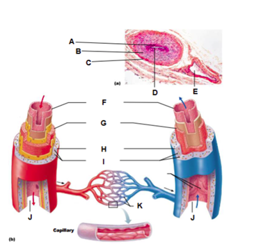 Label A | back 96 tunica intima |
front 97 Label B | back 97 tunica media |
front 98 Label C | back 98 tunica externa |
front 99 Label D | back 99 artery |
front 100 Label E | back 100 vein |
front 101 Label F | back 101 tunica intima |
front 102 Label G | back 102 tunica media |
front 103 Label H | back 103 tunica externa |
front 104 Label I | back 104 vasa vasorum |
front 105 Label K | back 105 capillary network |
front 106 Label J | back 106 Lumen |
front 107 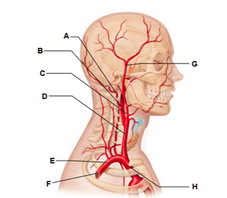 Label A | back 107 vertebral artery |
front 108 Label B | back 108 internal carotid artery |
front 109 Label C | back 109 external carotid artery |
front 110 Label D | back 110 common carotid artery |
front 111 Label E | back 111 subclavian artery |
front 112 Label F | back 112 axillary artery |
front 113 Label G | back 113 superficial temporal artery |
front 114 Label H | back 114 brachiocephalic trunk |
front 115 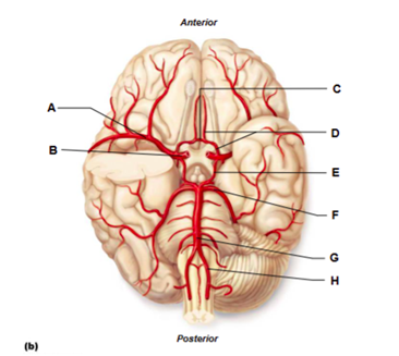 Label A | back 115 middle cerebral artery |
front 116 Label B | back 116 internal carotid artery |
front 117 Label C | back 117 anterior communicating artery |
front 118 Label D | back 118 anterior cerebral artery |
front 119 Label E | back 119 posterior communicating artery |
front 120 Label F | back 120 posterior cerebral artery |
front 121 Label G | back 121 basilar artery |
front 122 Label H | back 122 vertebral artery |
front 123 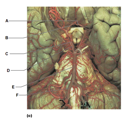 Label A | back 123 anterior communicating artery |
front 124 Label B | back 124 anterior cerebral artery |
front 125 Label C | back 125 posterior communicating artery |
front 126 Label D | back 126 posterior cerebral artery |
front 127 Label E | back 127 basilar artery |
front 128 Label F | back 128 vertebral artery |
front 129 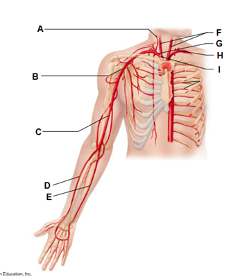 Label A | back 129 vertebral artery |
front 130 Label B | back 130 axillary artery |
front 131 Label C | back 131 brachial artery |
front 132 Label D | back 132 radial artery |
front 133 Label E | back 133 ulnar artery |
front 134 Label F | back 134 common carotid arteries |
front 135 Label G | back 135 right subclavian artery |
front 136 Label H | back 136 left subclavian artery |
front 137 Label I | back 137 brachiocephalic trunk |
front 138 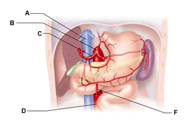 Label A | back 138 inferior vena cava |
front 139 Label B | back 139 celiac trunk |
front 140 Label C | back 140 common hepatic artery |
front 141 Label D | back 141 abdominal aorta |
front 142 Label F | back 142 superior mesenteric artery |
front 143 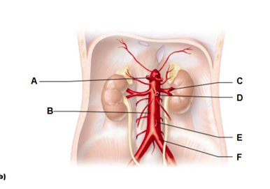 Label A | back 143 celiac trunk |
front 144 Label B | back 144 abdominal aorta |
front 145 Label C | back 145 renal artery |
front 146 Label D | back 146 superior mesenteric artery |
front 147 Label E | back 147 inferior mesenteric artery |
front 148 Label F | back 148 common iliac artery |
front 149 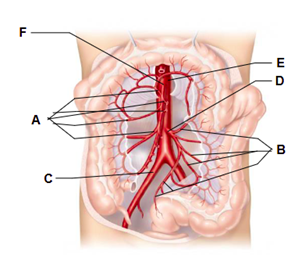 Label A | back 149 branches of the superior mesenteric artery |
front 150 Label B | back 150 branches of the inferior mesenteric artery |
front 151 Label C | back 151 right common iliac artery |
front 152 Label D | back 152 inferior mesenteric artery |
front 153 Label E | back 153 abdominal aorta |
front 154 Label F | back 154 superior mesenteric artery |
front 155 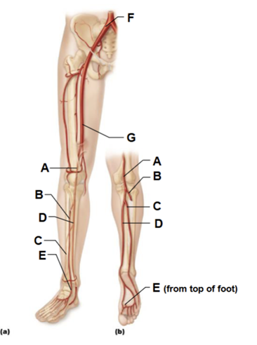 Label A | back 155 popliteal artery |
front 156 Label B | back 156 anterior tibial artery |
front 157 Label C | back 157 fibular artery |
front 158 Label D | back 158 posterior tibial artery |
front 159 Label E | back 159 dorsalis pedis artery (from top of foot) |
front 160 Label F | back 160 common iliac artery |
front 161 Label G | back 161 femoral artery |
front 162 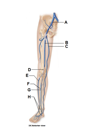 Label A | back 162 common iliac vein |
front 163 Label B | back 163 femoral vein |
front 164 Label C | back 164 great saphenous vein (superficial) |
front 165 Label D | back 165 popliteal vein |
front 166 Label E | back 166 small saphenous vein |
front 167 Label F | back 167 fibular vein |
front 168 Label G | back 168 anterior tibial vein |
front 169 Label H | back 169 dorsalis pedis vein |
front 170 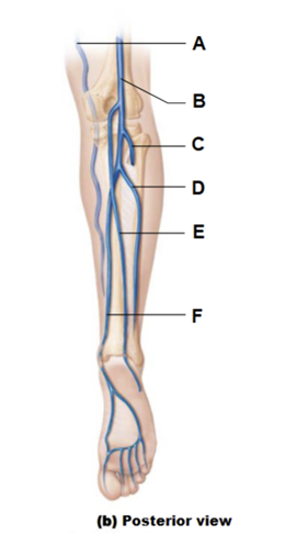 Label A | back 170 great saphenous vein |
front 171 Label B | back 171 popliteal vein |
front 172 Label C | back 172 anterior tibial vein |
front 173 Label D | back 173 fibular vein |
front 174 Label E | back 174 small saphenous vein (superficial) |
front 175 Label F | back 175 posterior tibial vein |
front 176 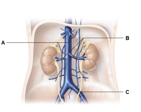 Label A | back 176 inferior vena cava |
front 177 Label B | back 177 renal veins |
front 178 Label C | back 178 common iliac vein |
front 179 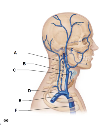 Label A | back 179 external jugular vein |
front 180 Label B | back 180 vertebral vein |
front 181 Label C | back 181 internal jugular vein |
front 182 Label D | back 182 brachiocephalic vein |
front 183 Label E | back 183 subclavian vein |
front 184 Label F | back 184 superior vena cava |
front 185 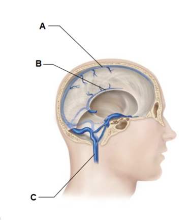 Label A | back 185 superior sagittal sinus |
front 186 Label B | back 186 inferior sagittal sinus |
front 187 Label C | back 187 internal jugular vein |
front 188 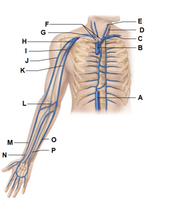 Label A | back 188 inferior vena cava |
front 189 Label B | back 189 superior vena cava |
front 190 Label C | back 190 left subclavian vein |
front 191 Label D | back 191 external jugular vein |
front 192 Label E | back 192 Internal jugular vein |
front 193 Label F | back 193 brachiocephalic veins |
front 194 Label G | back 194 right subclavian vein |
front 195 Label H | back 195 axillary vein |
front 196 Label I | back 196 brachial vein |
front 197 Label J | back 197 cephalic vein |
front 198 Label K | back 198 basilic vein |
front 199 Label L | back 199 median cubital vein |
front 200 Label M | back 200 cephalic vein |
front 201 Label N | back 201 radial vein |
front 202 Label O | back 202 basilic vein |
front 203 Label P | back 203 ulnar vein |
front 204  Label A | back 204 left pulmonary artery |
front 205 Label B | back 205 aortic arch |
front 206 Label C | back 206 pulmonary trunk |
front 207 Label D | back 207 right pulmonary artery |
front 208 Label E | back 208 three lobar arteries to right lung |
front 209 Label F | back 209 right pulmonary veins |
front 210 Label G | back 210 right atrium |
front 211 Label H | back 211 right ventricle |
front 212 Label I | back 212 two lobar arteries to left lung |
front 213 Label J | back 213 left pulmonary veins |
front 214 Label K | back 214 left atrium |
front 215 Label L | back 215 left ventricle |
front 216 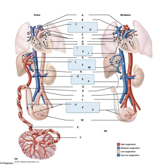 Label A | back 216 aortic arch |
front 217 Label B | back 217 Superior vena cava |
front 218 Label C | back 218 ductus arteriosus |
front 219 Label D | back 219 ligamentum arteriosum |
front 220 Label E | back 220 pulmonary artery |
front 221 Label F | back 221 pulmonary veins |
front 222 Label G | back 222 heart |
front 223 Label H | back 223 lung |
front 224 Label I | back 224 foramen ovale |
front 225 Label J | back 225 fossa ovalis |
front 226 Label K | back 226 liver |
front 227 Labal L | back 227 ductus venosus |
front 228 Label M | back 228 ligamentum venosum |
front 229 Label N | back 229 hepatic portal vein |
front 230 Label O | back 230 umbilical vein |
front 231 Label P | back 231 ligamentum teres |
front 232 Label Q | back 232 inferior vena cava |
front 233 Label R | back 233 umbilicus |
front 234 Label S | back 234 abdominal aorta |
front 235 Label T | back 235 common iliac artery |
front 236 Label U | back 236 umbilical arteries |
front 237 Lable V | back 237 medial umbilical ligaments |
front 238 Label W | back 238 urinary bladder |
front 239 Label X | back 239 umbilical cord |
front 240 Label Y | back 240 placenta |
front 241 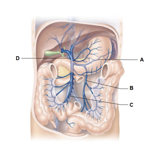 Label A | back 241 splenic vein |
front 242 Label B | back 242 inferior mesenteric vein |
front 243 Label C | back 243 superior mesenteric vein |
front 244 Label D | back 244 hepatic portal vein |
front 245 List the elements of the intrinsic conduction system in
order, | back 245 SA node → AV node → AV bundle (bundle of His) → left and right bundle
branches |
front 246 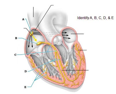 Label A | back 246 the sinoatrial (SA) node pacemaker generates impulses |
front 247 Label B | back 247 the impulses pause (0.1 sec) at the atrioventricular (AV) node |
front 248 Label C | back 248 the atrioventricular (AV) bundle conducts the impulses to the bundle branches |
front 249 Label D | back 249 the bundle branches conduct the impulses through the interventricular septum |
front 250 Label E | back 250 the subendocardial conducting network depolarizes the contractile cells of both ventricles |
front 251 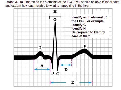 Label A | back 251 P-R interval |
front 252 Label B | back 252 Q wave |
front 253 Label C | back 253 S wave |
front 254 Label D | back 254 S-T segment |
front 255 Label E | back 255 Q-T interval |
front 256 Label F | back 256 T wave |
front 257 Label G | back 257 R wave |
front 258 Label H | back 258 QRS complex |
front 259 Label I | back 259 P wave |
front 260 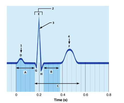 Label A | back 260 P-R interval |
front 261 Label B | back 261 S-T segment |
front 262 Label C | back 262 Q-T interval |
front 263 Label D | back 263 P wave |
front 264 Label E | back 264 R wave |
front 265 Label F | back 265 T wave |
front 266 Label G | back 266 Q wave |
front 267 Label H | back 267 S wave |
front 268 What is happening at 1 | back 268 atrial depolarization |
front 269 What is 2 | back 269 QRS complex |
front 270 What is happening at 3 | back 270 ventricular depolarization |
front 271 What is happening at 4 | back 271 ventricular repolarization |
front 272 immediately before the P wave: | back 272 The heart is in diastole |
front 273 during the P wave: | back 273 Depolarization of the atria |
front 274 immediately after the P wave: | back 274 Contraction of the atria. |
front 275 during the QRS complex: | back 275 Depolarization of the ventricles and |
front 276 immediately after the QRS complex (S-T segment): | back 276 Contraction of the ventricles |
front 277 during the T wave: | back 277 Repolarization of the ventricles. |
front 278 Enlarged R wave | back 278 enlarged ventricles |
front 279 prolonged P-R interval | back 279 First-degree heart block |
front 280 Prolonged Q-T interval | back 280 increased risk of ventricular arrhythmias |
front 281 S-T segment elevated from baseline | back 281 myocardial infarction (heart attack) |
front 282 WHAT IS 1 Complete the following statements. | back 282 lub-dup |
front 283 WHAT IS 2 Complete the following statements. | back 283 atrioventricular |
front 284 WHAT IS 3 Complete the following statements. | back 284 aortic and pulmonary (semilunar) |
front 285 WHAT IS 4 Complete the following statements. | back 285 ventricles |
front 286 WHAT IS 5 Complete the following statements. | back 286 atria |
front 287 WHAT IS 6 Complete the following statements. | back 287 atria |
front 288 WHAT IS 7 Complete the following statements. | back 288 ventricles |
front 289 Define pulse | back 289 Pressure surges in an artery occurring during each
contraction |
front 290 Identify the artery palpated at each of the pressure points
listed. | back 290 radial |
front 291 Identify the artery palpated at each of the pressure points listed. on the dorsum of the foot: | back 291 dorsalis pedis |
front 292 Identify the artery palpated at each of the pressure points listed. in front of the ear: | back 292 temporal |
front 293 Identify the artery palpated at each of the pressure points listed. at the side of the neck: | back 293 carotid |
front 294 When you were palpating the various pulse or pressure points, which
appeared to | back 294 carotid artery, The carotid arteries are the major arteries that deliver blood to the brain and they are closest to the heart. |
front 295 Assume someone has been injured in an auto accident and is
hemorrhaging badly. | back 295 femoral artery |
front 296 Assume someone has been injured in an auto accident and is
hemorrhaging badly. | back 296 popliteal artery |
front 297 Assume someone has been injured in an auto accident and is
hemorrhaging badly. | back 297 brachial artery |
front 298 Assume someone has been injured in an auto accident and is
hemorrhaging badly. | back 298 radial artery |
front 299 How could you tell by simple observation whether bleeding is arterial or venous? | back 299 If it spurts, it is arterial. It will flow evenly if it is venous blood |
front 300 Define blood pressure. | back 300 Pressure exerted by blood against the walls of the blood |
front 301 Identify the phase of the cardiac cycle to which each of the following applies: systolic pressure | back 301 systole (ventricular contraction) |
front 302 Identify the phase of the cardiac cycle to which each of the following applies: diastolic pressure | back 302 diastole (relaxation) |
front 303 What are the sounds of Korotkoff? | back 303 Sounds that can be auscultated over a partially |
front 304 What causes the systolic sound? | back 304 Sound of turbulent blood flow as it first begins to |
front 305 What causes the disappearance of the sound? | back 305 Blood is flowing freely; the artery is no longer constricted |
front 306 Define pulse pressure. | back 306 Systolic pressure minus diastolic pressure It indicates the actual working pressure (actual amount of blood forced out of the heart during systole) |
front 307 Explain why pulse pressure is different from pulse rate. | back 307 Pulse pressure is what generates the pulse felt calculated as the systolic pressure minus the diastolic pressure; pulse rate is the number of pulsations per minute |
front 308 How do venous pressures compare to arterial pressures? Why? | back 308 Venous pressures are lower. Veins are far removed from the pumping action of the heart |
front 309 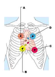 Label A | back 309 aortic valve sounds are heard in 2nd intercostal space at right sternal margin |
front 310 Label B | back 310 tricuspid valve sounds are heard in right sternal margin of 5th intercostal space variations include over sternum or over left sternal margin in 5th intercostal space |
front 311 Label C | back 311 mitral valve sounds are heard over heart apex, in 5th intercostal space in line with middle of clavicle |
front 312 Label D | back 312 pulmonary valve sounds are heard in 2nd intercostal space at left sternal margin |
front 313 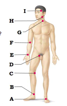 Label A | back 313 dorsalis pedis artery |
front 314 Label B | back 314 posterior tibial artery |
front 315 Label C | back 315 popliteal artery |
front 316 Label D | back 316 femoral artery |
front 317 Label E | back 317 radial artery |
front 318 Label F | back 318 brachial artery |
front 319 Label G | back 319 common carotid artery |
front 320 Label H | back 320 facial artery |
front 321 Label I | back 321 superficial temporal artery |