Instructions for Side by Side Printing
- Print the notecards
- Fold each page in half along the solid vertical line
- Cut out the notecards by cutting along each horizontal dotted line
- Optional: Glue, tape or staple the ends of each notecard together
pig practical
front 1 salivary glands | back 1 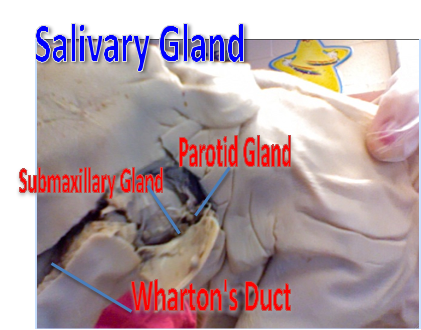 produce saliva; also helps break down carbohydrates (with salivary amylase; and lubricates the passage of food down from the oro-pharynx to the esophagus to the stomach. |
front 2 parotid gland | back 2 biggest producer of saliva; |
front 3 parotid duct | back 3 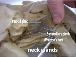 saliva travels through the gland into here |
front 4 submandibular gland | back 4 salivary gland ; less fatty than parotid |
front 5 opening to nasopharynx | back 5 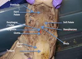 h allows a person to breathe through the nose. |
front 6 hard/ soft pallete | back 6 The soft palate is moveable; closing off the nasal passages during the act of swallowing, and also for closing off the airway. hard: help facilitate the movement of food backwards towards the larynx |
front 7 auditory (eustachian tubes) | back 7 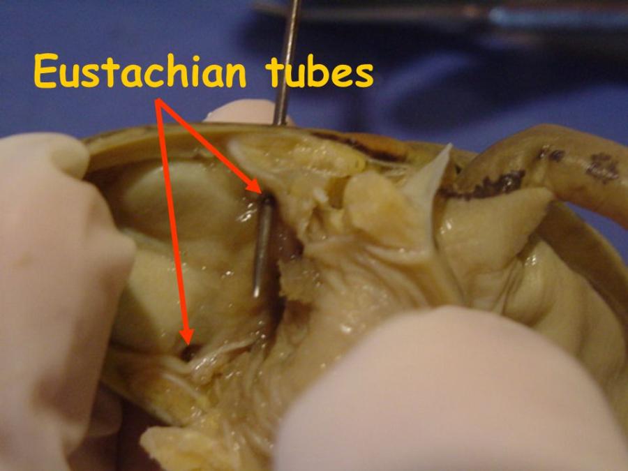 is to ventilate the middle ear space, ensuring that its pressure remains at near normal environmental air pressure |
front 8 esophagus | back 8  connects the pharynx (throat) to the stomach; functions as the conduit for food and liquids that have been swallowed into the pharynx to reach the stomach. |
front 9 larynx | back 9 houses the vocal folds, and manipulates pitch and volume, which is essential for phonation |
front 10 thymus | back 10  development of T-lymphocytes or T cells |
front 11 trachea | back 11  the vital function of providing air flow to and from the lungs for respiration. |
front 12 glottis | back 12  Sound production |
front 13 thyroid | back 13 hormones;Thyroid cells are the only cells in the body which can absorb iodine. |
front 14 diaphragm | back 14 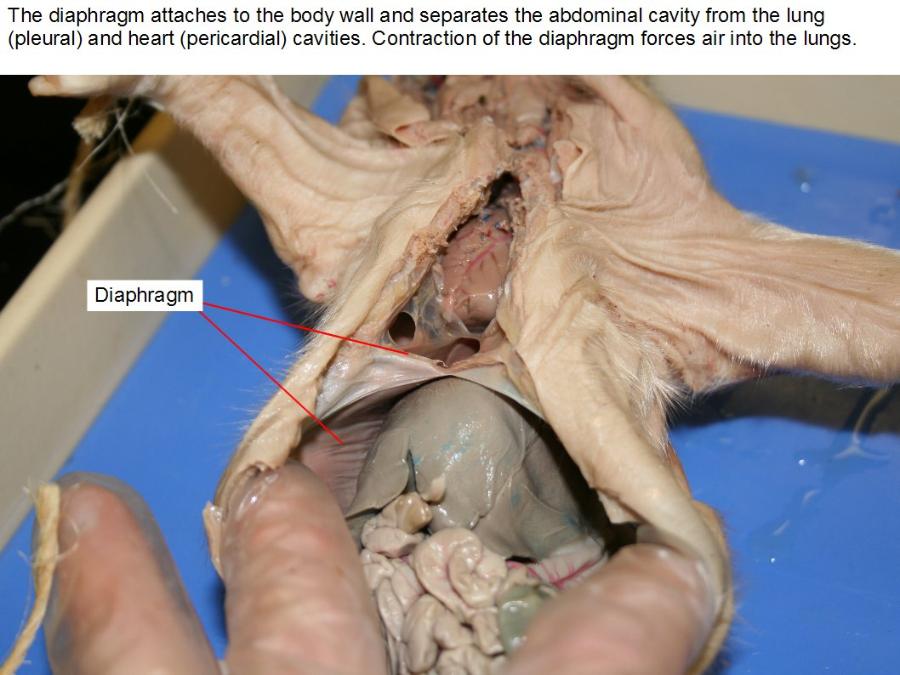 is the primary muscle used in the process of inspiration, or inhalation |
front 15 lungs | back 15 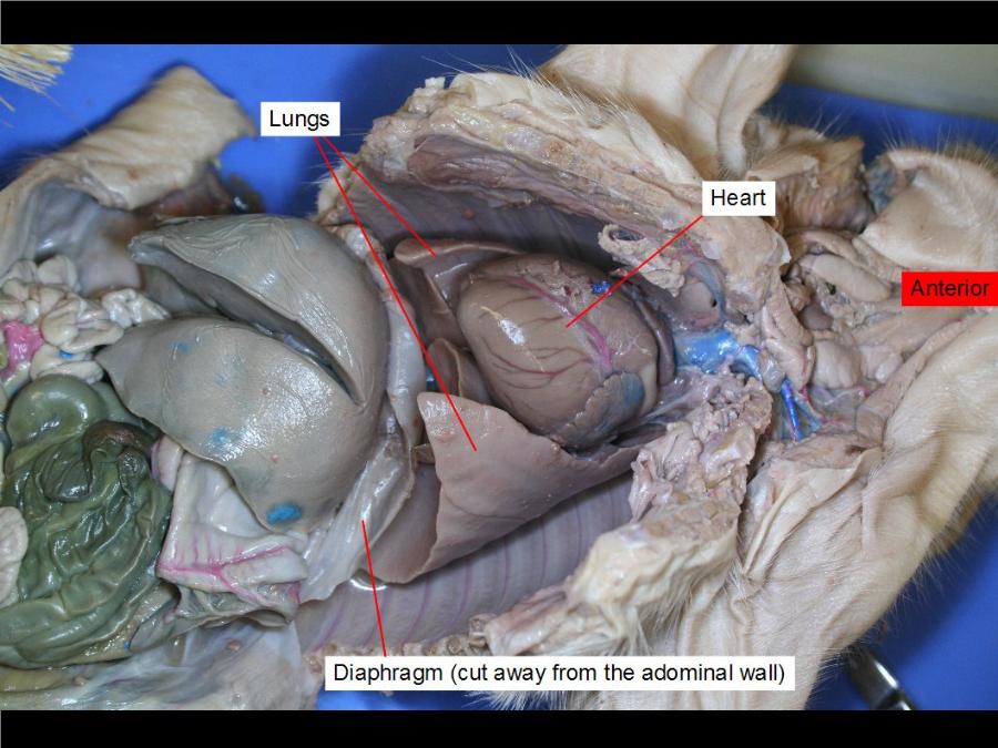 breathing bitch! o2 --> co2 |
front 16 peritoneum- parietal / visceral | back 16 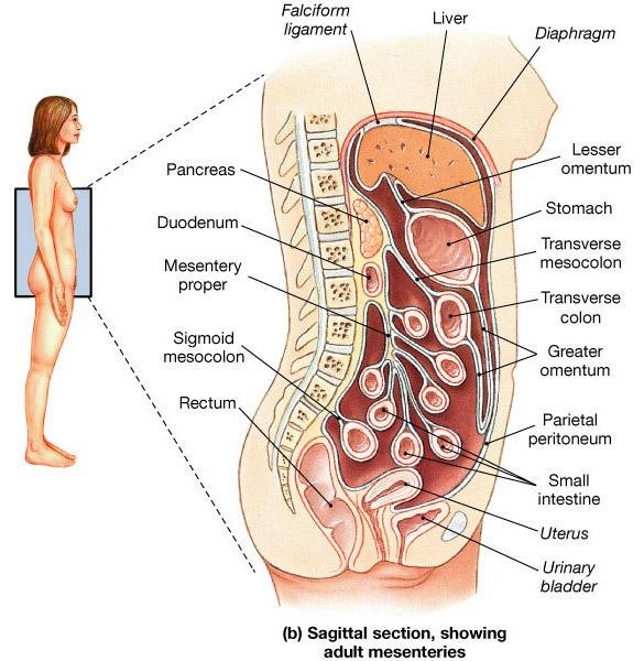 is that portion that lines the abdominal and pelvic cavities. |
front 17 pleura - parietal / visceral | back 17 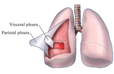 P: lines pleural cavity v: covers surface of the lungs |
front 18 bronchi | back 18 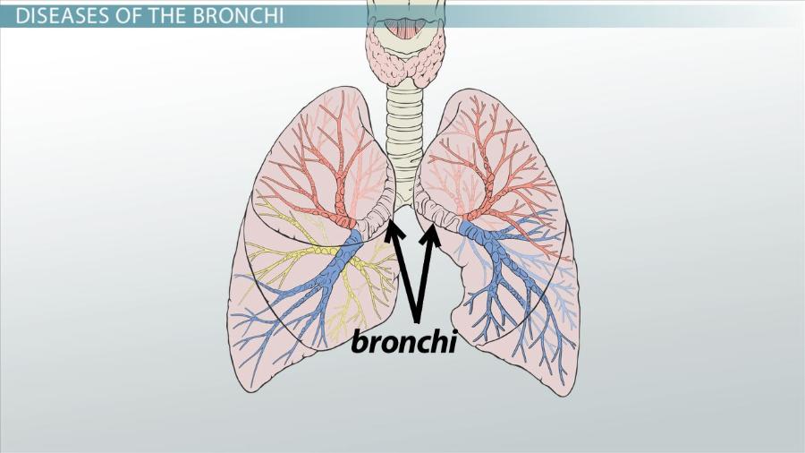 are the main passageway into the lungs |
front 19 liver | back 19 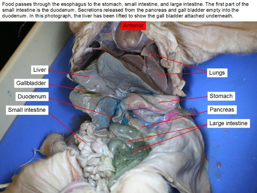 making proteins and blood clotting factors, manufacturing triglycerides and cholesterol, glycogen synthesis, and bile production. |
front 20 spleen | back 20 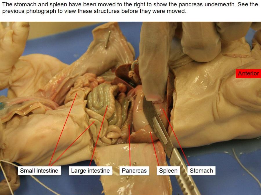 It acts as a filter for blood as part of the immune system. |
front 21 duodenum | back 21 plays a vital role in the chemical digestion of chyme in preparation for absorption in the small intestine. |
front 22 jejunum | back 22 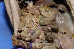 Most of the nutrients present in food are absorbed by the jejunum before being passed on to the ileum for further absorption. |
front 23 ileum | back 23 is mainly to absorb whatever products of digestion were not absorbed by the jejunum. |
front 24 pancreas | back 24 an exocrine function that helps in digestion and an endocrine function that regulates blood sugar. |
front 25 gall bladder | back 25 is to store and concentrate bile, a yellow-brown digestive enzyme produced by the liver |
front 26 common bile duct | back 26 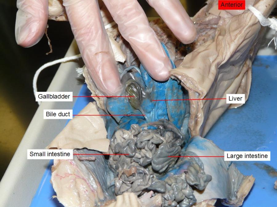 carry bile from thegallbladder and empty it into the upper part of the small intestine |
front 27 cystic duct | back 27 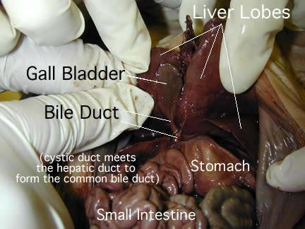 It then joins the common bile duct, which meets pancreatic duct before it empties into the duodenum |
front 28 hepatic duct | back 28 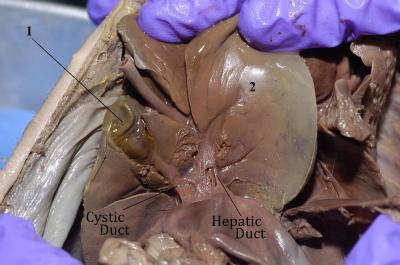 drains bile from the liver |
front 29 colon - all parts | back 29 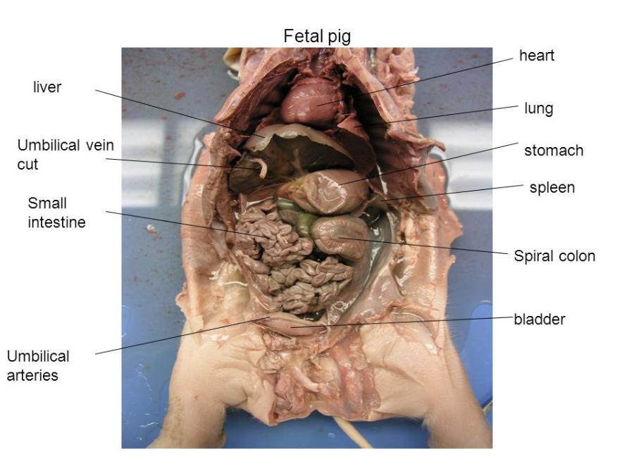 reabsorb fluids and process waste products from the body and prepare for its elimination |
front 30 caecum | back 30  absorb fluids and salts that remain after completion of intestinal digestion |
front 31 rectum | back 31  acts as a temporary storage site for feces |
front 32 anus | back 32  is the last part of the digestive tract. is specialized to detect rectal contents. It lets you know whether the contents are liquid, gas, or solid. |
front 33 large intestine | back 33 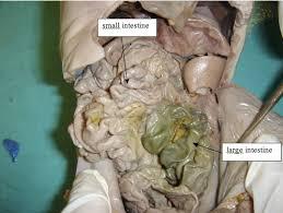 is to absorb water from the remaining indigestible food matter and transmit the useless waste material from the body |
front 34 small intestine | back 34 where 90% of the digestion and absorption of food occurs, the other 10% taking place in the stomach and large intestine. |
front 35 stomach - all parts | back 35 secretes acid and enzymes that digest food. Ridges of muscle tissue called rugae line thestomach. The stomach muscles contract periodically, churning food to enhance digestion. The pyloric sphincter is a muscular valve that opens to allow food to pass from the stomach to the small intestine |
front 36 ileo- cecal valve | back 36 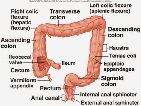 limit the reflux of colonic contents into the ileum |
front 37 pancreatic duct | back 37  supply pancreatic juice provided from the exocrine pancreas which aids in digestion |
front 38 pyloric sphincter | back 38 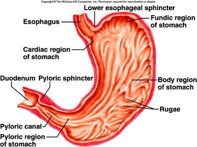 acts as a valve to controls the flow of partially digested food from the stomach to the small intestine |
front 39 mesentery | back 39 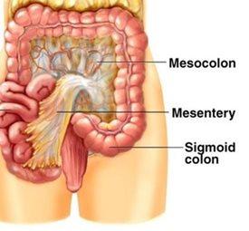 attaches your intestines to the wall of your abdomen and holds them in place |
front 40 anterior (superior, cephalic) vena cava | back 40 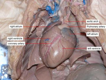 s- brings deox blood from body to heart. veins from head feed into it... then will empty into right atrium of heart |
front 41 posterior (inferior, caudial) vena cava | back 41 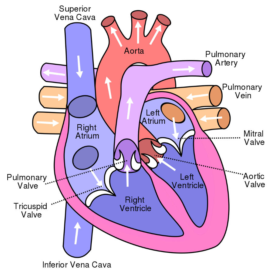 carries deox blood from lower body to heart |
front 42 subclavian artery and vein | back 42 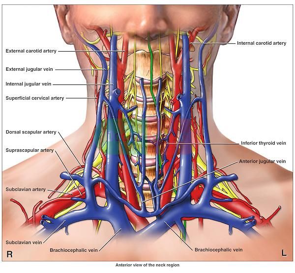 empty blood from the upper extremities and then carry it back to the heart |
front 43 internal thoracic artery and vein | back 43 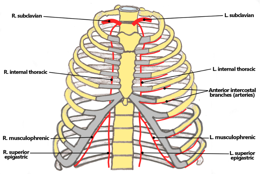 is an artery that supplies the anterior chest wall and the breasts |
front 44 brachiocephalic artery | back 44 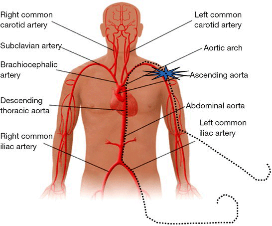 supplies oxygenated blood to the head, neck and arm regions of the body. |
front 45 coeliac artery | back 45 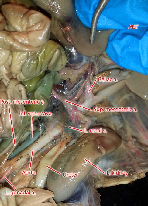 supplies oxygenated blood to the liver, stomach, abdominal esophagus, spleen and the superior half of both the duodenum and the pancreas. |
front 46 mesenteric artery | back 46 major blood vessel in the digestive system |
front 47 genital arteries | back 47 supplies ox blood to genitals/ bladder region |
front 48 external iliac artery | back 48 provides the main blood supply to the legs |
front 49 common iliac vein | back 49 They drain blood from the pelvis and lower limbs. |
front 50 hepatic portal vein | back 50  is a blood vessel that carries blood from the gastrointestinal tract, gallbladder, pancreas and spleen to the liver |
front 51 ductus venosus | back 51 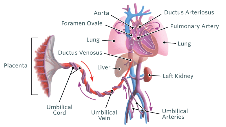 it allows oxygenated blood from the placenta to bypass the liver |
front 52 ductus arteriosus | back 52 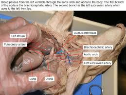 It allows most of the blood from the right ventricle to bypass the fetus's fluid-filled non-functioning lungs. |
front 53 jugular (external and internal) | back 53 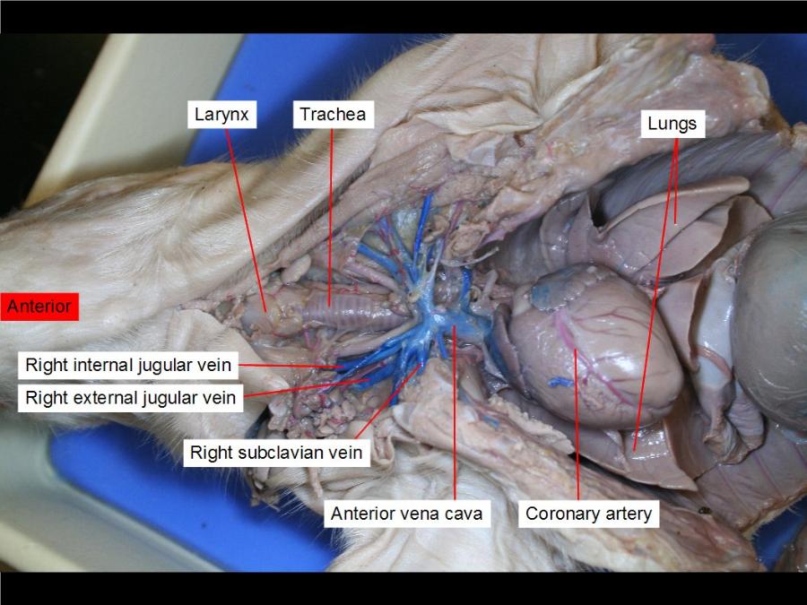 e-receives the greater part of the blood from the exterior of the craniumand the deep parts of the face i-collects blood from the brain and the superficial parts of the face and neck |
front 54 carotid (common, external, internal, and carotid body) | back 54 These arteries transfer blood to the structures inside and outside of the skull. |
front 55 brachial vien | back 55 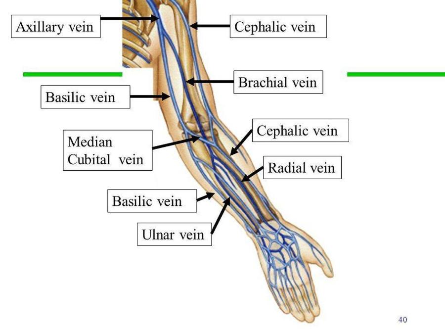 blood to bicep/tricep area |
front 56 renal artery and vein | back 56 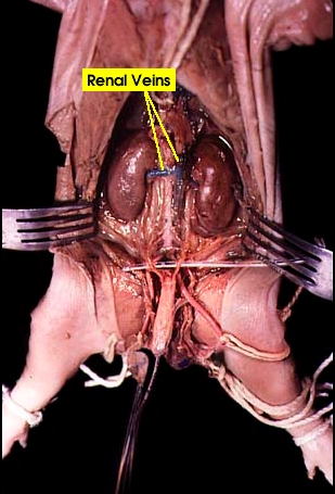 a-carries oxygenated blood to your kidneys. v-drain oxygen-depleted blood from the kidneys |
front 57 umbilical arteries and vein | back 57 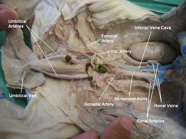 The umbilical vein carries oxygenated, nutrient-rich blood from the placenta to the fetus, and the umbilical arteries carry deoxygenated, nutrient-depleted blood from the fetus to the placenta |
front 58 aorta (arch and dorsal) | back 58 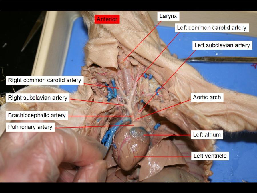 a-distributes blood from the left ventricle of the heart to the rest of the body d-give branches to the yolk-sac, and are continued backward through the body-stalk as the umbilical arteries to the villi of the chorion |
front 59 hemiazygos vein | back 59 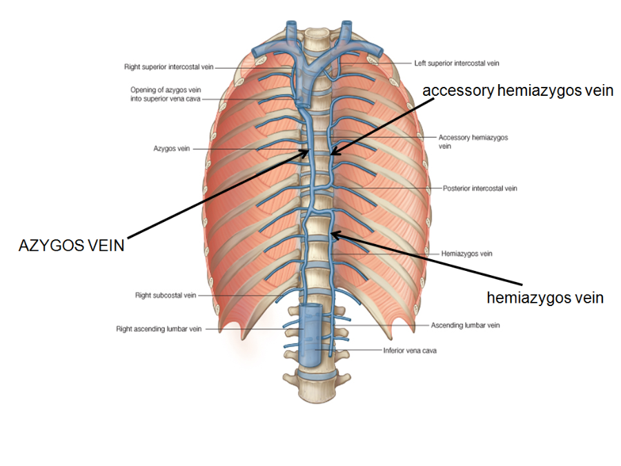 is a veinrunning superiorly in the lower thoracic region |
front 60 scattered lymph nodes | back 60 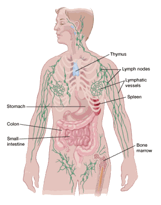 It is involved in protecting the body against infection, by delivering immune cells, known as lymphocytes |
front 61 pulmonary arteries and veins | back 61 a- carries blood from right ventircle to lungs |
front 62 coronary arteries and veins | back 62 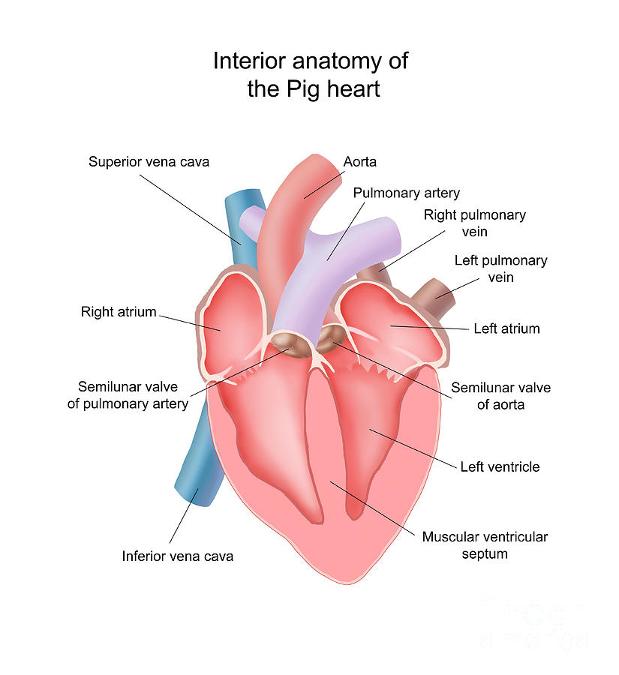 supply blood to/away the heart muscle |
front 63 right and left auricles (atria) | back 63 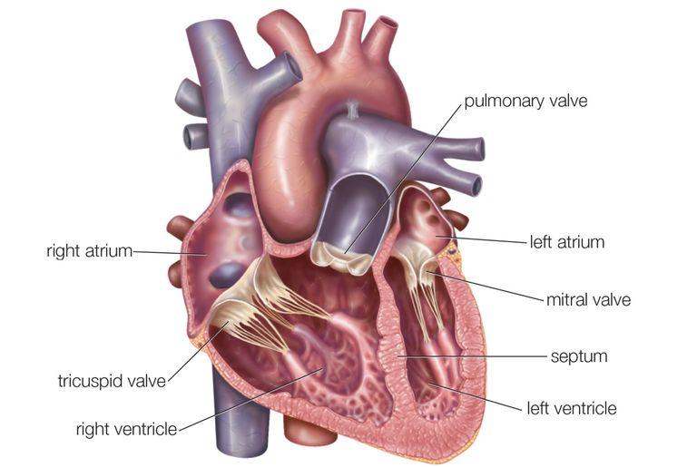 is the chamber where incoming blood from the major vein of the circulatory system (vena cava) brings deoxygenated blood into the heart. The right atrium (auricle) receives the blood and pushes it into the the right ventricle, which then transports the blood to the lungs for oxygenation. l-collect this blood and pump it to the left ventricle, which in turn pumps the blood to the body's tissues |
front 64 right and left ventricles | back 64 r- responsible for pumping deox blood to lungs l- recieves ox blood from left atrium |
front 65 pericardium- parietal and visceral | back 65 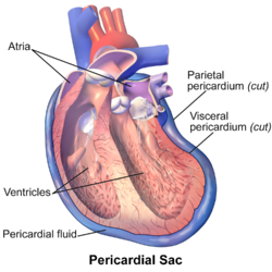 Both of these layers function in lubricating the heart to prevent friction during heart activity |
front 66 ovaries | back 66 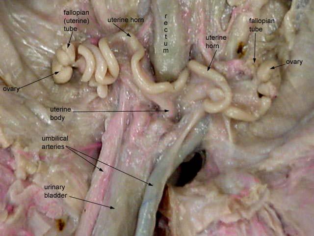 They produce oocytes (eggs) for fertilisation and they produce the reproductive hormones, oestrogen and progesterone. |
front 67 uterine (fallopian tubes) aka oviducts | back 67 is the tube that links the ovary to the uterus and which the ovulated oocyte travels down to become fertilised by sperm present in the female tract |
front 68 uterus (horns and body) | back 68 They are one of the points of attachment for the round ligament of uterus |
front 69 cervix | back 69 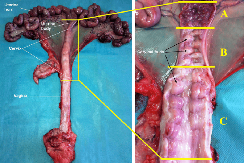 allow flow of menstrual blood from the uterus into the vagina, and direct the sperms into the uterus during intercourse |
front 70 vagina | back 70 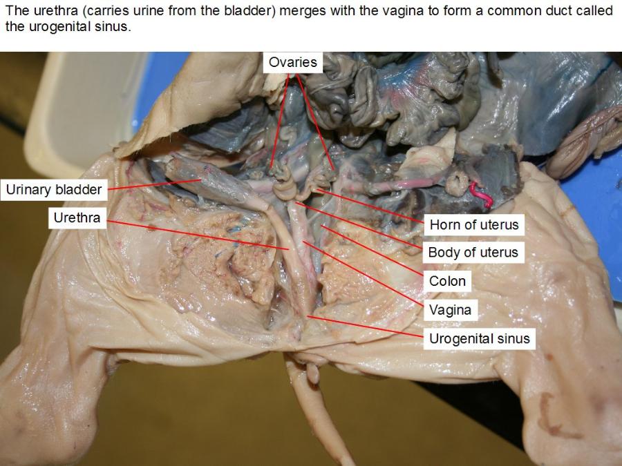 receives the penis during sexual intercourse and also serves as a conduit for menstrual flow from the uterus. During childbirth, the baby passes through the vagina |
front 71 urogenital sinus | back 71 he ventral part of the cloaca after its separation from the rectum, giving rise to the lower part of the bladder in both sexes, to the prostatic portion of the male urethra, and to the urethra and vestibule in the female. |
front 72 urogenital papilla | back 72 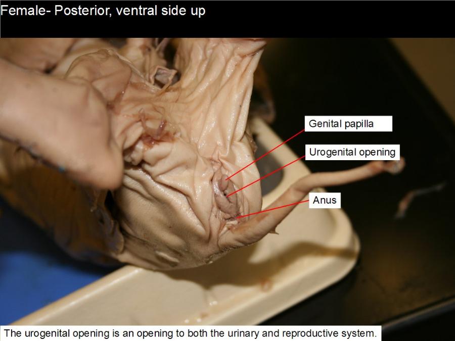 covers the opening of the vagina. |
front 73 testes | back 73 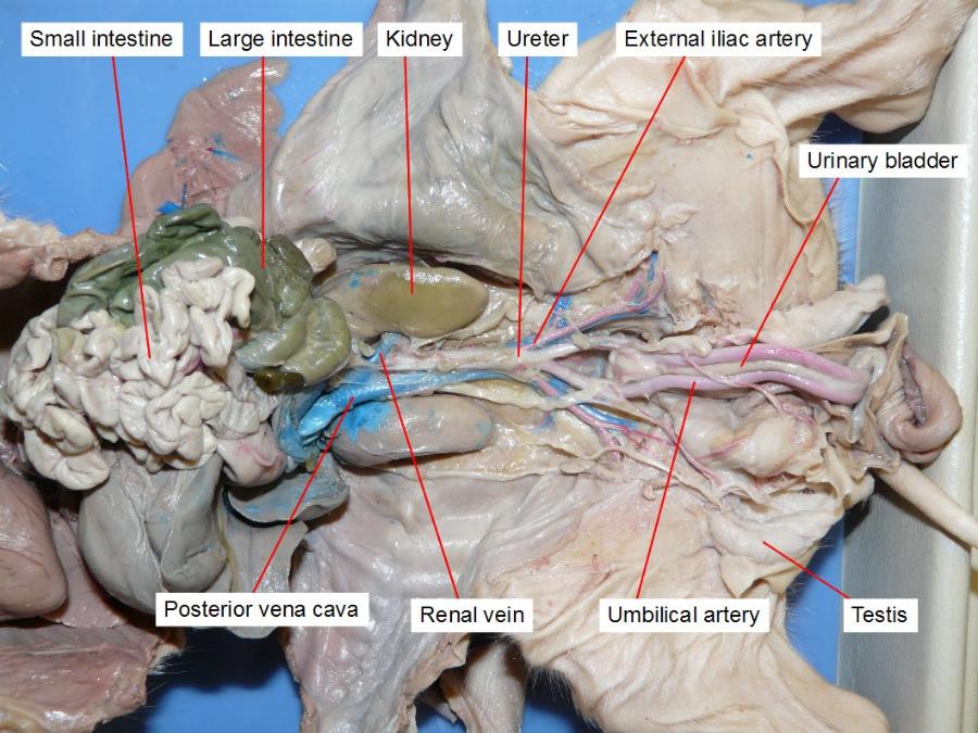 are the male gonads — the primary male reproductive organs. |
front 74 scrotal sac | back 74 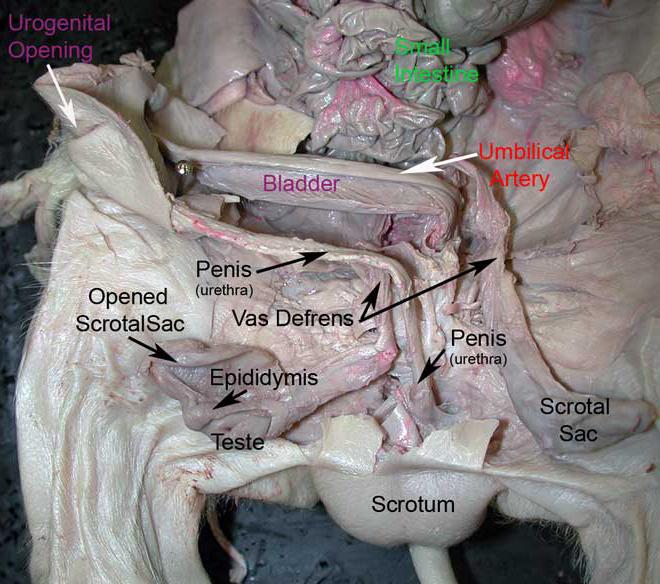 It contains the testicles (also called testes), as well as many nerves and blood vessels. The scrotum has a protective function and acts as a climate control system for the testes. |
front 75 gubernaculum | back 75 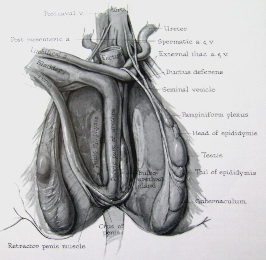 aids in the descent of the gonads (both testes and ovaries). |
front 76 epididymis | back 76 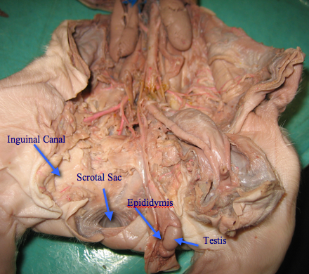 carries sperm from the testes to the ductus deferens in the male reproductive system |
front 77 vas (ductus) deferens | back 77 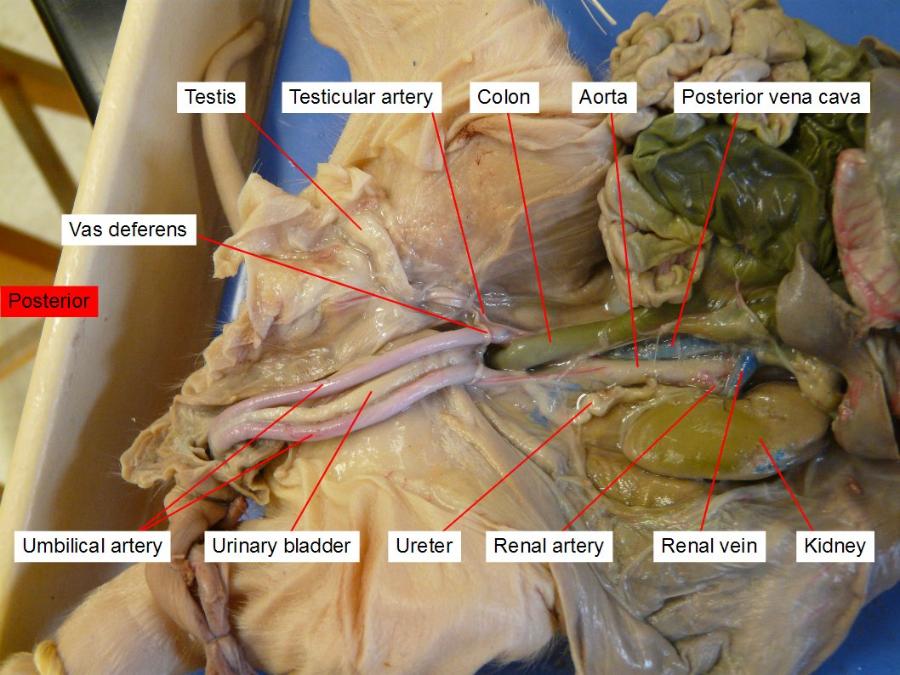 their purpose is to carry ejaculatory sperm out of the epididymis. To do this |
front 78 inguinal canal | back 78 Function. The structures which pass through the canals differ between males and females: in males: the spermatic cord and its coverings + the ilioinguinal nerve. in females: the round ligament of the uterus + the ilioinguinal nerve. |
front 79 prostate gland | back 79  The prostate secretes fluid that nourishes and protects sperm |
front 80 seminal vesicles | back 80 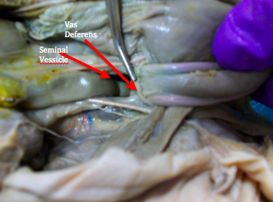 Semen combines fluid elements from the epididymis, seminal vesicles, prostate gland, and vas deferens. |
front 81 bulbo- urethral glands | back 81 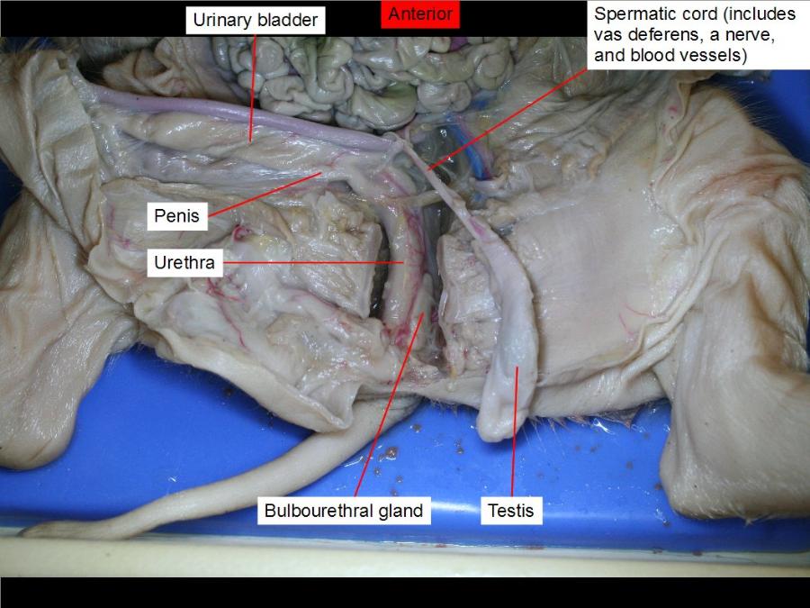 makes the precum |
front 82 penis | back 82 fuckinggggg |
front 83 urinary bladder | back 83 stores urine |
front 84 kidneys | back 84 filtration |
front 85 ureters | back 85 is a tube that carries urine from the kidney to the urinary bladder |
front 86 urethra | back 86 The urethra is the tube that carries urine from the bladder to outside of the body |
front 87 adrenal gland | back 87  Located at the top of each kidney, the adrenal glands produce hormones that help the body control blood sugar, burn protein and fat, react to stressors like a major illness or injury, and regulate blood pressure. |
front 88 head | back 88  |
front 89 nose | back 89 |
front 90 tongue | back 90 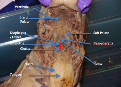 |
front 91 external ear | back 91 |
front 92 eye with lids | back 92 |
front 93 vestigal nictitating membrane | back 93 that protects and moisten the eye without losing visibility. |
front 94 cervical region | back 94 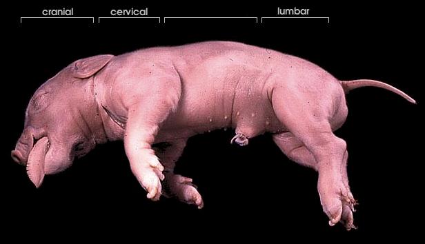 |
front 95 thoracic region | back 95 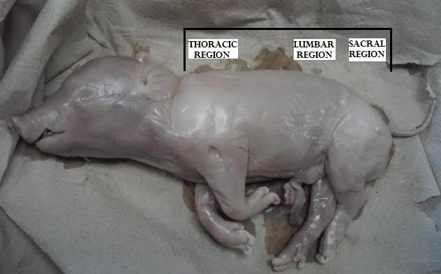 |
front 96 lumbar region | back 96 |
front 97 sacral region | back 97 |
front 98 caudal region | back 98 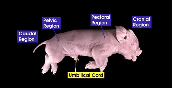 |
front 99 umbilical cord region | back 99 |