Instructions for Side by Side Printing
- Print the notecards
- Fold each page in half along the solid vertical line
- Cut out the notecards by cutting along each horizontal dotted line
- Optional: Glue, tape or staple the ends of each notecard together
Embryonic Development-Gastrulation
front 1 How does the blastula relate to clevage? | back 1 The blastula stage signals the end of the period in embryogenesis known as cleavage. |
front 2 Blastula anotomical description | back 2 The blastula can be defined in anatomical terms as a hollow stage in the embryo’s development, with a cavity (the blastocoele), surrounded by an epithelial cellular layer called the blastoderm. ***The teleosts (bony Fishes) have a blastoderm that does not surround the “Blastocoele” |
front 3 Blastula Formation | back 3  |
front 4 Blastula (single layer cell) Gastrula (Three cell layers- endoderm, mesoderm, ectoderm) | back 4  |
front 5 Gastrula | back 5
|
front 6 Gastrula 6 Primairy changes | back 6
Real New Mice Eat Qualil |
front 7 Frog Fate Map layers | back 7 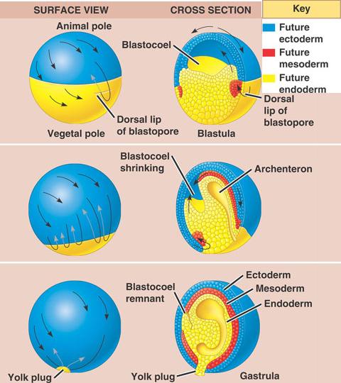 |
front 8 Frog fate map external | back 8 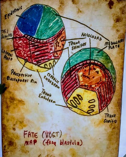 |
front 9 Bony Fish fate map | back 9  |
front 10 Primary Organizers | back 10
|
front 11 Micromeres- sea urchin | back 11  Primary Organizers Note: clevage is equal at 2 cell stage 16 cell stage early clevage not complete no green no orange but later these sections will become |
front 12 Gray crescent- frog | back 12 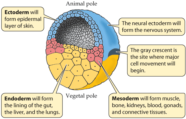 Primary Organizers |
front 13 Dorsal Lip of Blastopore- Amphibian, Frog* | back 13  Primary Organizers |
front 14 Primitive Streak- chick, human | back 14  Primary Organizers |
front 15 Primairy mesenchyme vs Secondary mesenchyme | back 15 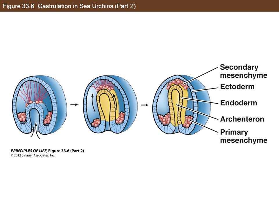 |
front 16 Teleosts/Bony fish Blastoderm/Blastocoele | back 16 Blastoderm does not surround the blastocoel, instead has cell death tube . Removed during excavation. |
front 17 Ectoderm Endoderm Mesoderm | back 17 Ectoderm Blue/Green Endoderm Yellow Mesoderm Red/Orange |
front 18 Ectoderm Endoderm Mesoderm become... | back 18  |
front 19 Prospective Significance | back 19 Prospective Fate of a cell or group of cells due to their anatomical position in the embryo |
front 20 Prospective Potency | back 20 Developmental Potential of a cell or group of cells under varying conditions, wherein new influences (inductive influences) are encountered. |
front 21 Induction | back 21 When one cell or group of cells influences the fate of another cell or group of cells without necessarily affecting itself |
front 22 Inductor | back 22 The cell or group of cells responsible for the influence mentioned above. |
front 23 Embryonic Competency | back 23 The ability of cells to respond to the influence of an inductor Period of competency is a fixed period of time. |
front 24 Determination | back 24 Fixing the fate of cells during development(narrowing of the prospective potency so that it more closely equals the prospecting significance) when the prospective potency and prospective significance are the same we say that cells are fully determined (happens during the s-period of the cell cycle) |
front 25 Differentiation | back 25 Physical (Anatomical) and Genetic/Biochemical (Physiological) expression of the fixed fate of cells. |
front 26 Spacial Differentiation | back 26 Spatially different parts of the embryo take on different anatomical structure. Morphogenesis/ Organogenesis |
front 27 Temporal Differentiation | back 27 Over a period of time different cells in an organ differentiates into different, specific cell types. Cytodifferentiation Histogenesis |
front 28 Morphogenesis/ Organogenesis | back 28 Spacial Differentiation |
front 29 Cytodifferentiation Histogenesis | back 29 Temporal Differentiation |
front 30 Morphogenetic (Formative) Movements | back 30 Invagination- pushing in of an epithelium Epiboly- Spreading of an epithelial layer so that it covers over epithelia Ingression (immigration)- breaking-up of an epithelium followed by inward migration of individual cells Involution- inward migration of an epithelium Separation of epithelial layers Local thickening followed by evacation |
front 31 Invagination , Ingression, involution | back 31 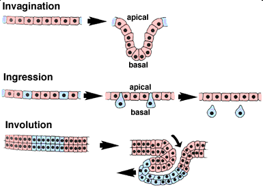 |
front 32 Epiboly | back 32 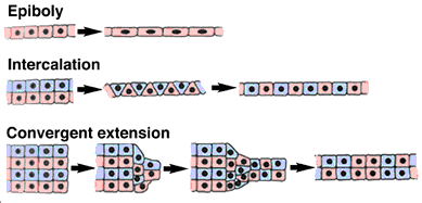 |
front 33 Additional Morphogenetic Formative movements | back 33
|
front 34 Draw Early Gastrulation | back 34  TOP Red- Non-notochordal mesoderm Orange- Chrdomesoderm Yellow- Endoderm Green- Neural Ectoderm Blue- Epidermal Ectoderm |
front 35 Draw Mid Gastrulation | back 35 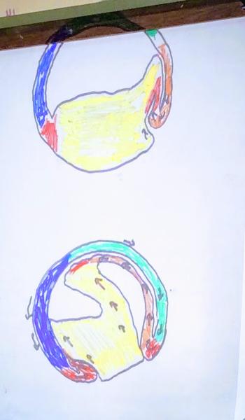 BOTTOM Red- Non-notochordal mesoderm Orange- Chrdomesoderm Yellow- Endoderm Green- Neural Ectoderm Blue- Epidermal Ectoderm |
front 36 Villus Arrangement | back 36 Diffuse Cotyledons Zonary Discoidal Bidiscoidal |
front 37 ICM=Inner Cell mass TB- Trophoblast HB- Hypoblast EB- Epiblast MES- mesoderm YS- Yolk Sac CH- Chorion | back 37 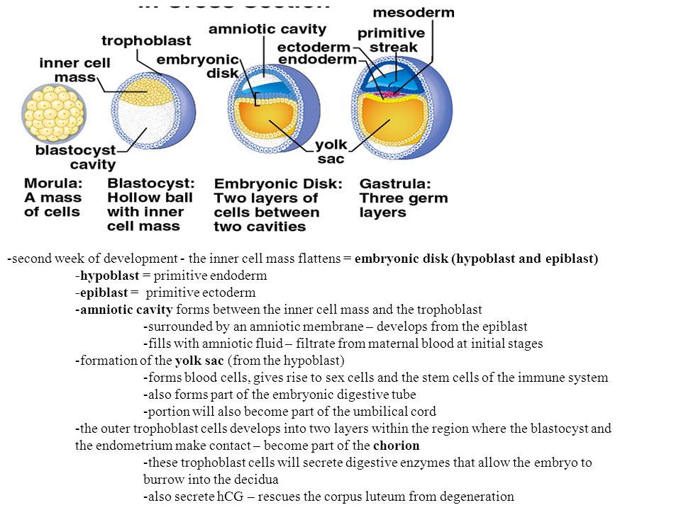 |
front 38 Chorion | back 38 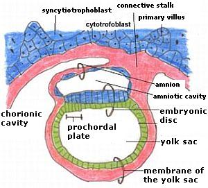 |
front 39 Diffuse Cotyledons Zonary Discoidal Bidiscodal | back 39 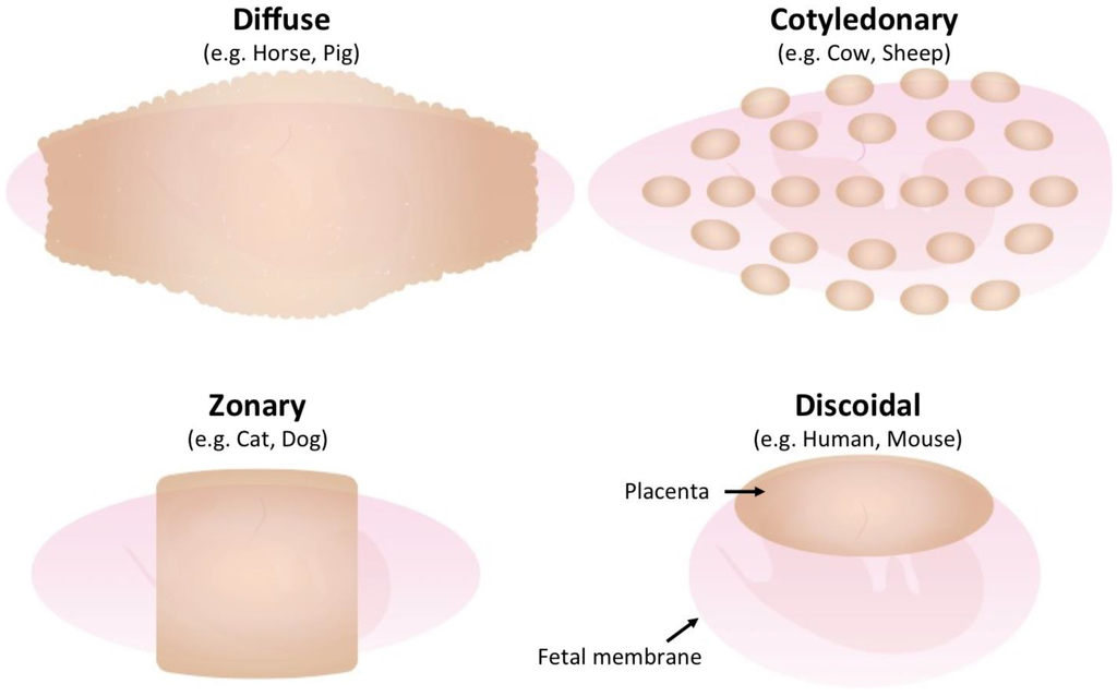 Bidiscodial-same as discoidal but with 2 disks |
front 40 Area Opeca Vasculosa Area Pellucida | back 40  |
front 41 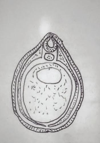 COLOR | back 41  |
front 42 mid gastrulation | back 42  |
front 43 When orange meets orange gastrulation stops. | back 43  |
front 44 blastula early gastrula late gastrula | back 44  |
front 45 Amnion Allantois Yolk Sac Chorion | back 45 |
front 46 Yolk Stalk | back 46 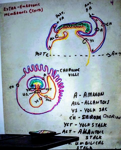 |
front 47 Heart rudiment | back 47 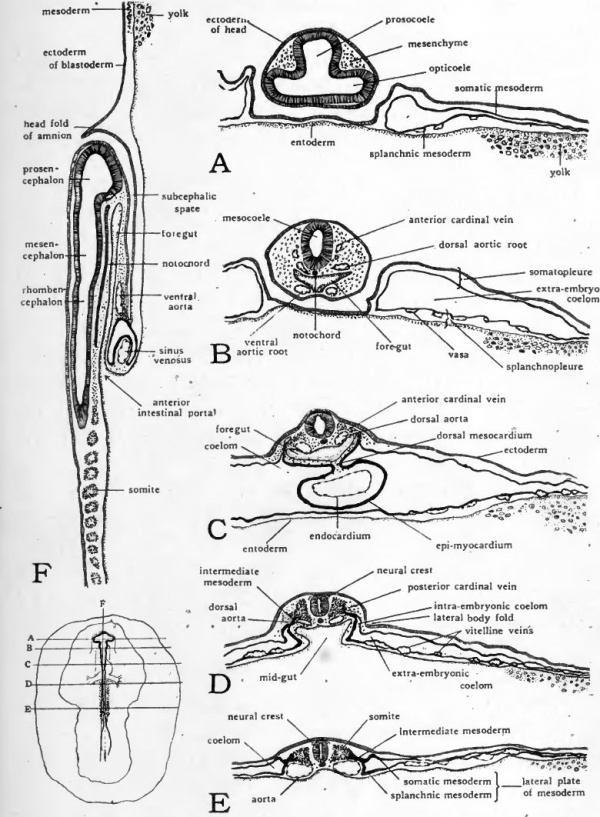 |
front 48 Yolk plug | back 48  |
front 49 Gastrulation in the Chick
| back 49 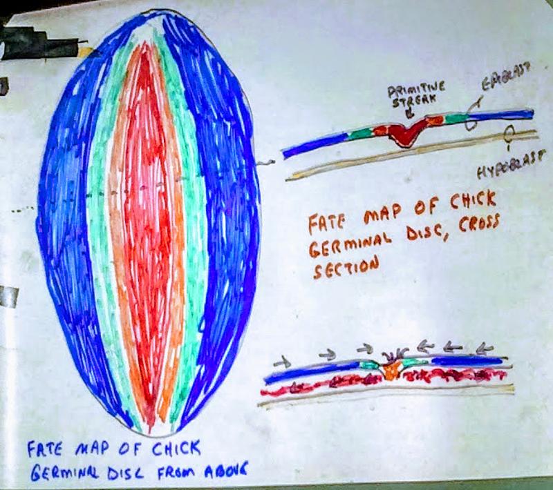 |
front 50 Primary Organ Rudiment Formation in the Chick | back 50 NO SUCH STUPID LUCK I BROUGHT APPLES ANYWAY
|
front 51 Primary Organ Rudiment Formation in the Chick
| back 51 Primary Organ Rudiment Formation in the Chick
|
front 52 Hilde mangold | back 52
Mangold ROL rude old lip |
front 53 A.S.G. Curtiss crescent | back 53 demonstrated this effect with the frog’s gray crescent (presumptive chordomesoderm) |
front 54 Determination | back 54 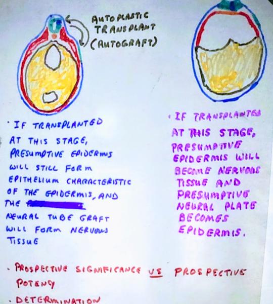 |
front 55 Determination vs Competency | back 55 ?what a cell becomes vs its ability to become something ? |
front 56 Types of Grafts | back 56 Always Hang Heavy Xrays
|
front 57 Autoplastic Transplant (autograft) | back 57 donor and host same individual |
front 58 Homolpastic transplant (homograft) | back 58 donor and host are different individuals of the same genus and species |
front 59 Heteroplastic transplant (hetrograft) | back 59 donor and host of same genus but different species |
front 60 Xenoplastic transplant (xenograft) | back 60 donor and host more distant than genus |