Instructions for Side by Side Printing
- Print the notecards
- Fold each page in half along the solid vertical line
- Cut out the notecards by cutting along each horizontal dotted line
- Optional: Glue, tape or staple the ends of each notecard together
A&P 2 Heart
front 1 Complete the following statement explaining the scheme of circulation
of blood in the human body. | back 1
|
front 2 What is the function of the fluid that fills the pericardial sac? | back 2 Allows Fluid for heart to beat |
front 3 The first heart sound is heard when the ______ valves close? | back 3 AV close |
front 4 List the correct sequence of parts that carry cardiac impulses from SA node to Purkinje fibers? | back 4 SA node, AV node, AV bundle, right and left bundle branches, Purkinje fibers |
front 5 What is the function of the valves found in the heart? | back 5 To prevent back flow |
front 6 What is the role of the chordae tendineae? | back 6 To hold the flaps of the valves so they dont swing back |
front 7 The pulmonary circuit sends ____________ blood to the lungs? | back 7 oxygen poor blood |
front 8 Deoxygenated blood is pumped by the right ventricle to lung via ___________ circuit? | back 8 Pulmonary circuit |
front 9 The heart wall is composed of ________ layers of tissue? | back 9 Three layers |
front 10 Name the layers of heart wall? | back 10
|
front 11 If the mitral valve does not close properly, which circulation is affected? | back 11 Systemic circulation |
front 12 Why might a thrombus (blood clot) in the anterior descending branch of the left coronary artery cause sudden death? | back 12 The coronary artery supplies blood to the heart |
front 13 Describe the unique anatomical features of cardiac muscle. What role does the unique structure of cardiac muscle play in its function? | back 13 It holds the myocytes together so that they do not pull apart when the heart contracts |
front 14 When the ventricular walls contract, which heart valves close? | back 14 AV Valves |
front 15 Even though cardiac muscle has an inherent ability to beat, the nodal system plays a critical role in heart physiology. What is that role? | back 15 Generate action potentials at a greater frequency than other cardiac muscle cells, they are the pacemaker of the heart. Therefore, it sets the heart rate. |
front 16 The valve located between right atrium and right ventricle is the ….valve? | back 16 Tricuspid Valve |
front 17 Define the following terms: tachycardia; bradycardia; fibrillation? | back 17 Tachycardia: Abnormally Rapid Heart rate. Bradycardia: An abnormally Slow heart rate. Fibrillation: A muscular Twitching. |
front 18 The aortic and pulmonary valves opens when the ventricles_____? | back 18 Contract |
front 19 Which would be more serious, atrial or ventricular fibrillation? Why? | back 19
|
front 20 Of the following layers of the heart wall, which consumes the most energy: Epicardium; Myocardium; Endocardium? Why? | back 20 Myocardium: Because it provides a scaffolding for the heart chambers, assisting in contraction and relaxation of the cardiac walls so that blood can pass between the chambers |
front 21 The right atrium receives blood directly from______________? | back 21 Inferior/Superior vena cava |
front 22 What purpose does the coronary circuit serve? | back 22 it delivers 1/20 of the body's blood supply to the heart muscle itself. |
front 23
| back 23 SA node, AV node, AV bundle, right and left bundle branches, Purkinje fibers |
front 24
| back 24 Left Ventricle Left Atrium Bicuspid Valves |
front 25
| back 25 Calcium. |
front 26
| back 26 Bradycardia |
front 27
| back 27 Pacemaker |
front 28 Aortic semilunar valve-permits one-way blood flow from the _________to the________? | back 28
|
front 29 The function of an atrium is to ________? | back 29 Receive blood |
front 30 In a normal heart, which of the following structures is responsible for setting the heart’s pace? Bicuspid valve is also called________valve? | back 30
|
front 31 The pacemaker cells of the heart are located in the? | back 31 Top of Right Atrium |
front 32 Depolarization of ventricles is represented by which waves in EKG (ECG)? | back 32 QRS complex |
front 33 The “lub-dup” heart sounds are produced by? | back 33 -Lub: Atrioventricular Valves -Dub: Semilunar Valves |
front 34 The left and right pulmonary arteries carry blood to the? | back 34 Lungs |
front 35 Abnormally high heart rate is termed? | back 35 Tachycardia |
front 36 Mitral valve-permits one-way blood flow from the _________to the________? | back 36
|
front 37 In terms of blood flow, why is it important that atrial diastole occurs just as ventricular systole begins? | back 37 when ventricular systole begins atrial diastole is important because the atrial needs to accept the new blood that comes in. Both can be contracted |
front 38 The myocardium is primarily composed of ________ tissue? | back 38 Cardiac Muscle |
front 39 Cardiac output is determined by________ and _________? | back 39
(heart rate*stroke volume) |
front 40 The heart is roughly the size of ________? | back 40 Your Fist |
front 41 What is stroke volume? | back 41 Volume of blood pumped by one ventricle with each heartbeat |
front 42 What is rate? | back 42 Beats per 1 min |
front 43 Depolarization of the atria corresponds directly to the EKG's? | back 43 P wave |
front 44 The inner lining of the heart is called? | back 44 Endocardium |
front 45 The pain of angina pectoris comes from a blockage in an artery that supplies the? | back 45 Heart |
front 46 Your heart seems to “pound” after you hear a sudden, loud noise. This increased contractility is due to? | back 46 -Epinepherin -Neuroepinepherin |
front 47 In cardiac muscle, the depolarization phase of the action potential is the result of increased membrane permeability to? | back 47 increased membrane permeability to sodium ions. |
front 48 What is the nature of acetylcholine’s inhibitory effect on heart rate? | back 48 Acetylcholine causes opening of potassium channels in the SA node, thereby hyperpolarizing it. |
front 49 Which of the following layers of the heart wall contracts and this contraction force blood out of the heart? | back 49 Myocardium: muscular contractions |
front 50 Which of the following layers of the heart wall is a protective inner lining of the heart chambers and valves? | back 50 Endocardium |
front 51 Why is high blood pressure damaging to the heart? | back 51 Coronary artery disease affects the arteries that supply blood to your heart muscle. Arteries narrowed by coronary artery disease don't allow blood to flow freely through your arteries. |
front 52 The T wave on an ECG tracing represents? | back 52 Ventricular Repolarization |
front 53 Cardiac muscle tissue is found in which layer of the heart wall? | back 53 Myocaridum |
front 54 Follow a drop of blood from the vena cava to the lungs and then back from the lungs to the heart and then from the heart to the cells and back to the heart. Use the following key:
| back 54 Vena Cava → RA → Tricuspid Valve → RV → Pulmonary Semilunar Valve → Pulmonary Trunk → Pulmonary Arteries → Lungs → Pulmonary Veins → LA → Bicuspid (Mitral) Valve → LV → Aortic Semilunar Valve → Aorta |
front 55 Name the valve that is found between the left atrium and the left ventricle? | back 55 Bicuspid (Mitral) Valve. |
front 56 Blood is carried to the heart by which vessels? Arteries or Veins? | back 56 Veins |
front 57 The heart is innervated by ________ nerves? | back 57 Somatic Motor Nerves |
front 58 Blood is carried away from the heart by which vessels? Arteries or Veins? | back 58 Arteries |
front 59 The ________ ventricle has a greater workload than the ________ventricle? Why? | back 59
|
front 60 In cardiac muscle, the plateau phase of the action potential is the result of? | back 60 Calcium Ions remaining open |
front 61 All oxygenated blood returns to the heart via which vein? | back 61 Pulmonary Veins |
front 62 Depolarization of atria is represented by which waves in EKG (ECK)? | back 62 P wave |
front 63 Blood being pumped out of the left ventricle enters the….? | back 63 System circuit |
front 64 Blood returning to the heart from the systemic circuit first enters the? | back 64 Right Atrium |
front 65 Blood is prevented from flowing back into the left ventricle by the…? | back 65 Aortic Semi lunar Valve. |
front 66 Which area of the heart is known as the pacemaker of the heart? | back 66 SA node |
front 67 The right ventricle pumps blood to which organ? | back 67 Lungs |
front 68 Atrial repolarization occurs during which period of time, seen on an EKG? Hint: remember that repolarization of atrium should occur at the same time as depolarization of ventricles | back 68 QRS Complex |
front 69 Contractions of the papillary muscles prevent the _____valves from reversing into the atria? | back 69 Atroiventricular |
front 70 The lub-dup heart sounds heard during auscultation of the heart are associated with……? | back 70 Closing of the valves. Lub: AV Closing Dub: SV Closing |
front 71
| back 71 Isovolumetric Ventricular |
front 72 The atrioventricular valve on the side of the heart that receives the superior vena cava is the ________ valve? | back 72 Tricuspid Valve |
front 73 The amount of blood pumped out of each ventricle in one minute is called? | back 73 Cardiac Output |
front 74 Abnormalities of heart valves can be detected more accurately by auscultation than by electrocardiography. Why is this so? | back 74 Extra heart sounds are produce and you can hear them with a stethoscope. |
front 75
| back 75 Electrocardiograpy: Device used to show hearts electrical activity |
front 76 Draw an ECG wave form representing one heartbeat. Label the P, QRS, and T waves; the P-R interval; the S-T segment; and the Q–T interval? | back 76 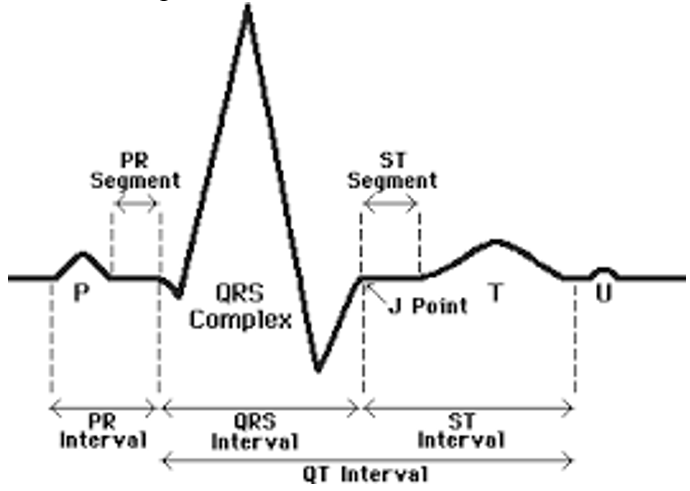 |
front 77 The volume of blood ejected from each ventricle during a contraction is called the? | back 77 Cardiac Output |
front 78 When the semilunar valves close, the AV valves ________? | back 78 Open |
front 79 Describe what happens in the cardiac cycle in the following situations:
| back 79
|
front 80 Why does heart rate increase during running? | back 80 More oxygen is needed for muscles and organs. So the heart needs to pump more. |
front 81 List the elements of the intrinsic conduction system in order, starting from the SA node?
| back 81
|
front 82 Differentiate clearly between the roles of the pulmonary and systemic circulations? | back 82 Pulmonary: Between heart and Lungs. Systemic: Between Heart and Organs. |
front 83 Depolarization of the ventricles is represented on an electrocardiogram by the? | back 83 QRS Complex |
front 84 Use the key and match them to the correct descriptions provided below. Key: ventricles, coronary sinus , epicardium, atria, Mediastinum, myocardium
| back 84
|
front 85  An anterior view of the heart is shown here. Use the key (1 to 24)and label the diagram bellow. | back 85
|