Instructions for Side by Side Printing
- Print the notecards
- Fold each page in half along the solid vertical line
- Cut out the notecards by cutting along each horizontal dotted line
- Optional: Glue, tape or staple the ends of each notecard together
Activity 1: Identifying Respiratory System Organs-Upper and Lower Respiratory System Structures
front 1 The trachea is also commonly known as what? | back 1 The windpipe |
front 2 Air entering the trachea from the larynx travels down to the level of what angle? | back 2 Sternal angle |
front 3 The sternal angle is located where judging from the thoracic vertebrae? | back 3 Located between the fourth and fifth thoracic vertebrae |
front 4 The trachea divides into right and left at which level? | back 4 At the level between the fourth and fifth thoracic vertebrae |
front 5 Between the fourth and fifth thoracic cavity, the trachea divides into what 2 sections? | back 5 1. Right primary bronchi 2. Left primary bronchi |
front 6 The right and left primary bronchis plunge into their respective lungs at which area? | back 6 hilum |
front 7 How is the right primary bronchus different from the left in 3 ways? | back 7 1. Wider 2. Shorter 3. More vertical |
front 8 Why is the right primary bronchus structural differences beneficial? | back 8 It is more likely to trap foreign objects |
front 9 The trachea is lined with what type of cell? | back 9 Pseudostratified ciliated columnar epithelium |
front 10 Mucus is produced by what type of cells? | back 10 Goblet cells |
front 11 What is the purpose of mucus in the trachea? | back 11 Traps dust particles, debris, and bacteria |
front 12 What is the purpose of cilia in the trachea? | back 12 Propels mucus that is trapped with dust particles, debris, and bacteria toward the throat where it can be swallowed |
front 13 The walls of the trachea are reinforced with what type of rings? | back 13 C-shaped rings |
front 14 The C-shaped rings of the trachea walls are made out of what material? | back 14 Cartilage |
front 15 The C-shaped rings of the trachea walls differ posteriorly in which way? | back 15 They are incomplete, meaning they don't fully connect |
front 16 What are the 2 functions of the C-shaped rings? | back 16 1. Incomplete parts allow the esophagus to expand anteriorly for a food bolus to pass 2. The solid parts reinforce the trachea walls to keep it open regardless of the pressure changes during breathing |
front 17 Which muscle allows the trachea to expand? | back 17 Trachealis muscle |
front 18 What are the 8 subsequent divisions of the main bronchi? | back 18 1. Lobar 2. Segmental 3. Bronchioles 4. Terminal bronchioles 5. Respiratory bronchioles 6. Alveolar ducts 7. Alveolar sacs 8. Alveoli |
front 19 Lobar and segmental are also known as what? (Hint: think of the main bronchi being also being called "primary") | back 19 Lobar=secondary Segmental=tertiary |
front 20 Each bronchiole further divides into what? | back 20 Terminal bronchioles |
front 21 Each terminal bronchiole further divides into what? | back 21 Respiratory bronchioles |
front 22 All of the branches of the main bronchi, except for the smallest branches, have what specific type of reinforcement in their walls? | back 22 Hyaline cartilage reinforcement |
front 23 As the respiratory tubes get smaller and smaller, what happens to the amount of smooth muscle vs the amount of cartilage? | back 23 The amount of smooth muscle gradually increases while the amount of cartilage continuously decreases until there is none. |
front 24 The continuous branching of the respiratory passageways in the lungs if often referred to as what? | back 24 Bronchial tree |
front 25 The respiratory bronchioles further subdivide into what? | back 25 Alveolar ducts |
front 26 The alveolar ducts terminate in what type of structures? | back 26 Alveolar sacs |
front 27 Alveolar sacs then subdivide into what? | back 27 Alveoli |
front 28 Alveoli are composed of what type of cells? | back 28 Squamous epithelium |
front 29 Alveoli are covered with what type of blood vessels? | back 29 Capillaries |
front 30 What 3 walls form the respiratory membrane? | back 30 1. Alveolar walls 2. Capillary walls 3. ***And the fused basement membranes of the above two |
front 31 The respiratory membrane is also called what? | back 31 Blood air barrier |
front 32 The respiratory membrane is the site of what exchange? | back 32 Gas exchange |
front 33 What 3 structures make up the respiratory zone structures? | back 33 1. Alveolar sacs 2. Alveolar ducts 3. Respiratory bronchioles |
front 34 Why are the alveolar sacs, alveolar ducts, and respiratory bronchioles referred to as the respiratory zone structures? | back 34 Because gas exchanges occur by simple diffusion across the respiratory membrane |
front 35 What are conducting zone structures generally? | back 35 All other respiratory passageways from the nasal cavity to the terminal bronchioles |
front 36 How do conducting zone structures get their general name? | back 36 Because they simply serve as access or exit routes to and from the gas exchange chambers of the respiratory zone structures; there is no diffusion |
front 37 Conducting zone structures are also collectively called what? | back 37 Anatomical dead space |
front 38 The paired lungs occupy which cavity except around what structure? | back 38 Occupy the entire thoracic cavity except for the mediastinum |
front 39 The mediastinum house what 3 example organs? | back 39 1. Heart 2. Bronchoi 3. Esophagus |
front 40 Each lung is connected to the mediastinum by what? | back 40 A root |
front 41 A root which connects each lung to the mediastinum contains what 2 attachments? | back 41 1. Bronchial attachments 2. Vascular attachments |
front 42 The structures of the root enter or leave the lung via what medial indentation? | back 42 Hilum |
front 43 All structures distal to the main bronchi are found where? | back 43 Within the lung substance |
front 44 The lung's apex lies deep to what bone? | back 44 Clavicle |
front 45 The lung's base rests on which organ? | back 45 Diaphragm |
front 46 What 3 surfaces make up the costal surface? | back 46 1. Anterior lung surface 2. Posterior lung surface 3. Lateral lung surface |
front 47 Why are the anterior, posterior, and lateral lung surfaces referred collectively to as the costal surface? | back 47 They are in close proximity with the ribs |
front 48 The medial surface of the left lung exhibits what notch? | back 48 Cardiac notch |
front 49 What is the function of the cardiac notch? | back 49 Accommodates the heart |
front 50 Fissures divide the lungs into what? | back 50 Lobes |
front 51 How many lobes are in the left lung? | back 51 2 |
front 52 How many lobes are in the right right lung? | back 52 3 |
front 53 The lungs are mostly made out of what type of tissue? | back 53 Elastic connective tissue |
front 54 Elastic connective tissue composition provides what functional benefit to the lungs? | back 54 Allows them to recoil passively during expiration |
front 55 Each lung is enclosed in what serous membrane? | back 55 Pleura |
front 56 Describe the pleura structurally. (Hint: type of membrane and type of sac) | back 56 Double-layered sac of serous membrane |
front 57 What is the outer covering layer of the lungs? | back 57 Parietal pleura |
front 58 The parietal pleura is attached to what wall and organ? | back 58 Attached to the thoracic wall and the diaphragm. |
front 59 The inner layer of the lungs is what? | back 59 Visceral pleura |
front 60 The visceral pleura covers what type of tissue? | back 60 Lung tissue |
front 61 The parietal and visceral pleura are separated by what? | back 61 Pleural cavity |
front 62 The pleural cavity is filled with what? | back 62 Pleural fluid |
front 63 The pleural fluid is produced by what? | back 63 Pleurae |
front 64 What is the function of the pleural fluid? | back 64 Allows the lungs to glide without friction over the thoracic wall during breathing |
front 65 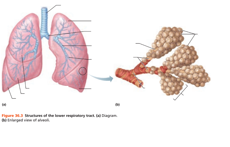 | back 65 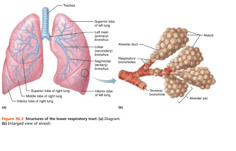 |
front 66 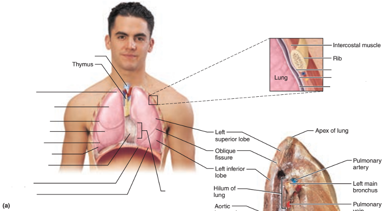 | back 66 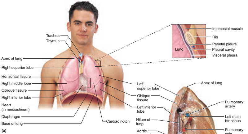 |
front 67 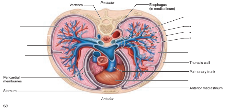 | back 67  |
front 68  | back 68 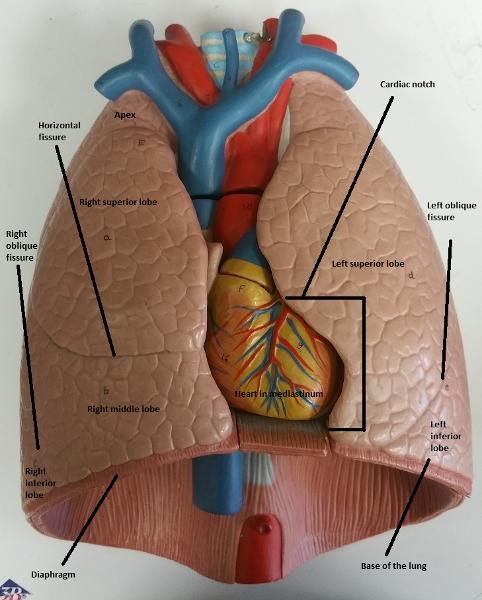 |
front 69 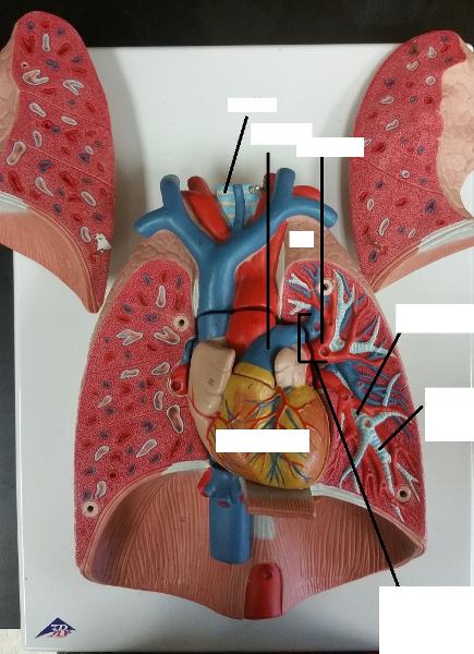 | back 69 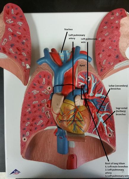 |
front 70 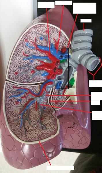 | back 70 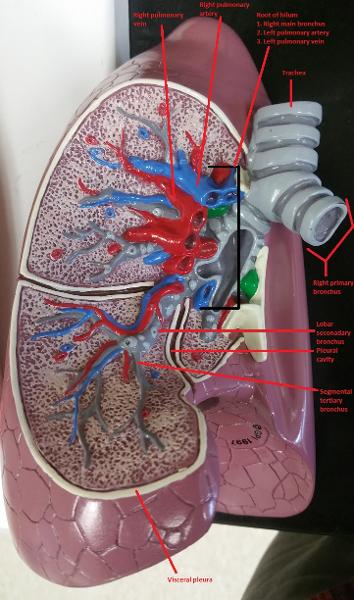 |