Instructions for Side by Side Printing
- Print the notecards
- Fold each page in half along the solid vertical line
- Cut out the notecards by cutting along each horizontal dotted line
- Optional: Glue, tape or staple the ends of each notecard together
autonomic nervous system
front 1 daw a flowchart of the organization of the nervous system | back 1 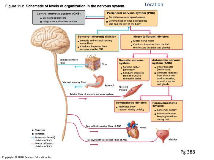 |
front 2 Effector of somatic ns Effector of autonomic ns | back 2 SNS: skeletal muscles ANS: smooth muscle, cardiac muscle, and glands |
front 3 Efferent pathway of SNS Efferent pathway of ANS | back 3 one neuron system two neuron system (pre- and post- synaptic) |
front 4 neurotransmitter and response of target organ of somatic nervous system | back 4 acetylcholine - excitatory |
front 5 neurotransmitter and response of target organ of autonomic nervous system | back 5 presynaptic neuron - acetylcholine - excitatory; postsynaptic neuron - sympathetic - norepinephrine and epinephrine postsynaptic neuron - parasympathetic - acetylcholine |
front 6 where are motor neuron cell bodies located? sensory neuron cell bodies? | back 6 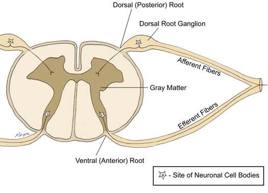 ventral horn of spinal cord; dorsal root ganglia |
front 7 parasympathetic cranial nerves? | back 7 3,7,9,10 |
front 8 spinal cord levels of sympathetic nervous system? | back 8 T1-L2 |
front 9 spinal cord levels of parasympathetic nervous system? | back 9 S2-s4 |
front 10 origin of presynpatic neuron cell body in SNS origin of presynaptic neuron cell body in PNS (think not super specific) | back 10 thoracolumbar region of the spinal cord (called thoracolumbar region) brain and sacral spinal cord (called craniosacral division) |
front 11 pre and post synaptic fiber lengths of SNS and PNS | back 11 SNS - presynaptic is short and postsynaptic is long PNS presynaptic is long and postsynaptic is short |
front 12 Why is the presynaptic fiber length short and postsynaptic long in the SNS? | back 12 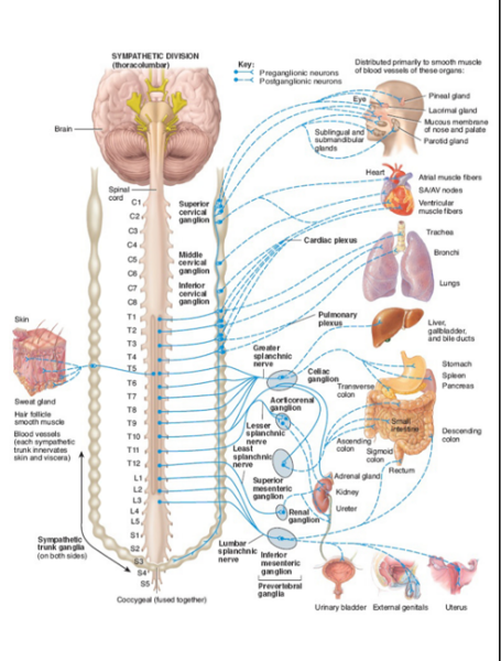 b/c presynpatic goes to the sympathetic chain of white rami communicnte - ganglia is close to spinal cord. and then postsynaptic has to travel all the way to effector organ |
front 13 Why is the presynaptic fiber length long and postsynaptic fiber short in the PNS? | back 13 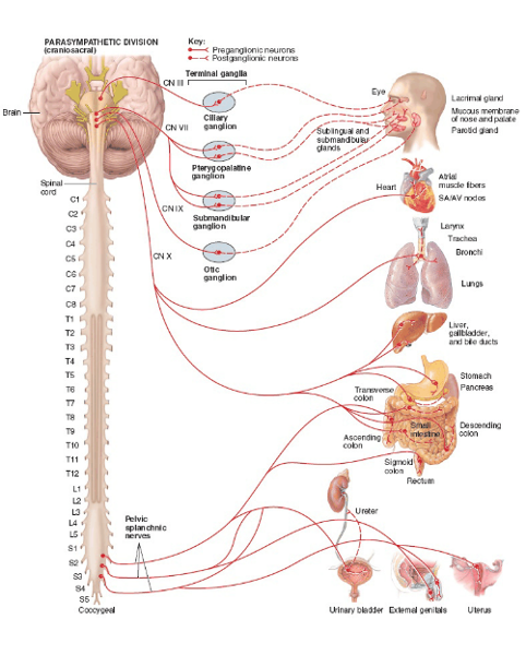 ganglia is located in the effector organ |
front 14 what is the only major body function not inhibited by PNS? | back 14 digestion |
front 15 what are the three options when presynaptic fiber joins sympathetic chain via white rami? | back 15 synapse at same level of chain entrance ascend or descend to different level pass through chain without synapsing - splanchnic nerve |
front 16 which are the only splanchnic nerves that carry parasympthetic fibers? | back 16 pelvic splanchnic nerves |
front 17 presynaptic fibers enter sympathetic chain via _______ and exit via ____ | back 17 white rami; grey rami |
front 18 Where are presynaptic neuron cell bodies located in parasympathetic nervous system? | back 18 nuclei for cranial nerves III, VII, IX, X and S2-S4 spinal cord levels |
front 19 where is the synapse located between neurons in parasympathetic nervous system? | back 19 terminal ganglia -named ganglia like ciliary, pterygopalatine, submandibular, and otic -unnamed intramural gnaglia (in or near the wall of effector organ) |
front 20 Presynaptic fibers running via the oculomotor nerve (III) innervates what? facial nerve? glossopharangyeal? vagus? | back 20 smooth muscle in eye lacrimal, submandibular and sublingual glands parotid gland almost every organ in the thoracic and abdominal cavity |
front 21 which organs receive parasympathetic communication via the vagus nerve? | back 21 heart, lungs, liver, gallbladder, stomach, small intestine, pancreas, and proximal half of large intestine |
front 22 what is the pathway of cranial outflow of PNS? sacral outflow? | back 22 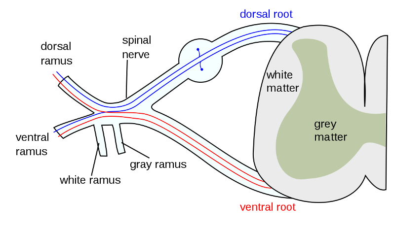 presynaptic fibers run via the four parasympathetic cranial nerves presynaptic fibers run through the ventral root --> spinal nerve --> ventral rami --> exit ventral rami as pelvic splanchnic nerves |
front 23 what innervates distal portion of large intestine? proximal? | back 23 pelvic splanchnic nerves; vagus nerve |
front 24 where do pelvic splanchnic nerves synapse and what do they innervate? | back 24 synapse in intramural ganglia and innervate distal half of large intestine and pelvic viscera (urinary bladder, uterus, and reproductive organs) |
front 25 where are presynaptic neuron cell bodies located in the SNS? | back 25 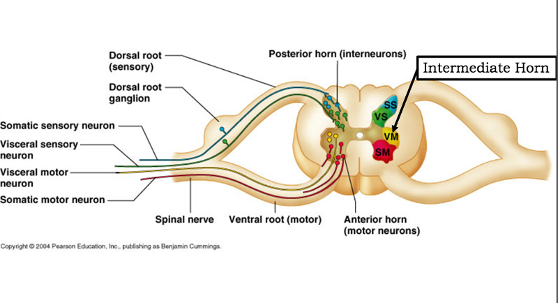 thoracolumbar region of spinal cord (T1-L2) and the intermediate horn |
front 26 where is the synapse between the two neurons in the SNS? | back 26 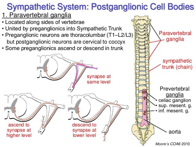 paravertebral ganglia on the sympathetic chain or prevertebral/collateral ganglia on the abdominal aorta |
front 27 the sympathetic chain extends from _____ to _____ | back 27 cranial base; coccyx |
front 28 how are the paravertebral ganglia joined to the spinal nerve? | back 28 by white and grey rami communicantes |
front 29 what is the pattern of thoracolumbar outflow in the SNS? | back 29 presynaptic fibers exit through ventral root --> spinal nerve--> white ramus--> paravertebral ganglia on the sympathetic chain |
front 30 if preganglionic fibers do not synapse on the sympathetic chain in SNS, how do they exit the chain? | back 30 as splanchnic nerves |