Instructions for Side by Side Printing
- Print the notecards
- Fold each page in half along the solid vertical line
- Cut out the notecards by cutting along each horizontal dotted line
- Optional: Glue, tape or staple the ends of each notecard together
Chapter 6
front 1 Chapter 6: | back 1 Bones and skeletal tissue |
front 2 Skeletal System: | back 2 1. Composed of bones, cartilages, ligaments and tendons
|
front 3 1ST Function of Bone and Skeletal System: | back 3 1. Supports the body
|
front 4 2ND Function of Bone and Skeletal System: | back 4 1. Protects important internal organs
|
front 5 3RD Function of Bone and Skeletal System: | back 5 1. Assistance in Movement
|
front 6 Mineral Homeostasis: | back 6 1. Bone tissue store several minerals
|
front 7 Blood Cell Production (Hemopoesis): | back 7 Red bone marrow produces Red blood cells, White blood |
front 8 Triglyceride Storage: | back 8 1. Yellow bone marrow
|
front 9 Bones in Human skeleton: | back 9 206 named bones |
front 10 Bones of the skeleton are grouped into two principal divisions: | back 10 1. Axial skeleton
|
front 11 Axial skeleton: | back 11 Bones along the longitudinal axis of the human body
|
front 12 Appendicular skeleton : | back 12 Bones of the upper and lower limbs (extremities)
|
front 13 Long Bones: | back 13 Greater length than width, slightly curved for strength
|
front 14 Short bones: | back 14 Cube-shaped
|
front 15 Flat bones: | back 15 Thin, two parallel plates of compact bone tissue enclosing spongy bone tissue
|
front 16 Irregular bones: | back 16 Complex shapes that cannot be grouped into any of the previous categories
|
front 17 Sesamoid bones: | back 17 Protect tendons from excessive wear and tear
|
front 18 Sutural bones: | back 18 Small bones located in sutures of cranial bones |
front 19 Surface markings: | back 19 Structural features adapted for specific functions |
front 20 Two major types of surface markings: | back 20 1. Depressions and openings
|
front 21 Depressions and openings: | back 21 1. Allow the passage of blood vessels and nerves
|
front 22 Projections/Processes: | back 22 1. Projections or outgrowths that form joints
|
front 23 Diaphysis: | back 23 Shaft |
front 24 Epiphysis : | back 24 distal and proximal ends |
front 25 Metaphysis: | back 25 Epiphyseal growth plate (hyaline cartila) |
front 26 Articular cartilage: | back 26 Hyaline cartilage |
front 27 Periosteum: | back 27 1.Membrane surrounding bone
|
front 28 Medullary cavity: | back 28 yellow marrow |
front 29 Epiphyseal line: | back 29 membrane lining canals and trabeculae (osteoblasts, osteoclasts) |
front 30 Type of cells: | back 30 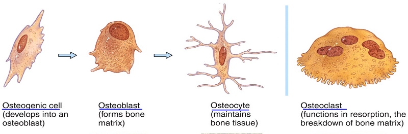 Osteogenic cells
|
front 31 Extracellular surrounding widely separated cells: | back 31 Matrix
|
front 32 How Many Types Of Cells In Bone Tissue: | back 32 Four |
front 33 Osteogenic cells: | back 33 1. Undergo cell division
|
front 34 Osteoblasts: | back 34 "Bone-building cells"
|
front 35 Osteocytes: | back 35 1. Mature bone cells, “trapped” in mineralized extracellular matrix
|
front 36 Osteoclasts: | back 36 1. Release enzymes and acids that digest the protein and mineral components of bone matrix (resorption)
|
front 37 Two Bone Categories: | back 37 1. Compact
|
front 38 Compact Bone: | back 38 (80% of skeleton)
|
front 39 Compact Bone PART 2: | back 39 1. Resists the stresses produced by weight and movement
|
front 40 Spongy Bone: | back 40 1. Internal to Compact bone
|
front 41 Spongy Bone PART 2: | back 41 1. Lacks osteons
|
front 42 Osteon: | back 42 1. Canals tha run through bones
|
front 43 Ossification / osteogenesis: | back 43 Bone Formatic
|
front 44 Bone formation occurs in four situations: | back 44 - Formation of bone in an embryo
|
front 45 Calciification: | back 45 hardening of tissue, occurs during ossification |
front 46 Bone’s flexibility depends on: | back 46 collagen fibers |
front 47 Two Types Of Ossification: | back 47 1. Intramembranous
|
front 48 Intramembranous ossification: | back 48 direct laying down of bone into the primitive connective tissue (mesenchyme REPLACED by BONE)
|
front 49 Endochondrial / intracartilaginous ossification: | back 49 (involves chartilage) MORE COMMON
|
front 50 Endochondral ossification/intracartilaginous(prenatal): | back 50 1. First osteoblasts appear at Bone collar.
|
front 51 Growth in Length (postnatal): | back 51 1. Interstitial growth at the epiphyseal plate: chondrocytes keep dividing and secreting ECM at the epiph side
|
front 52 Growth in Thickness (postnatal): | back 52 1. Occurs by appositional growth:
|
front 53 Bone REMODELLING: | back 53 1. Adult bone tissue continually renews itself
|
front 54 Normal bone metabolism depends on several factors
| back 54 1. Negative feedback that maintains Ca2+
|
front 55 Bone’s Role in Calcium Homeostasis: | back 55 1. Bone is the body’s major calcium reservoir (99% of total body Ca)
|
front 56 Parathyroid hormone (PTH): | back 56 produced in parathyroid glands
|
front 57 Calcitonin : | back 57 1. secreted by the thyroid gland
|
front 58 The main mechanical stresses on bone are those that result from the : | back 58 pull of skeletal muscles and the pull of gravity. |
front 59 Minerals: | back 59 Large amounts of calcium and phosphorus and smaller amounts of magnesium, fluoride, and manganese are required for bone growth and remodeling |
front 60 Vitamins: | back 60 - Vitamin A: stimulates activity of osteoblasts
|
front 61 GH: | back 61 Growth Hormone |
front 62 Giantism: | back 62 ^^^ GH |
front 63 Dwarfism: | back 63 vvv GH |
front 64 Acromegaly: | back 64 ^^^ GH in adulthood |
front 65 Types Of Fractures: | back 65 1. Bone penetrates skin or not
|
front 66 Bone penetrates skin or not: | back 66 - Simple (closed) fracture
|
front 67 Completeness of the break: | back 67 Complete, incomplete (greenstick) |
front 68 Position of the bone ends after fracture: | back 68 Non displaced, displaced (2 ends not lined up), comminuted (many pieces) |
front 69 Treatment: | back 69 1. Closed reduction: alignment by manipulation
|
front 70 The repair of a bone fracture involves: | back 70 1. Formation of fracture hematoma
|
front 71 Formation of fracture hematoma: | back 71 Blood leaks from the torn ends of blood vessels |
front 72 Fibrocartilaginous callus formation: | back 72 Fibroblasts invade the fracture site and produce collagen fibers which bridge the broken ends of the bone (takes ~3 weeks |
front 73 Bony callus formation: | back 73 Osteoblasts begin to produce spongy bone |
front 74 Bone remodeling | back 74 Compact bone replaces spongy bone |
front 75 Homeostatic imbalances: | back 75 Imbalances between bone formation and resorption
|
front 76 Osteomalacia/Rickets: | back 76 - adults/children
|
front 77 Paget’s disease: | back 77 - Excessive remodelling (bone deposition and resorbtion)
|
front 78 Osteoporosis: | back 78 - Bones become fragile, bone mass is reduced
|