Instructions for Side by Side Printing
- Print the notecards
- Fold each page in half along the solid vertical line
- Cut out the notecards by cutting along each horizontal dotted line
- Optional: Glue, tape or staple the ends of each notecard together
Pathology
front 1 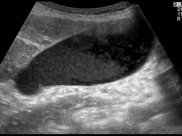 | back 1 Courvoisier's GB Distension without wall thickening due to obstruction distal to the cystic duct *Panncreatic head mass * Duodenal papilla mass *CBD mass |
front 2 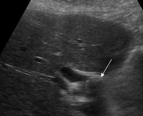 RUQ pain Jaundice recurrent cholangitis | back 2 Mirizzi SYndrome impacted stone in the cystic duct or GB neck presence of two tubular structures representing the bile duct above the level of the cystic duct |
front 3 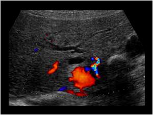 | back 3 Mirizzi SYndrome impacted stone in the cystic duct or GB neck presence of two tubular structures representing the bile duct above the level of the cystic duct |
front 4  Patient has been fed intravenously for 3 days. | back 4 Sludge highly concentrated bile |
front 5 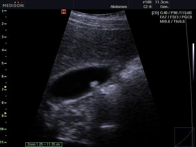 Gravity dependent | back 5 Cholelithiasis Mobile, hyperechoic, round or triangular structures casting a well defined posterior acoustic shadowing Will change when patient’s body position changes (Gravity dependent) |
front 6 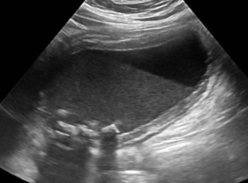 RUQ pain fever leukocytosis Positive Murphy's sign | back 6 Acute cholecystitis inflamation of the GB wall w/ stone |
front 7 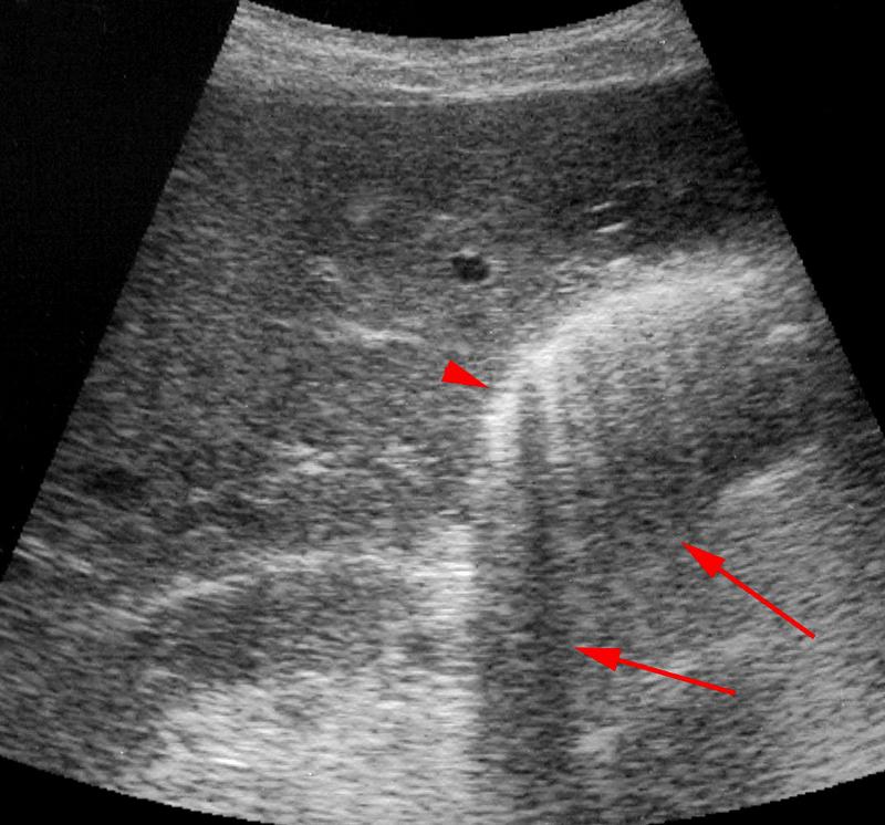 Diabetic RUQ pain fever leukocytosis | back 7 Emphysematous Cholecystitis Gas forming bacteria in gallbladder wall yields to high intensity echoes and comet tail artifact |
front 8 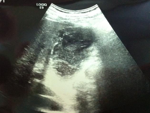 RUQ pain fever leukocytosis | back 8 Empyema of the gallbladder |
front 9 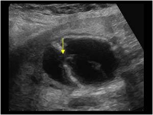 | back 9 Perforation in the gallbladder wall |
front 10 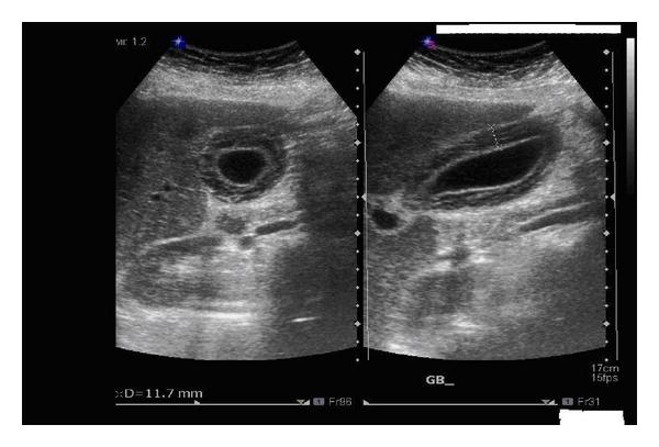 Typically secondary event in critically ill | back 10 Acalculus Cholecystitis Inflammation of GB wall without stones |
front 11 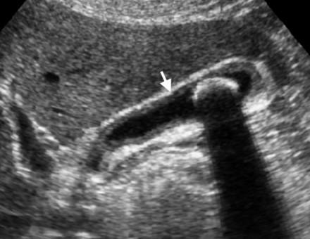 | back 11 Chronic Cholecystitis Contracted gallbladder with acoustic shadowing from cholelithiasis Thick hyperechoic gallbladder wall greater than 4 – 5 mm Sludge may be present |
front 12 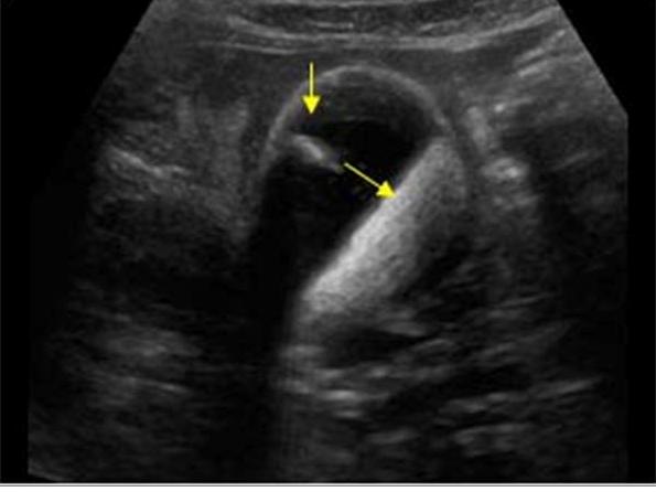 patient present with chronic Cholecytitis | back 12 Milk of calcium Bile (Limy bile) |
front 13 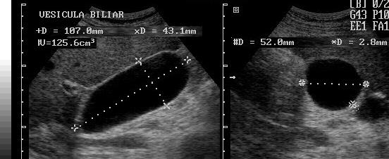 Asymptomatic with palpable RUQ mass | back 13 Hydrops GB Greater than 4cm in diameter |
front 14 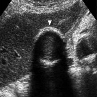 Female patient with Chronic Cholecystitis | back 14 Porcelain GB Calcification of GB wall due to chronic cholecytitis |
front 15 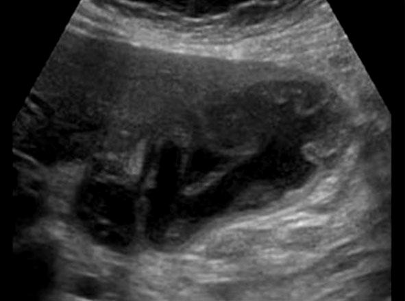 | back 15 Gangrenous Cholecystitis |
front 16 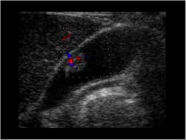 | back 16 GB Ademoma epithelial tumor overgrowth of the lining |
front 17 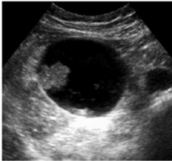 | back 17 GB Ademoma epithelial tumor overgrowth of the lining |
front 18 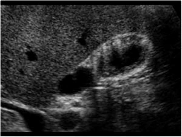 Asymptomatic | back 18 Gallbladder Adenomyomatosis diverticulum of the GB Focal, segmental or diffuse smooth muscle proliferation with exaggerated diverticular appearance of the Rokitansky – Aschoff sinuses into the muscular wall |
front 19 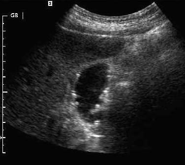 | back 19 Gallbladder Cholesterolosis Non shadowing, hyperechoic, polyp Strawberry GB Lipids |
front 20 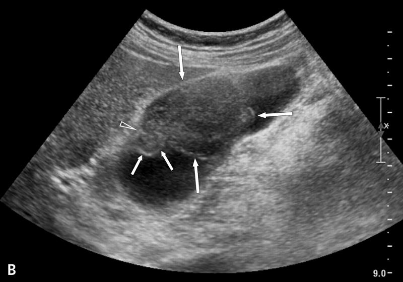 60 year old with long standing cholecystitis and porcelain GB | back 20 Gallbladder Carcinoma Most commonly a mass from the gallbladder fossa replaces the gallbladder and invades adjacent liver. Focal or diffuse irregular gallbladder wall thickening Polypoid intramural lesions with irregular borders |
front 21 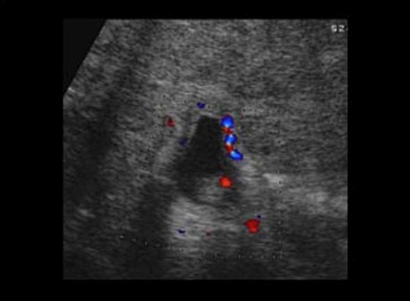 Stage 4 colon cancer | back 21 Gallbladder Metastasis commonly from stomach, pancreas and bile ducts |
front 22 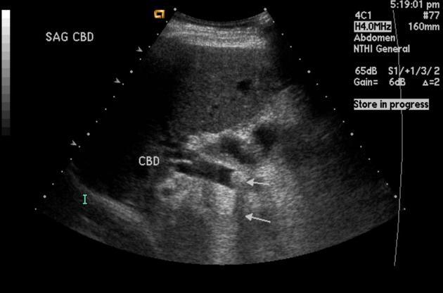 RUQ pain Jaundice Elevated Alkaline phosphatase Elevated conjugated bilirubin Elevated Gamma gluamyl transpeptidase | back 22 Choledocholithiasis obstrution by biliary stone |
front 23 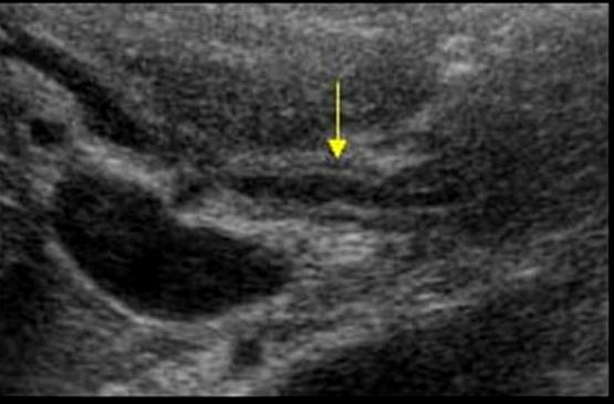 RUQ pain Fever Jaundice Elevated Conjugated bilirubin Elevated Alkaline phosphatase ALP Elevated GGT Elevated amylase and lipase Elevated white blood count | back 23 Cholangitis inflamation of the duct walls |
front 24 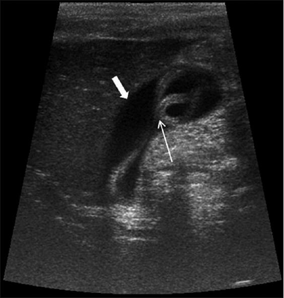 asymptomatic biliary colic cholangitis Poor hygiene | back 24 Ascariasis infection of round worms |
front 25 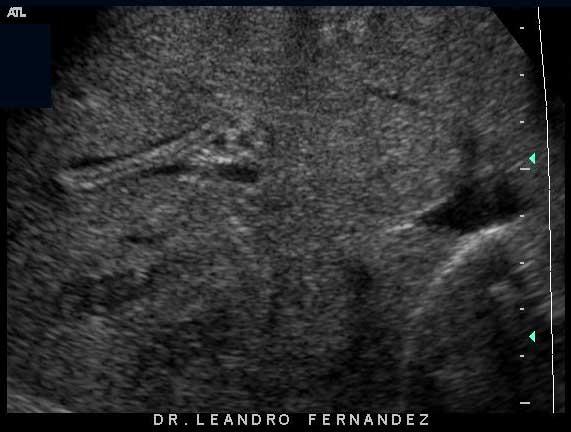 asymptomatic biliary colic cholangitis Poor hygiene | back 25 Ascariasis infection of round worms |
front 26  Pain Hematemesis caused by procedures or biopsy | back 26 Hemobilia blood in the biliary tree caused by procedures or biopsy |
front 27 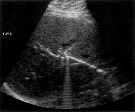 Patient recently had ERCP | back 27 pneumobilia Air within the biliary tree |
front 28 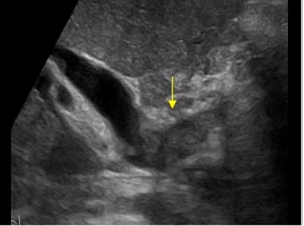 Jaundice weight loss abdominal pain | back 28 Cholangiocarcinoma typically originate within the extrahepatic bile ducts |
front 29 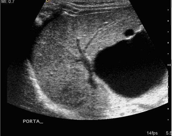 Neonate presents with Jaundice for 14+ days | back 29 Biliary Atresia Congenital cystic formation without dilated interhepatic ducts |
front 30 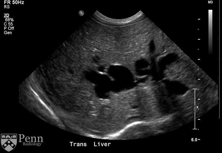 congenital hepatic fibrosis Portal hypertension | back 30 Caroli's Disease congenital cystic dilatation of the intrahepatic biliary tree |
front 31 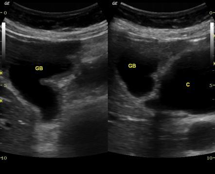 9 year old Japanese patient | back 31 Choledochal Cyst Congenital cystic dilation of the extrahpatic biliary tree |
front 32 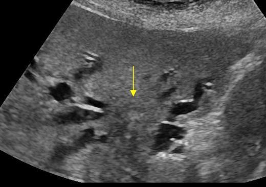 | back 32 Klatskin Tumor malignant tumor arising between the left and right hepatic ducts |
front 33 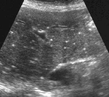 Patient in Great Lakes basin | back 33 Liver granulomas |
front 34  Hypoechoic renal cortex obese Type II diabetes | back 34 Fatty infiltration |
front 35 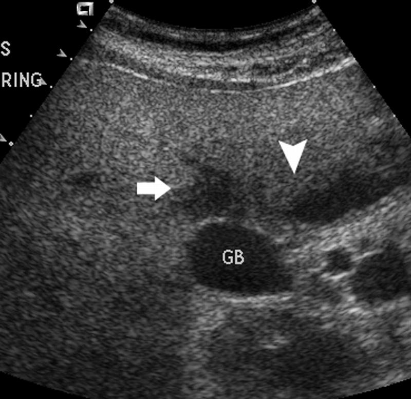 | back 35 Fat Sparing |
front 36 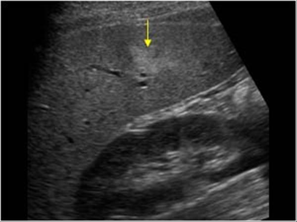
| back 36 focal fatty infiltration |
front 37 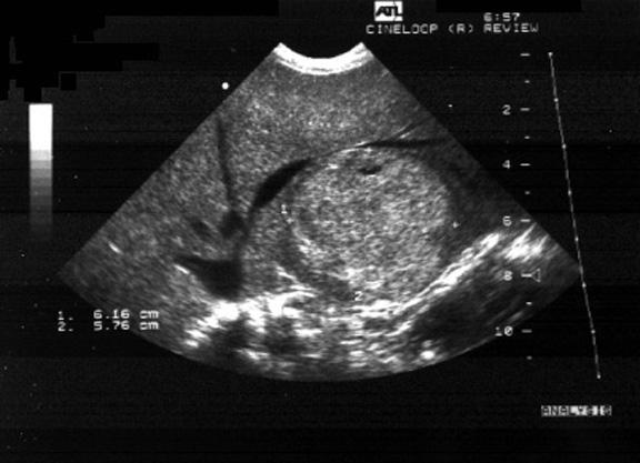 Infant impaired growth hypoglycemia CHF delayed puberty osteoporosis | back 37 Glycogen Storage Disease Autosomal recessive disorder of carb metabolism found in infants seen with adenomas |
front 38 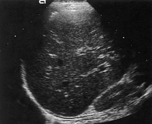 Elevated ALT AST Bilirubin con & un | back 38 Acute Hepatitis Generally normal but can have portal cuffing (starry sky) |
front 39 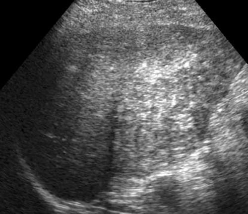 Elevated ALT AST Bilirubin con & un | back 39 Chronic Hepatitis course texture increased echogenicity |
front 40 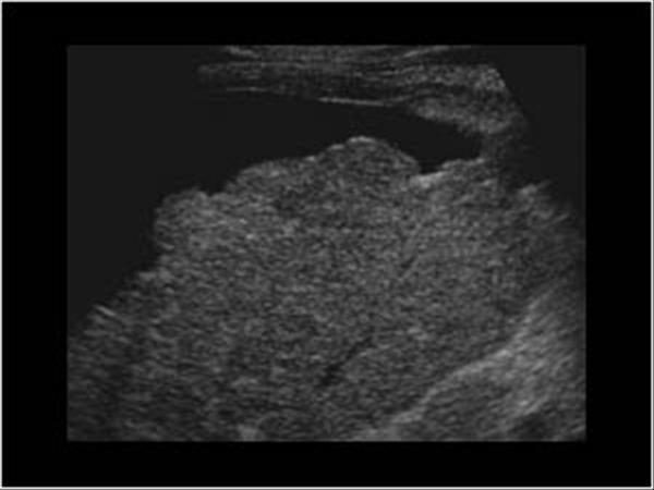 chronic hepatitis alcoholism elevated AST ALT GGT LDH conjugated bilirubin | back 40 Cirrhosis nodular and course heterogenic Ascites |
front 41 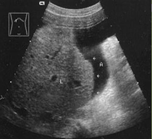 | back 41 Cirrhosis nodular and course heterogenic Ascites |
front 42 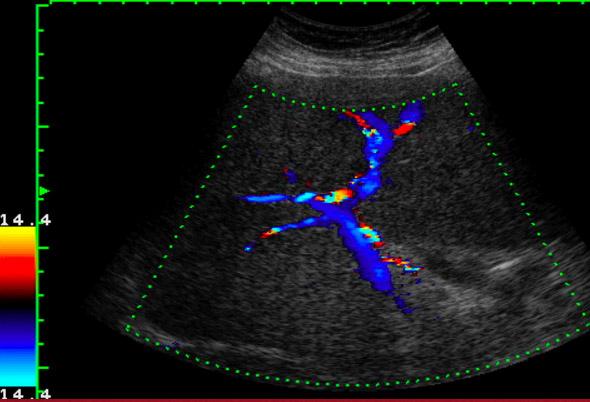 asymptomatic sudden painless upper GI hemorrhage due to ruptures esophageal varices | back 42 Portal Hypertension |
front 43 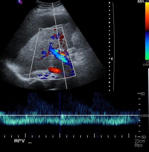 | back 43 Portal Hypertension |
front 44 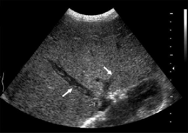 | back 44 Budd-Chiari Life threatening emergency |
front 45 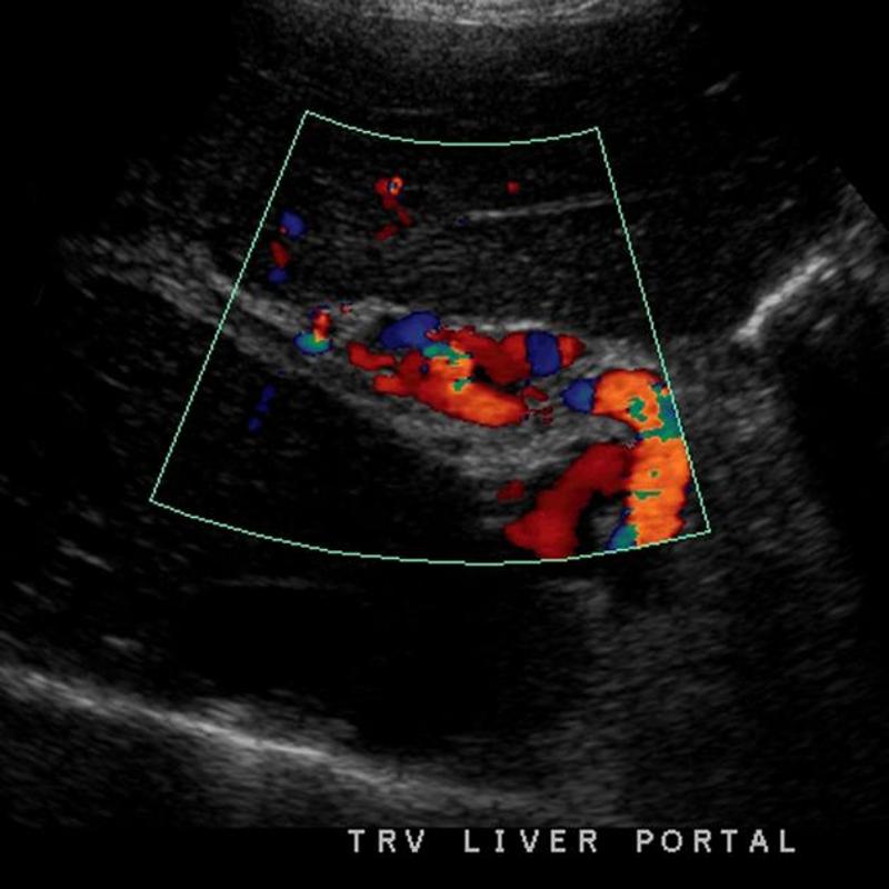 | back 45 Portal vein Thrombosis - cavernous transformation replacement of the normal single channel portal vein with numerous tortuous venous channels. |
front 46 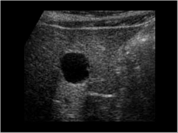 50+ | back 46 Cysts True cysts are congenital other cysts lack an epithelial lining and are not true cysts |
front 47 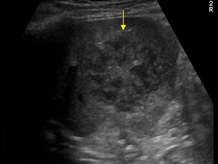 Presentation Fever pain N&V Leukocytosis Elevated LFTs | back 47 Pyogenic (bacterial) abscess |
front 48 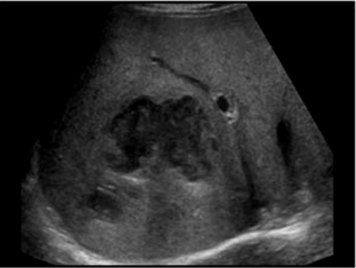 Presentation fever pain diarrhea Leukocytosis Elevated LFTs | back 48 Amebic abscess contaminated food or water usually in colon but can invade the liver through the portal vein fever pain diarrhea |
front 49 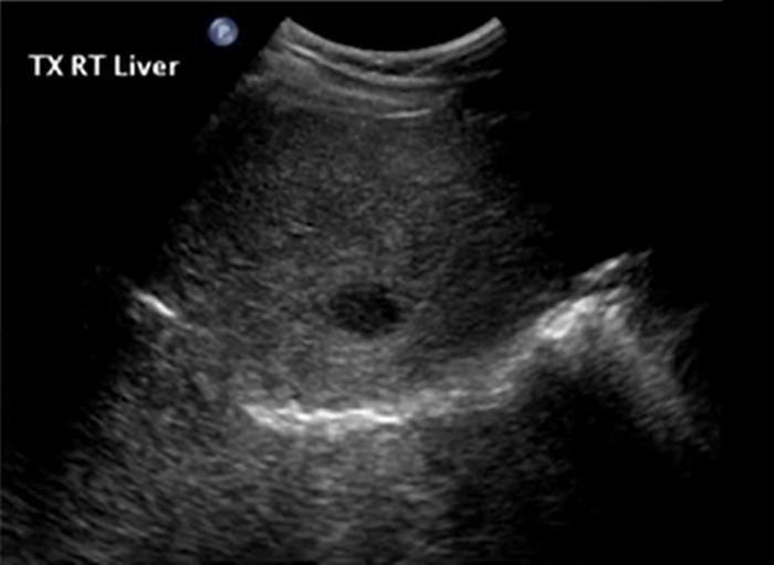 May also present wheel within a wheel Bull's eye Echogenic focus | back 49 Candidiasis early wheel within wheel later hypoechoic |
front 50 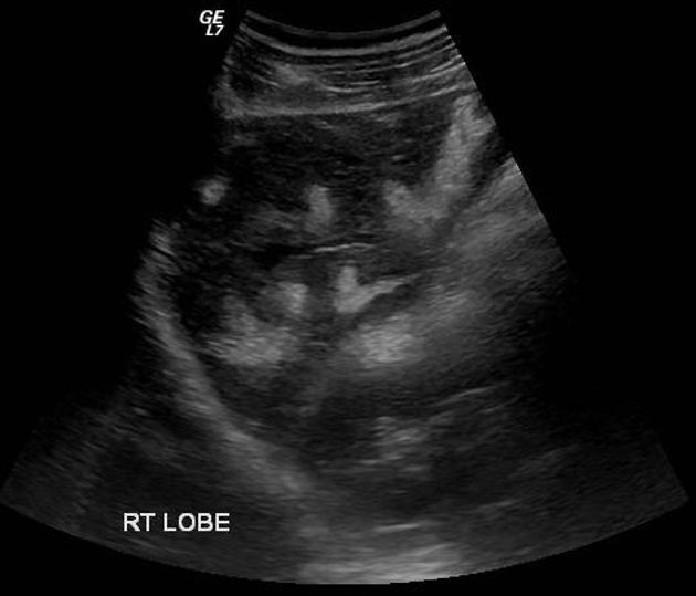 # cause of portal hypertension | back 50 Schistosomiasis #1 cause of portal hypertension in the world. Not common in US but estimated 400,000 infected people have immigrated parasitic infection mainly in Egypt
|
front 51 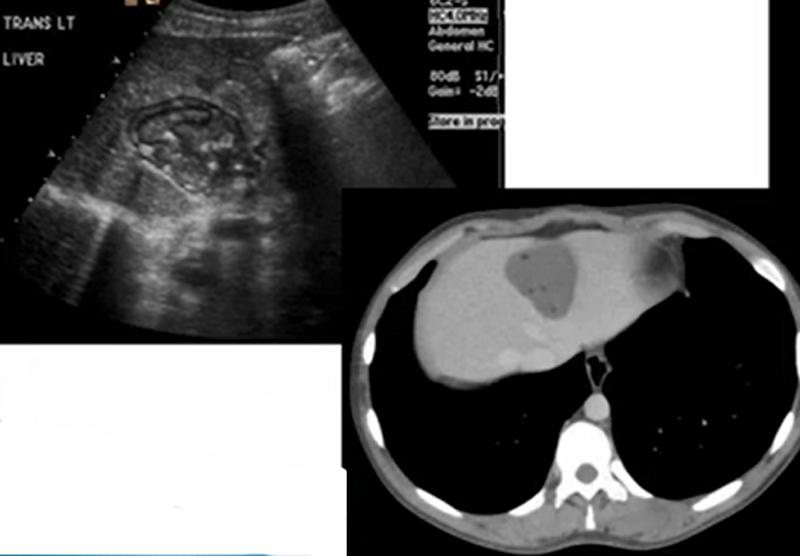 Sheep herder | back 51 Echinococcal Cyst Sheep Herders AKA Hydatid Cyst |
front 52 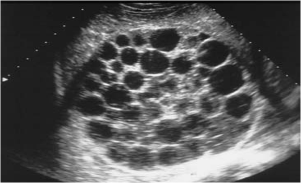 Sheep herder | back 52 Echinococcal Cyst Sheep Herders AKA Hydatid Cyst |
front 53 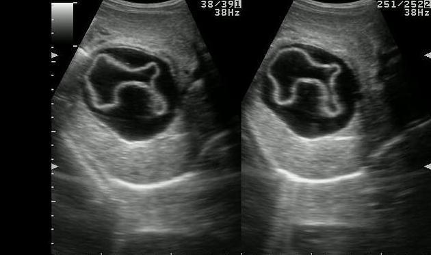 Water lily Sheep herder | back 53 Echinococcal Cyst Sheep Herders AKA Hydatid Cyst |
front 54 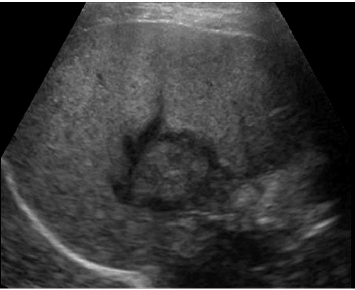 24 year old female on birth control pills Pain | back 54 Liver Adenoma Females taking birth control *Hypoechoic *Hyperechoic *Isoechoic *Mixed |
front 55 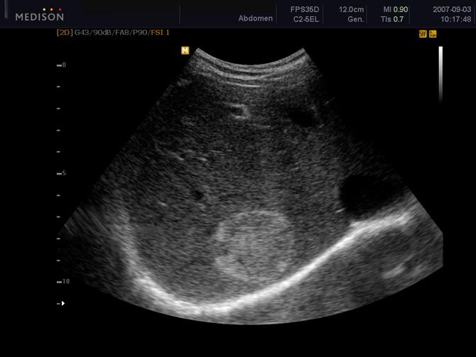 enlarge with pregnancy | back 55 Cavernous Hemangioma |
front 56 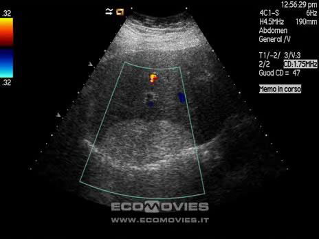 | back 56 Hepatic lipoma |
front 57 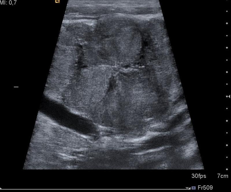 Asymptomatic | back 57 Focal Nodular Hyperplasia abnormally arranged hepatocytes Second most common benign liver mass |
front 58 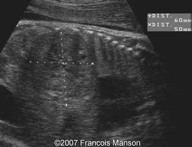 6 month old female abdominal mass CHF | back 58 Hemangioendothelioma Benign condition of overgrowth of endothelium of capillaries Found in infants Usually seen as hepatic lesions that are predominantly hypoechoic; however, hepatic lesions can also have mixed echotexture or be predominantly hyperechoic. |
front 59 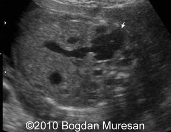 Two year old with palpable mass on right side | back 59 Mesenchymal hamartoma rare developmental cystic tumor of the lover complex mass more common in rt lobe |
front 60 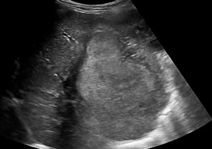 3 year old abdominal enlargement weight loss nausea and vomiting marked elevation of AFP | back 60 Hepatoblastoma malignant germ cell tumor most common malignant liver tumor of children under 3 associated with Beckwith Wiedermann |
front 61 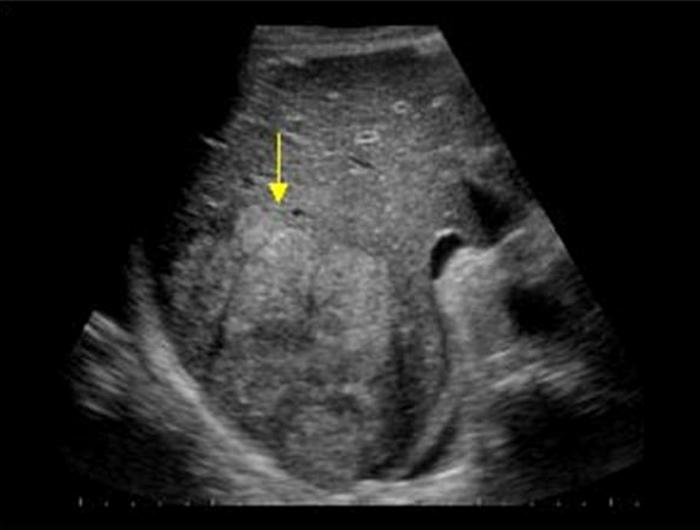 Patient with cirrhosis | back 61 Hepatocellular Carcinoma solid multiple diffuse |
front 62 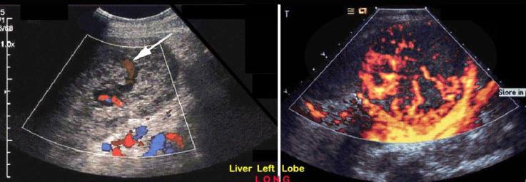 | back 62 Hemangiosarcoma rare in 60 - 80 related to exposure of thorotrast, arsenic or polyvinyl chloride large mixed mass |
front 63 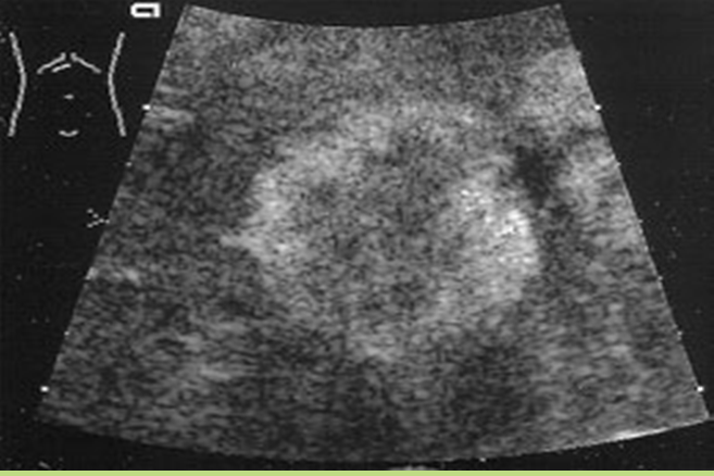 Liver mets from ? | back 63 Metastasis to the liver colon Most common |
front 64 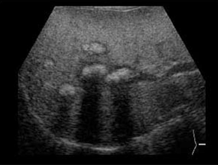 Liver mets from ? | back 64 Metastasis to the liver Breast |
front 65 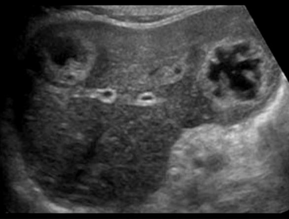 Liver mets from ? | back 65 Metastasis to the liver Lung |
front 66 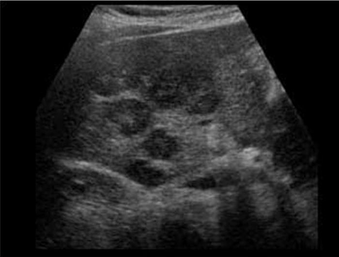 Liver mets from ? | back 66 Lymphoma |
front 67 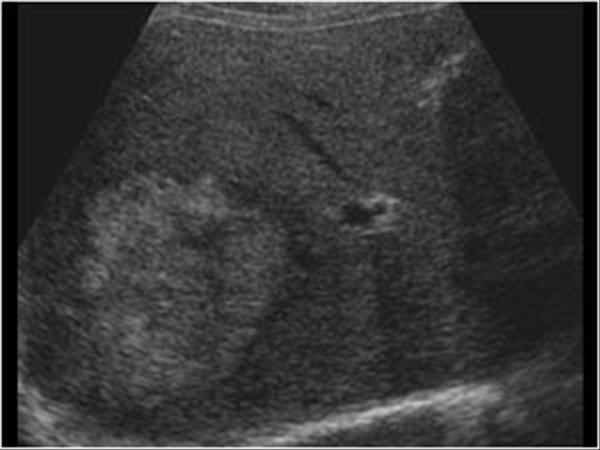 Pain hypertension after car accident | back 67 Liver hematoma |
front 68 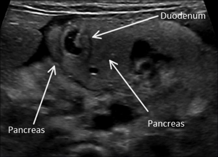 | back 68 Annular Pancreas |
front 69 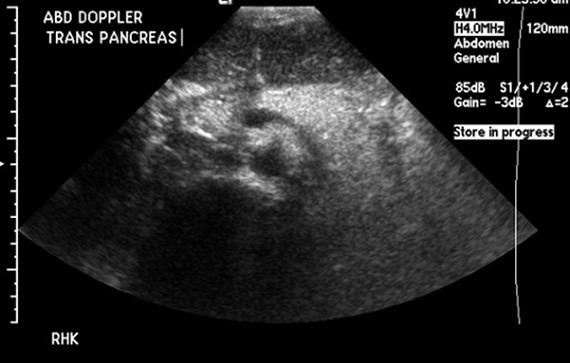 6 year old present | back 69 Cystic Fibrosis autosomal Recessive Pancreas appears hyperechoic due to microcystic changes, increased fibrotic and fat |
front 70 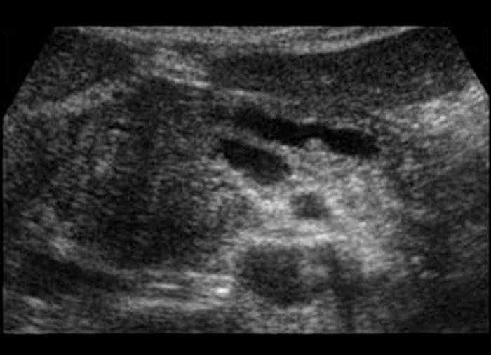 severe constant, intense pain radiating to the back N & V fever sweating paralytic ilius elevated amylase 48 - 78 hours elevated lipase 5 - 7 days WBC | back 70 Acute Pancreatitis |
front 71 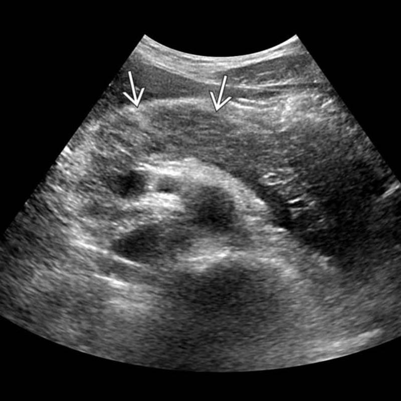 severe constant, intense pain radiating to the back N & V fever sweating paralytic ilius elevated amylase 48 - 78 hours decreased hematocrit & calcium | back 71 Hemorrhagic Pancreatitis Type of acute pancreatitis 2% to 5% significant fat necrosis that results in rupture of pancreatic vessels and secondary hemorrhage |
front 72 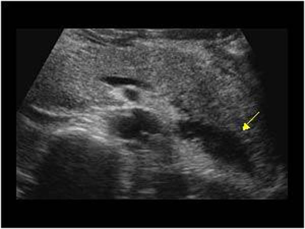 N & V fever sweating paralytic ilius elevated amylase 48 - 78 hours elevated lipase 5 - 7 days WBC | back 72 Phlegmonous Pancreatitis Type of acute pancreatitis 18% enlarged solid inflammatory mass with retroperitoneal fat necrosis usually lesser sac is involved |
front 73 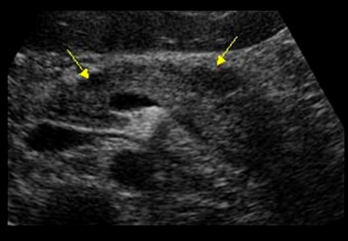 | back 73 Pancreatic Abscess infection of necrotic pancreatic and retroperitoneal fat |
front 74 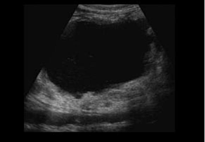 persistently elevated amylase and lipase | back 74 Pancreatic Psydocyst Spherical fluid collection of pancreatic enzymes that arise from inflamatory, necrotic and hemorrhage processes of the pancrreas |
front 75 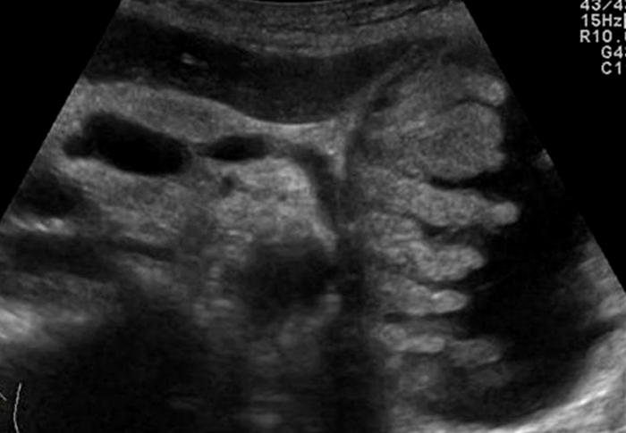 persistently elevated amylase and lipase | back 75 Pancreatic Psydocyst Spherical fluid collection of pancreatic enzymes that arise from inflamatory, necrotic and hemorrhage processes of the pancrreas |
front 76 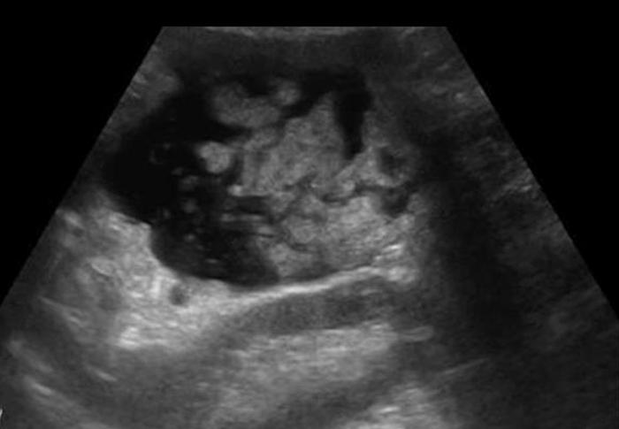 persistently elevated amylase and lipase | back 76 Pancreatic Psydocyst Spherical fluid collection of pancreatic enzymes that arise from inflamatory, necrotic and hemorrhage processes of the pancrreas |
front 77 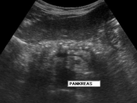 N & V flatulence weight loss | back 77 Chronic Pancreatitis Ongoing inflammation that results in permanent damage |
front 78 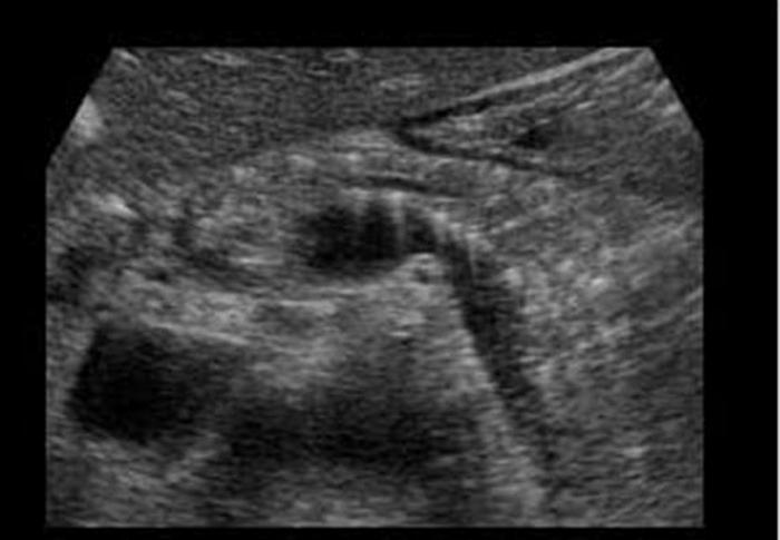 N & V flatulence weight loss | back 78 Chronic Pancreatitis Ongoing inflammation that results in permanent damage |
front 79 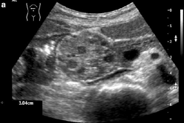 Mid epigastric Pain weight loss jaundice palpable mass | back 79 Cystadenoma multiple cystic masses that contain secreted material |
front 80 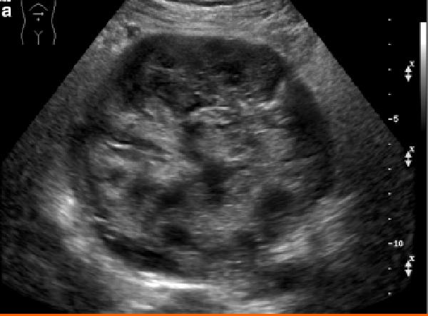 Mid epigastric Pain weight loss jaundice palpable mass | back 80 Mucinous Cystic / Cystadenocarcinoma malignant tumor from glandular tissue in which secretions are oobtained |
front 81 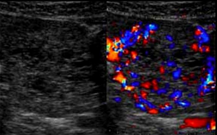 25 year old female with vague abdominal pain | back 81 Serious Cystadenoma / Microcystic adenoma type of serous cystadenoma lobulated mass of numerous small cysts |
front 82 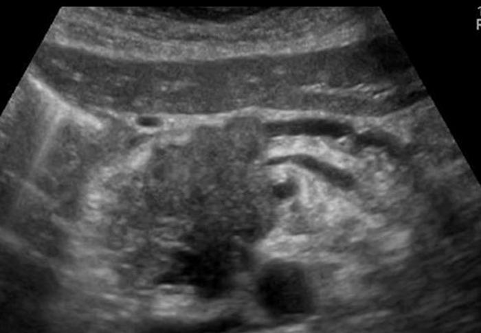 Most common cause of malignant neoplasm abdominal / back pain jaundice weight loss | back 82 Adenocarcinoma arises from the epithelium and involves the exocrine portion of the pancreas |
front 83 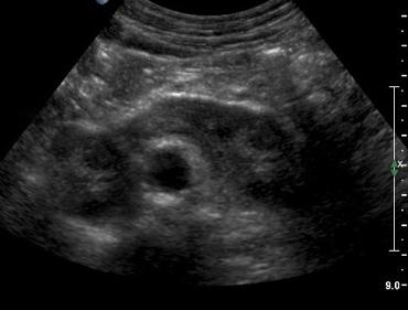 The most common form of fusion anomaly of the kidneys | back 83 Horseshoe Kidneys |
front 84 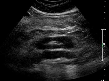 The most common form of fusion anomaly of the kidneys | back 84 Horseshoe Kidneys |
front 85 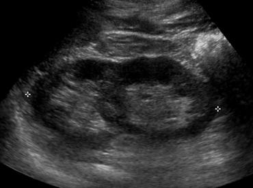 | back 85 Crossed fused Renal Ectopia |
front 86 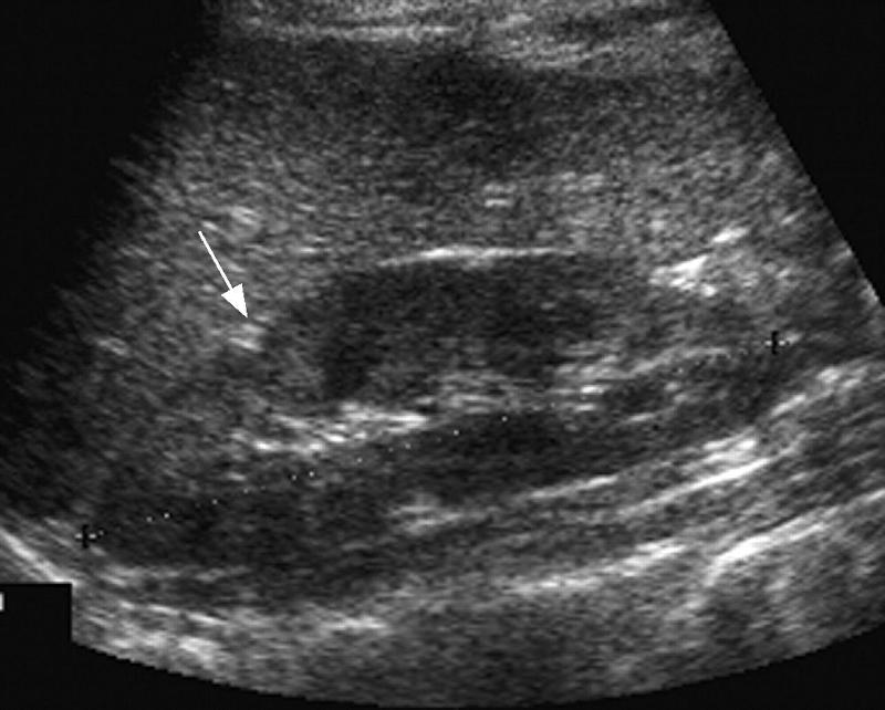 Right kidney | back 86 Junctional Parenchymal Defect |
front 87 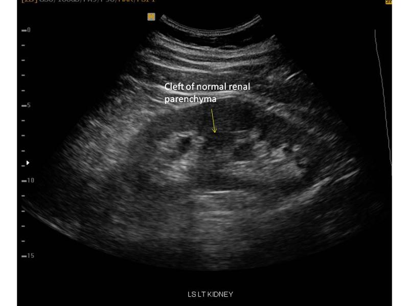 often seen with hydronephrosis in the upper pole | back 87 Duplex Kidney Seen in 15% of population |
front 88 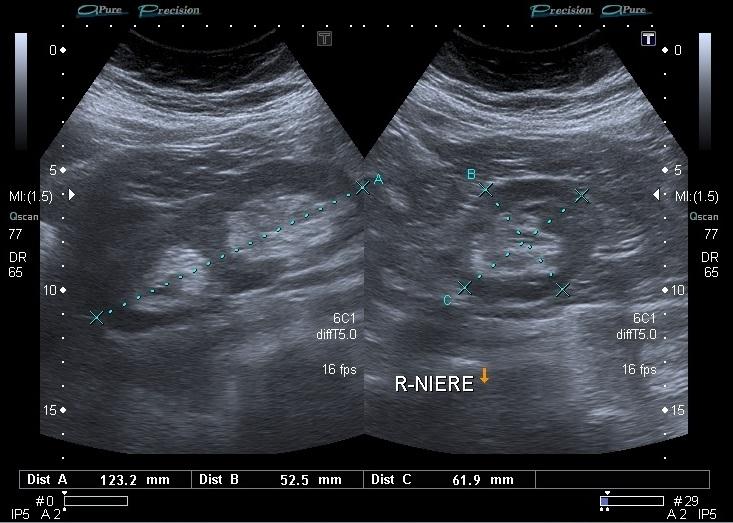 | back 88 Duplex Kidney |
front 89 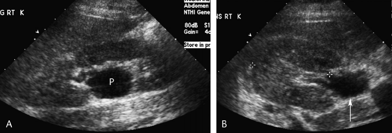 | back 89 Extrarenal Pelvis |
front 90  Male neonate | back 90 Posterior Urethral valves |
front 91 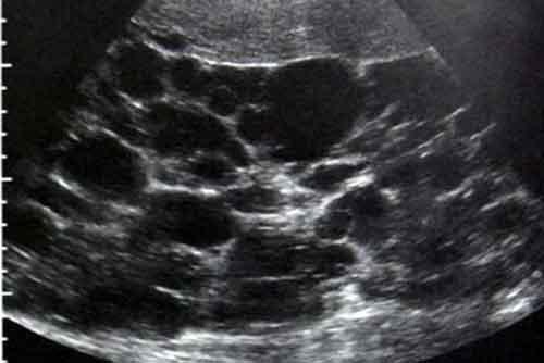 30 year old male presents for a renal ultrasound liver & spleen cysts | back 91 Adult Polycystic Kidney Disease |
front 92  infant presents with renal dysfunction | back 92 Infantile Polycystic Kidney Disease results from cystic dilation of the collecting tubules |
front 93 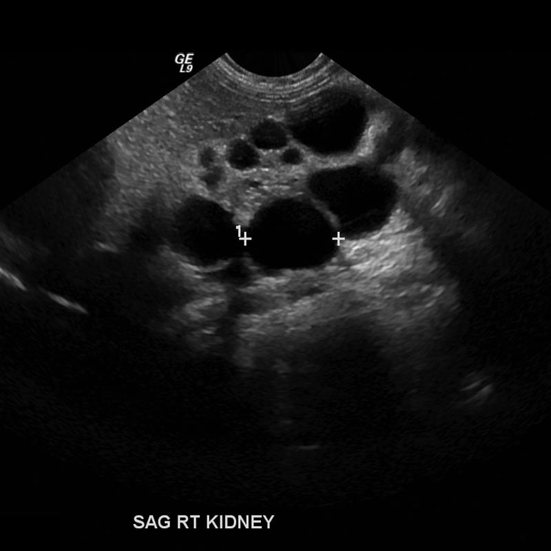 Most common cause of abdominal mass in newborns unilateral | back 93 Multicystic Dysplastic Kidney |
front 94 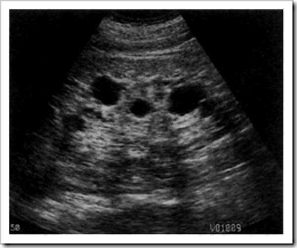 chronic renal failure hemodialysis | back 94 Aquired Cystic Disease multiple cysts in failing kidney during long term hemodialysis hemorrhage often occurs |
front 95 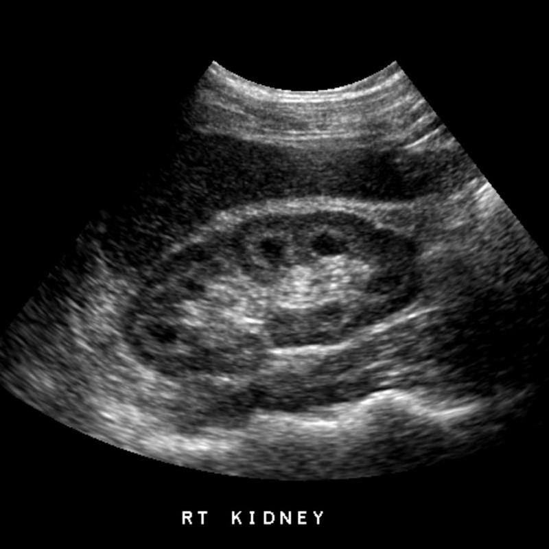 | back 95 Medullary Sponge Kidney congenital dysplastic dilatation of the medullary pyramids due to tubular ectasia or dysplasia medullary neprocalcinosis |
front 96 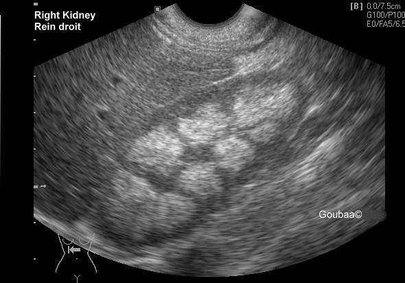 | back 96 medullary neprocalcinosis |
front 97 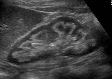 hypercalcemia hypercalciuria | back 97 Nephrocalcinosis |
front 98 20 year old presents with visual impairments | back 98 Von Hippel-Lindau Disease inhearited usually present in 2nd to 3rd decade |
front 99 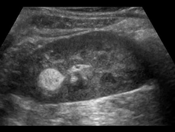 right kidney | back 99 Angiomyolipoma benign fatter renal tumor 80% involve right kidney echogenicity is gretr than of equal to the renal sinus |
front 100 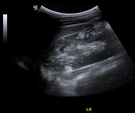 right kidney | back 100 Angiomyolipoma benign fatter renal tumor 80% involve right kidney echogenicity is gretr than of equal to the renal sinus |
front 101 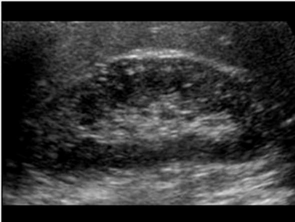 seizures mental retardation facial angiofibroma bilateral Angiomyolipomas | back 101 Tuberous Sclerosis genetic |
front 102 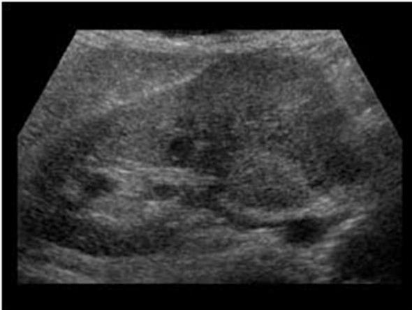 Most common solid renal mass in adults hematuria flank pain palpable mass lung mets | back 102 Renal Cell Carcinoma |
front 103 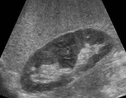 Most common solid renal mass in adults hematuria flank pain palpable mass lung mets | back 103 Renal Cell Carcinoma |
front 104 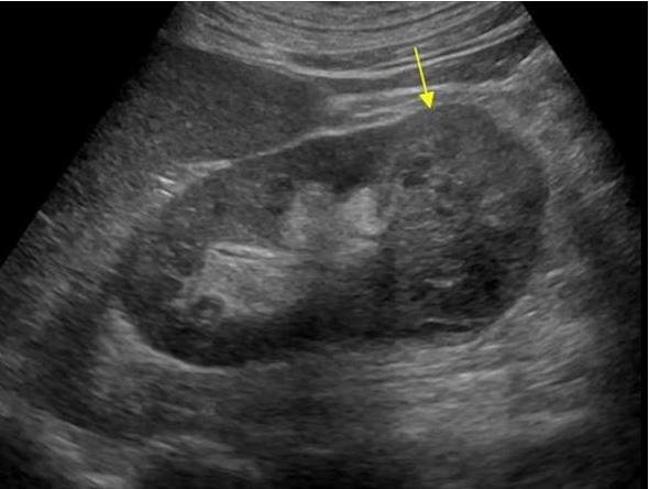 generally appear as hypoechoic or diffuse enlargement | back 104 Renal Metastases |
front 105 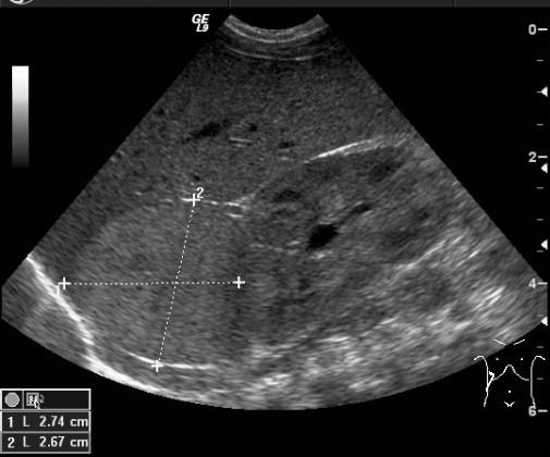 3 years large asymptomatic flank amss hypertension fever hematuria | back 105 Wilm's Tumor 3 years 90% survival |
front 106 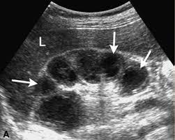 | back 106 Acute Pyelonephritis travel from bladder AKA acute focal bateria nephritis / lobar nephritis |
front 107 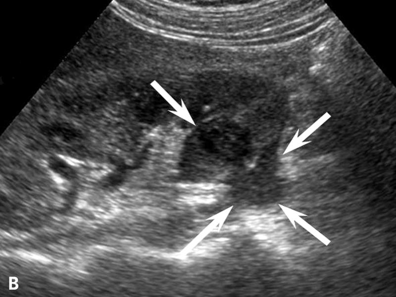 | back 107 Acute Pyelonephritis travel from bladder AKA acute focal bateria nephritis / lobar nephritis |
front 108 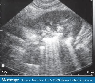 diabetic | back 108 Emphysematous pyelonephritis Gas in the kidney nephroectomy is usually required |
front 109 small hyperechoic kidneys with cortical thinning | back 109 Chronic Pyelonephritis due to recurrent renal infection |
front 110 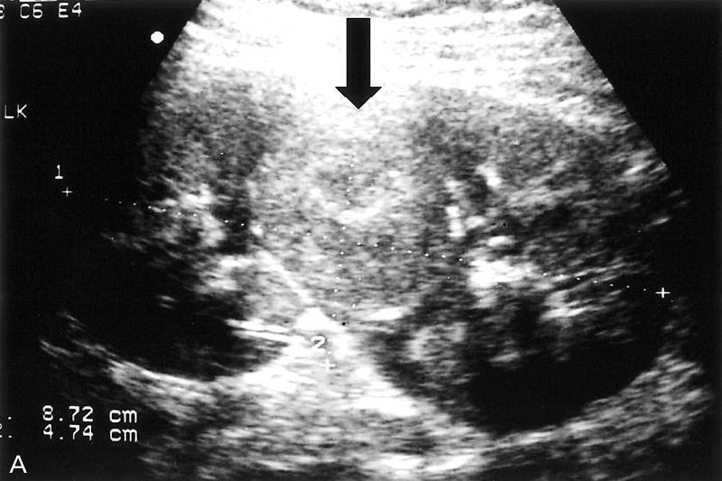 type of Chronic Pyelonephritis | back 110 Xanthogranulomatous Pyelonephritis XGPN Chronic Pyelonephritis due to stone |
front 111  type of Chronic Pyelonephritis | back 111 Xanthogranulomatous Pyelonephritis XGPN Chronic Pyelonephritis due to stone |
front 112 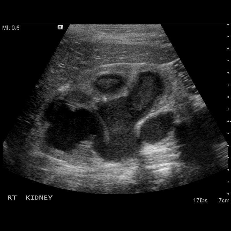 due to secondary infection from renal obstrution | back 112 Pyonephrosis |
front 113 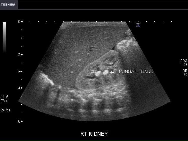 | back 113 Mycetoma (fungal ball) candidiasis is the most common Non shadowing hyperechoic mass |
front 114 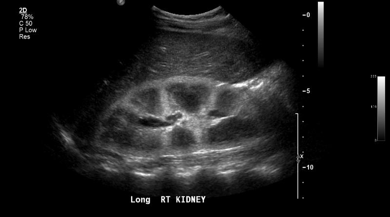 Most common cause of acute renal failure prolonged drugs or contrast agents | back 114 Acute Tubular Necrosis (ATN) |
front 115 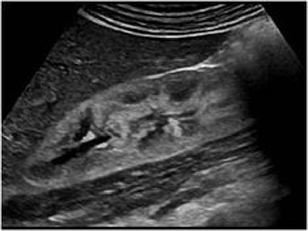 hematuria proteinuria azotemia red blood cell casts in urine | back 115 Acute Glomerulonephritis gomerular damage caused by autoimmune infection toxins |
front 116  anagesic abuse diabetes mellitus UTI and obstruction sickle cell CHF | back 116 Papillary Necrosis |
front 117 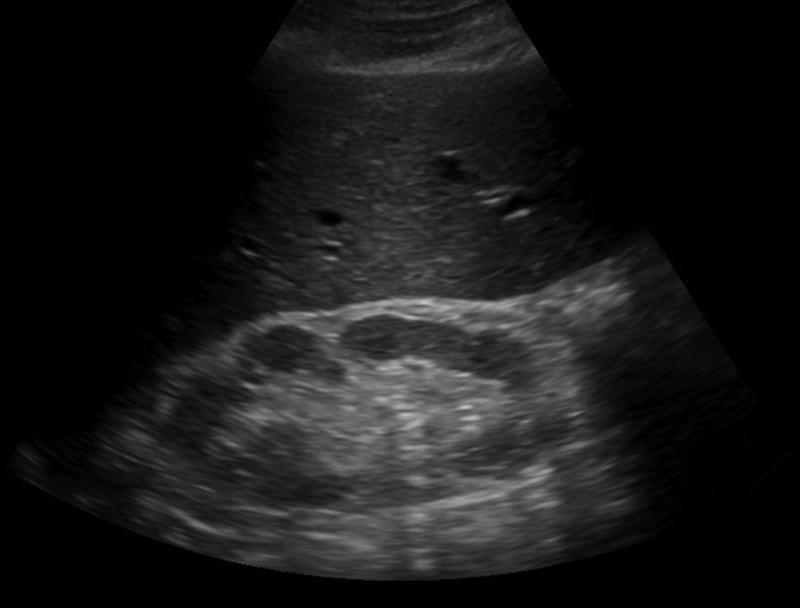 | back 117 Renal Sinus Lipomatosis |
front 118 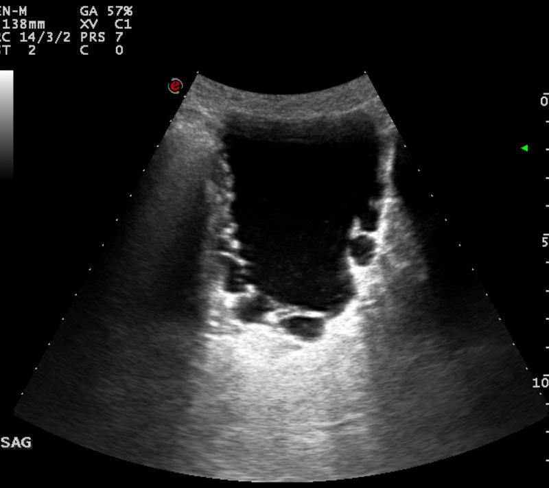 benign prostatic hypertrophy | back 118 Bladder diverticula |
front 119 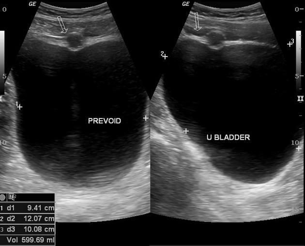 | back 119 Urachal Cyst |
front 120 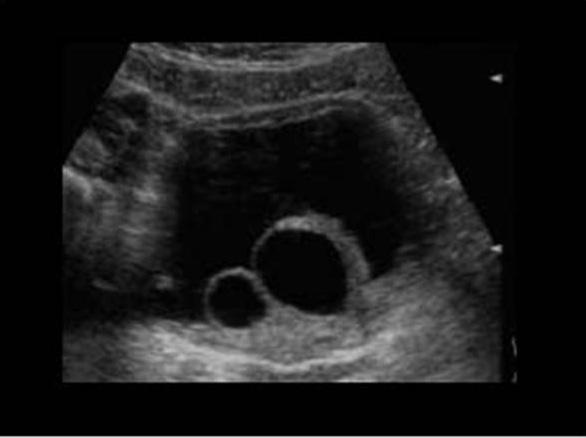 | back 120 Ureteroceles |
front 121 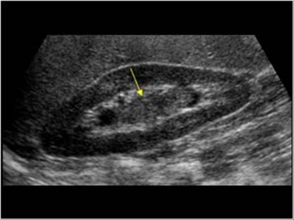 Most common bladder neoplasm hematuria hydronephrosis | back 121 Transitional Cell Carcinoma |
front 122 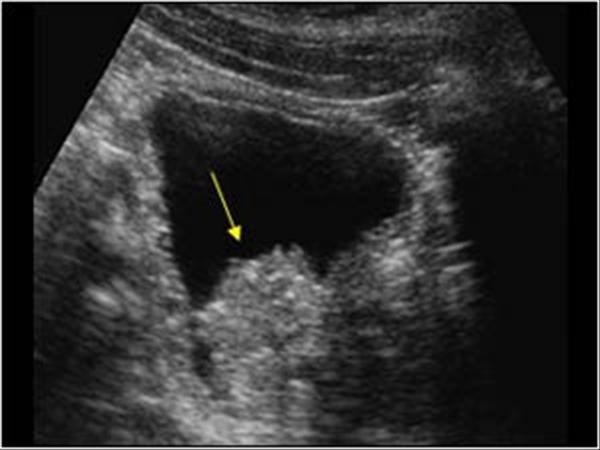 Most common bladder neoplasm hematuria hydronephrosis | back 122 Transitional Cell Carcinoma |
front 123 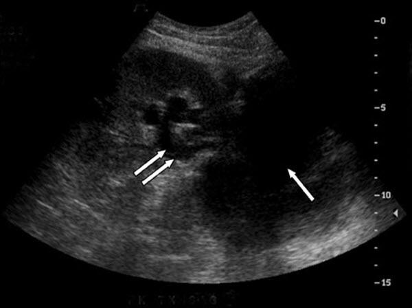 | back 123 Lymphocele |
front 124  most common germ cell tumor white male smoker infertility | back 124 Seminoma |
front 125 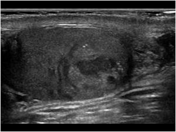 25 - 35 year olds most aggressive testicular cancer elevated beta-hcg elevated AFp | back 125 Embryonal Cell carcinoma |
front 126 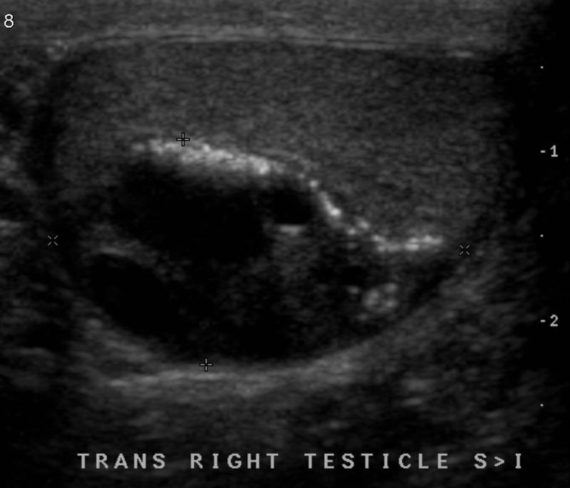 common in infants 25 - 35 year olds | back 126 Testicular Teratoma |
front 127 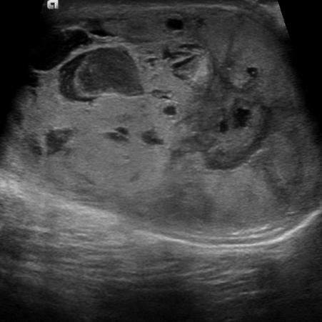 most common testicular tumor in infants and young children elevated afp | back 127 Yolk sac tumors |
front 128 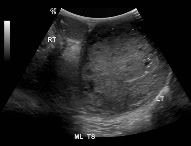 20 - 30 year old elevated beta-Hcg | back 128 Choriocarcinoma |
front 129 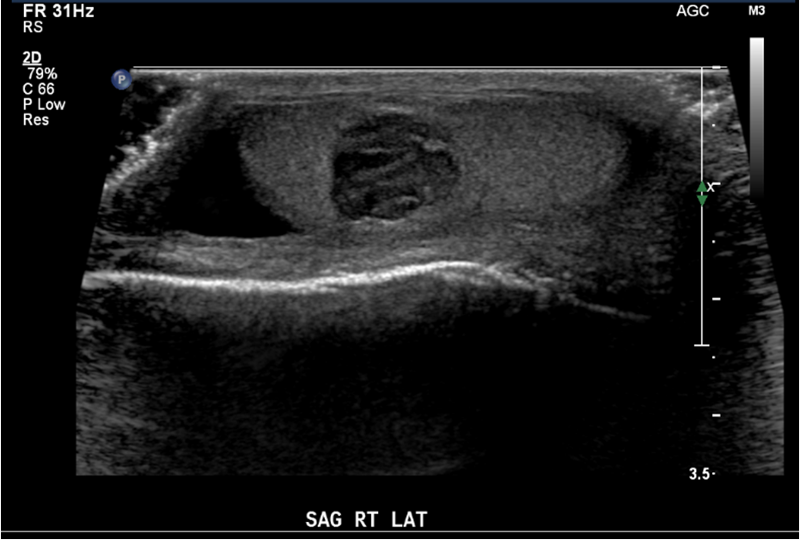 5-10 year old - benign precocious puberty feminizing features (gynecomastia) | back 129 Leydig Cell tumors |
front 130 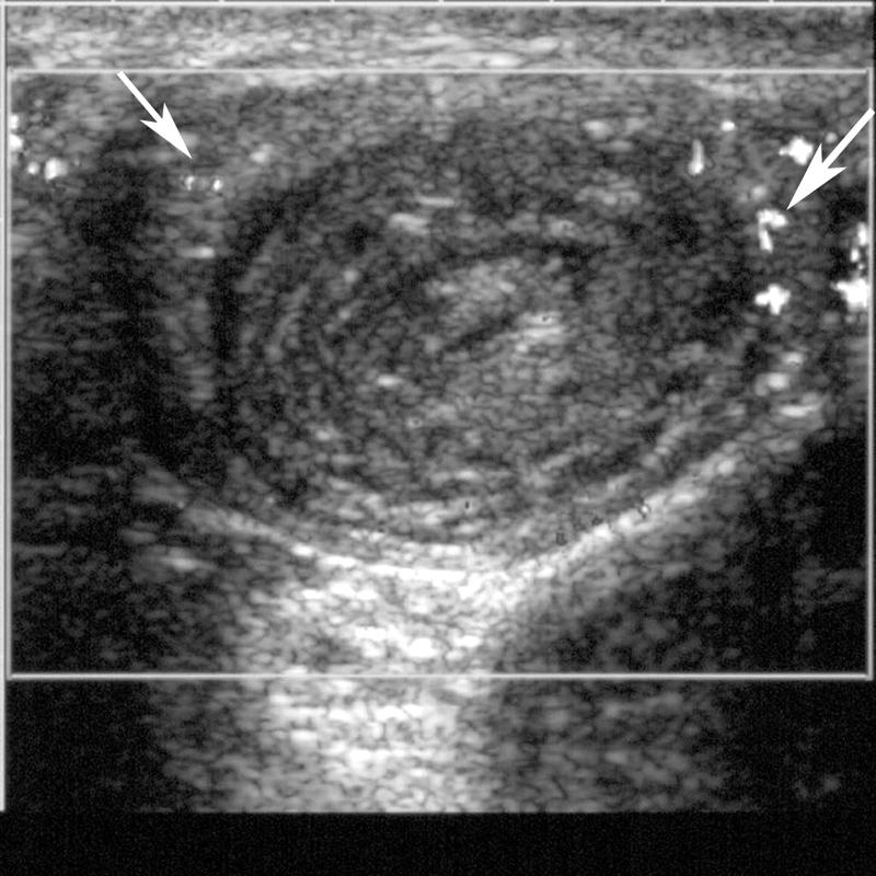 appears in 10% of population | back 130 Epidermoid Cysts benign testicular cyst |
front 131 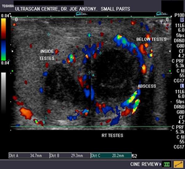 fever scrotal pain swelling untreated orchitis | back 131 Testicular abscess co |
front 132 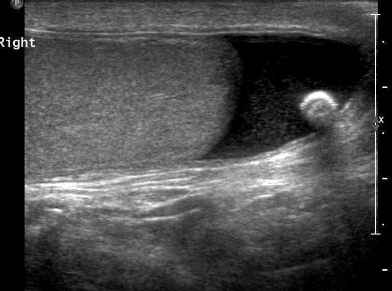 | back 132 Testiular Pearls testicular calcification |
front 133 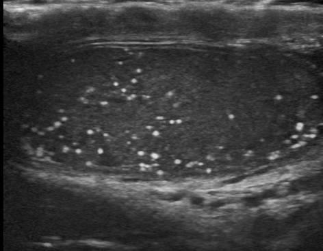 40% have neoplasm asscoiation | back 133 Microlithiasis |
front 134  trauma torsion | back 134 Testicular infarct |
front 135 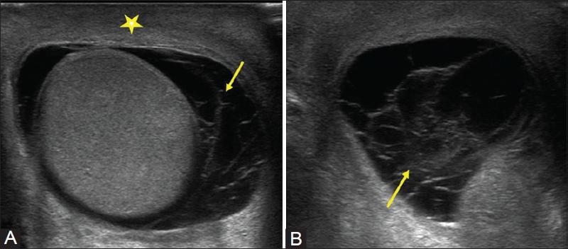 trauma pain | back 135 scrotal Hematocele |
front 136 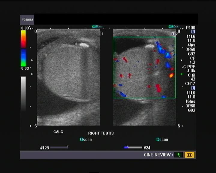 trauma pain swelling fever leukocytosis | back 136 scrotal pyocele |
front 137 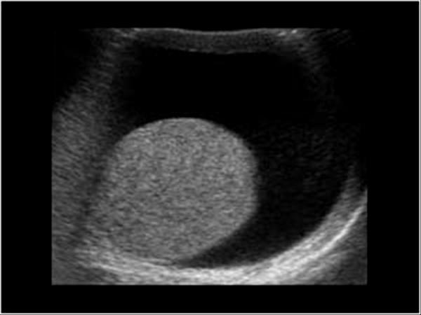 most common fluid collection of the testicle | back 137 Hydrocele serious fluid between the tunica vaginalis |
front 138  left side most common correctible cause of male infertility | back 138 Varicocele |
front 139 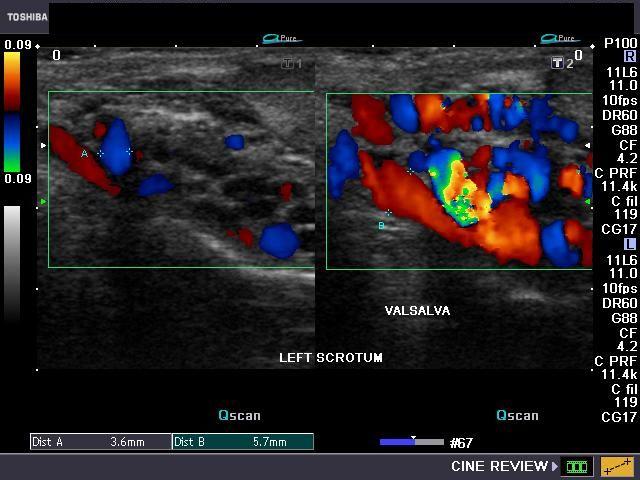 left side most common correctible cause of male infertility | back 139 Varicocele |
front 140 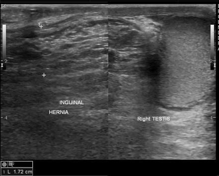 swollen scrotum persistent or intermittent palpable mass abdominal pain blood in stool | back 140 Scrotal Hernia |
front 141 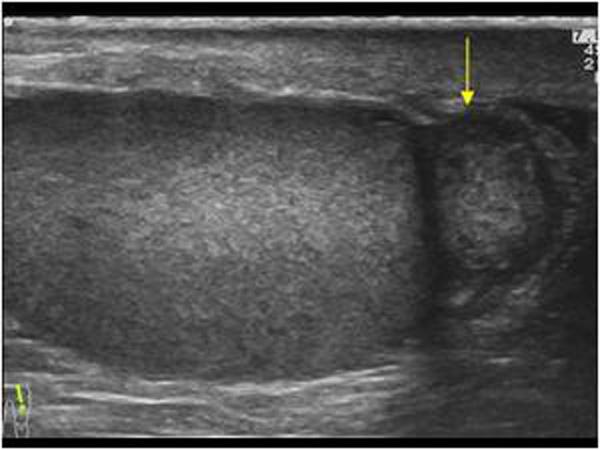 most common extratesticular tumor | back 141 adenomatoid tumor |
front 142 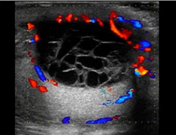 | back 142 Spermatoceles AKA Epididymal Cysts result of dilation of the epididymal tubules |
front 143 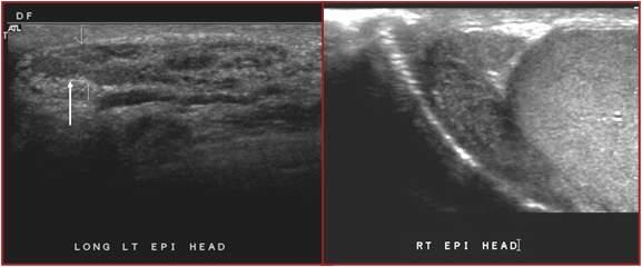 most common condition that causes scrotal pain possible fever pyuria STD UTI | back 143 Epididymitis inflammation of epididymis usually due to UTI |
front 144 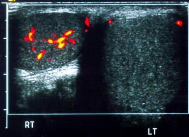 pain usually during rest or sleep N & V | back 144 torsion less than 6 hours 80% + salvage 6 - 12 hours 70% salvage 12 + hours :( |
front 145 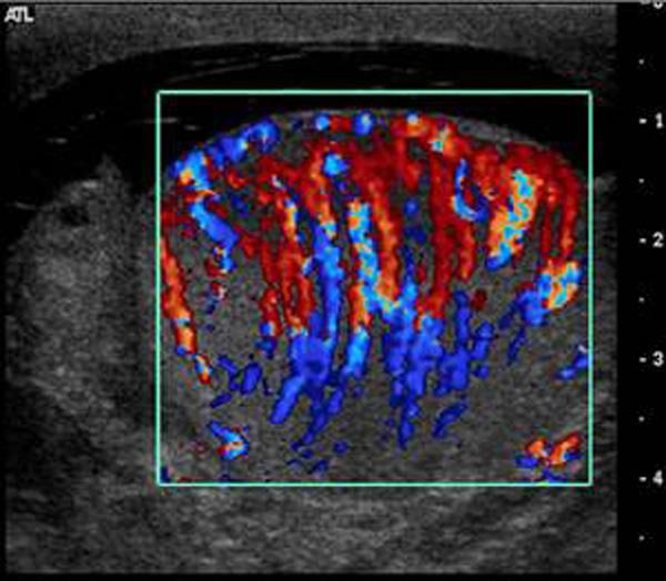 most common cause of mender 35 is chlamidia pain elevated WBC | back 145 Orchitis |
front 146 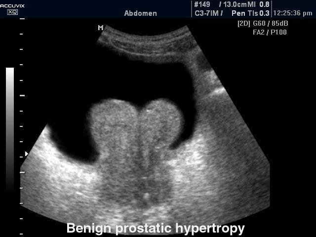 50+ year old man difficult voiding urinary frequency small stream | back 146 Benign Prostatic Hyperplasia usually in transitional zones |
front 147 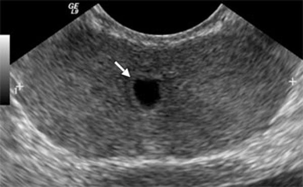 | back 147 Mullerian Duct Cyst usually midline |
front 148 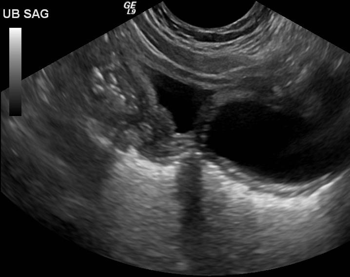 | back 148 Utricle Cysts usually midline |
front 149  | back 149 Retention cyst usually lateral |
front 150 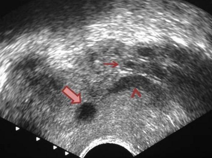 | back 150 Ejaculatory duct cyst Usually lateral & central |
front 151  pain rectal and prostate tenderness Fever | back 151 Prostatitis |
front 152 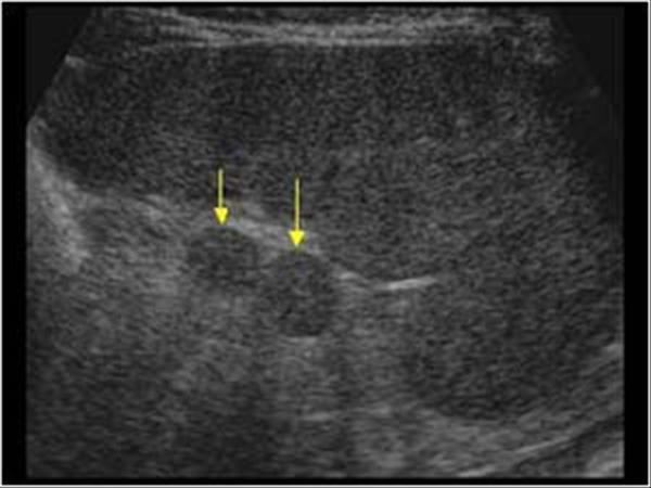 | back 152 Accessory Spleen |
front 153 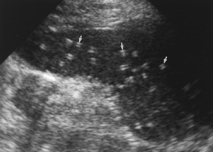 | back 153 Splenic granulomas focal lesions resulting from previous infection |
front 154 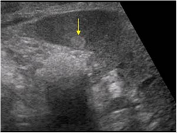 | back 154 Hemangioma |
front 155 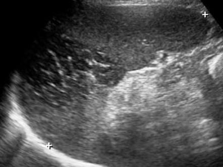 | back 155 Splenic infarct common in patients with bacterial endocarditis and splenic artery aneurysms |
front 156 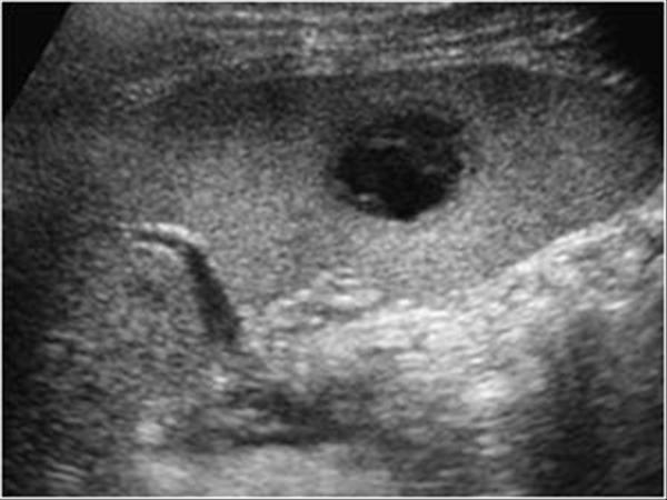 general abdominal sepsis | back 156 Splenic Abscess |
front 157 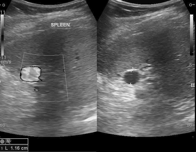 | back 157 Splenic Artery Aneurysm |
front 158 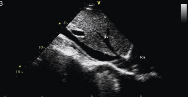 | back 158 Crus of the diaphragm |
front 159 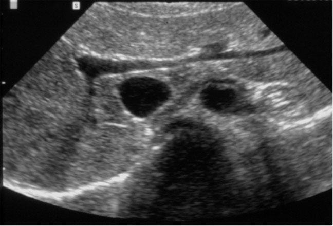 | back 159 Crus of the diaphragm |
front 160 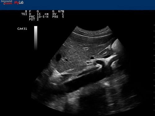 | back 160 Crus of the diaphragm |
front 161 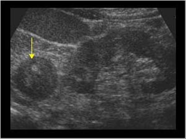 asymptomatic cushing's conn's | back 161 Adreanal Adenoma |
front 162 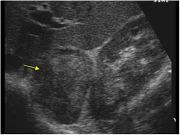 cushing's | back 162 Adrenal Cortical Carcinoma |
front 163 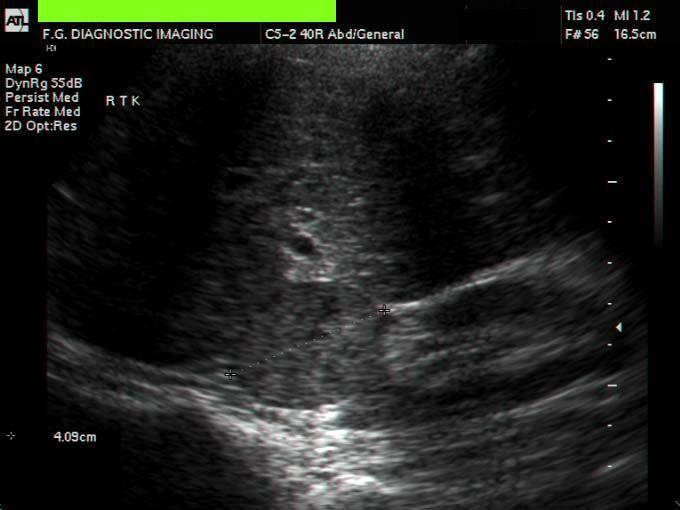 hypertension headache palpitations tachycardia anxiety excessive persperation | back 163 Pheochromocytoma tumor arising from the adrenal medusa |
front 164 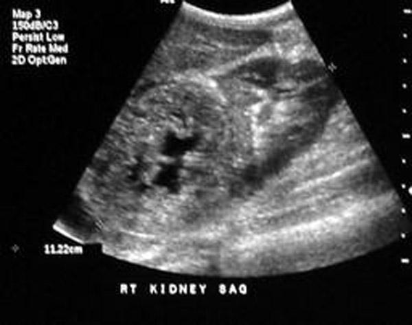 most common childhood adrenal mass | back 164 Adrenal Neuroblastoma |
front 165 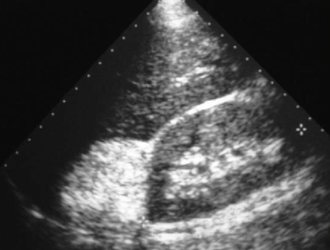 | back 165 Myelolipoma benign, nonfunctioning adrenal masses that contain fat |
front 166 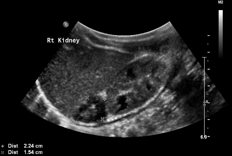 large neonate difficult birth | back 166 Adrenal Hemorrahage |
front 167 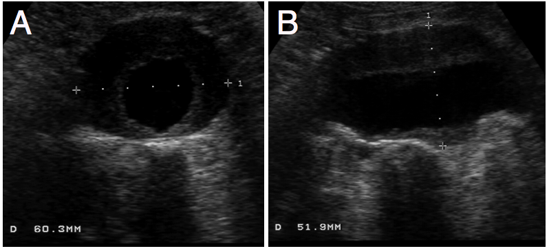 Caucasian male 75 year old hypertension abdominal, back and leg pain palpable abdominal mass | back 167 Fusiform aneurysms |
front 168 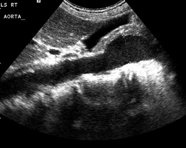 | back 168 Sacular aneurysms rare |
front 169 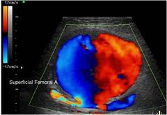 | back 169 Pseudoaneurysm |
front 170 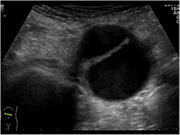 tearing back pain shock headache abdominal pain | back 170 Aortic Dissection |
front 171 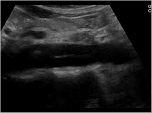 tearing back pain | back 171 Aortic Dissection |
front 172 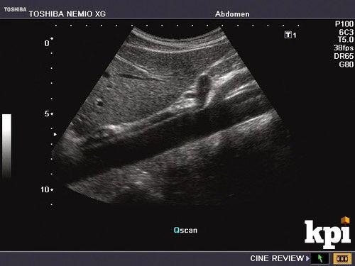 | back 172 Gastoesophageal Junction |
front 173 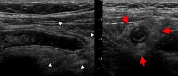 Pain over McBurney's point umbilical pain shifting to RLQ loss of appetite leukocytosis rebound tenderness | back 173 Acute appendicitis |
front 174 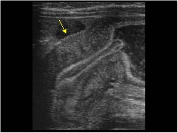 3 - 6 months old projectile vomiting palpable olive shaped abdominal mass | back 174 Hypertrophic Pyoric Stenosis |
front 175 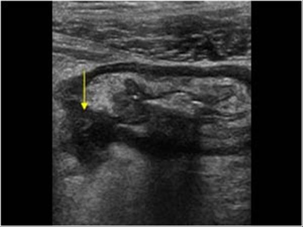 Fever leukocytosis LLQ pain | back 175 Diverticulitis |
front 176 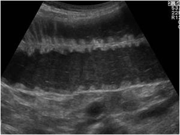 abdominal distension pain vomiting hypotension leukocytosis | back 176 Small bowel Obstruction |
front 177 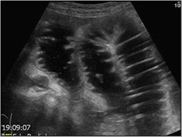 | back 177 Colon obstruction |
front 178  vomiting abdominal pain rectal bleeding | back 178 Intussception |
front 179 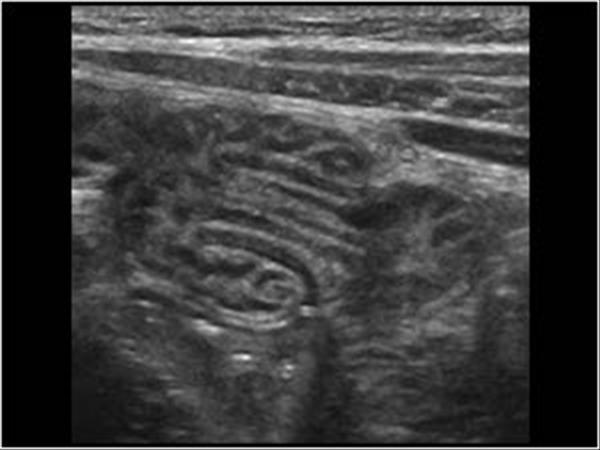 vomiting | back 179 Intussception |
front 180 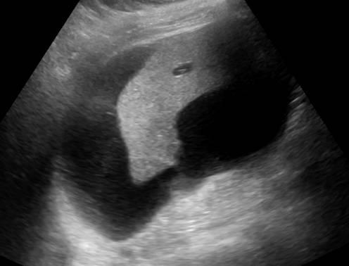 | back 180 Biloma |
front 181 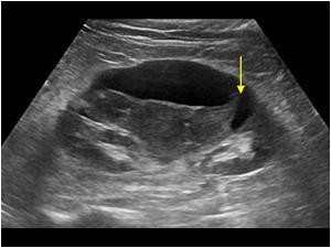 renal trauma renal transplant | back 181 Urinoma |
front 182 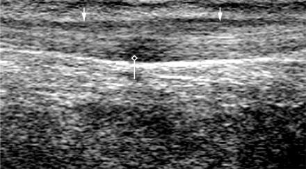 failed thompson test | back 182 Achilles Tendon Rupture |
front 183 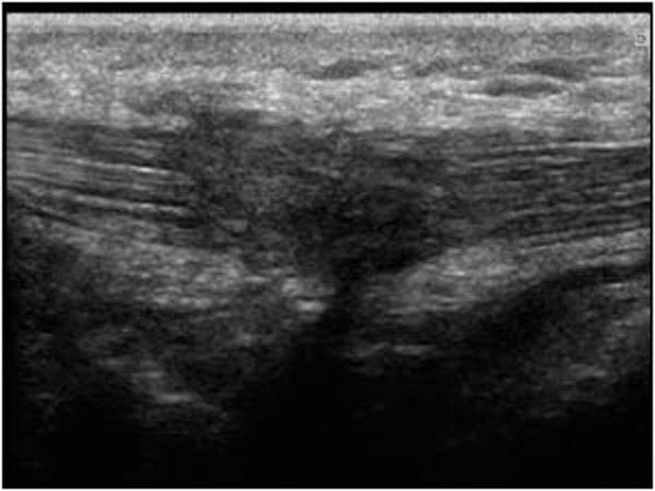 inability to extend knee | back 183 Patellar tendon Rupture |
front 184 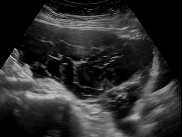 matted bowel loops malignant ascites | back 184 Pseudomyxoma Peritonel |
front 185 no data | back 185 Hematoma |
front 186 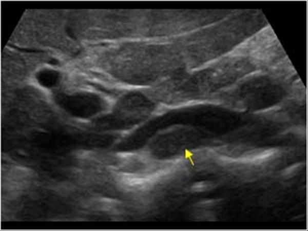 sandwich sign | back 186 Lymphadenopathy |
front 187  carpet layer | back 187 Baker's Cyst |
front 188  2nd most common tumor of the hand and wrist | back 188 Giant Cell tumor (ganglion cyst) |
front 189 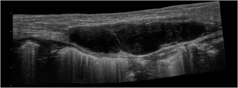 | back 189 Rectus Sheath Hematoma |