Instructions for Side by Side Printing
- Print the notecards
- Fold each page in half along the solid vertical line
- Cut out the notecards by cutting along each horizontal dotted line
- Optional: Glue, tape or staple the ends of each notecard together
Exercise 10: The Axial Skeleton
front 1 Frontal | back 1 Forehead bone |
front 2 Zygomatic | back 2 Cheekbone |
front 3 Mandible | back 3 Lower jaw bone |
front 4 Nasals | back 4 Bridge of nose |
front 5 Palatines | back 5 Posterior part of hard plate |
front 6 Parietals | back 6 Much of the lateral and superior cranium |
front 7 Occipital | back 7 Most posterior part of cranium |
front 8 Sphenoid | back 8 Single, irregular, bat- shaped bone, forming part of the cranial floor |
front 9 Lacrimals | back 9 Tiny bones, bearing tear ducts |
front 10 Maxillae | back 10 Anterior part of hard plate |
front 11 Ethmoid | back 11 Superior and middle nasal conchae formed from its projections |
front 12 Temporals | back 12 Site of mastoid process |
front 13 Sphenoid | back 13 Site of sella turcica |
front 14 Ethmoid | back 14 Site of cribriform plate |
front 15 Mandible | back 15 Site of mental foremen |
front 16 Temporals | back 16 Site of styloid process |
front 17 Ethmoid, Frontal, Maxillae, Sphenoid | back 17 Four bones, containing paranasal sinuses |
front 18 Occipital | back 18 condyles articulate with the atlas |
front 19 Occipital | back 19 Foramen magnum contained here |
front 20 hyoid | back 20 small U-shaped bone in neck, where many tongue muscles attach |
front 21 Temporals | back 21 Middle ear found here |
front 22 Vomer | back 22 Nasal septum |
front 23 Ethmoid | back 23 Bears an upward protrusion, the " cock's comb", or crista galli |
front 24 mandible, maxilla | back 24 contain alveoli bearing teeth |
front 25 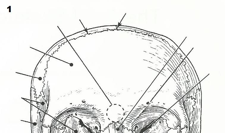 | back 25  |
front 26 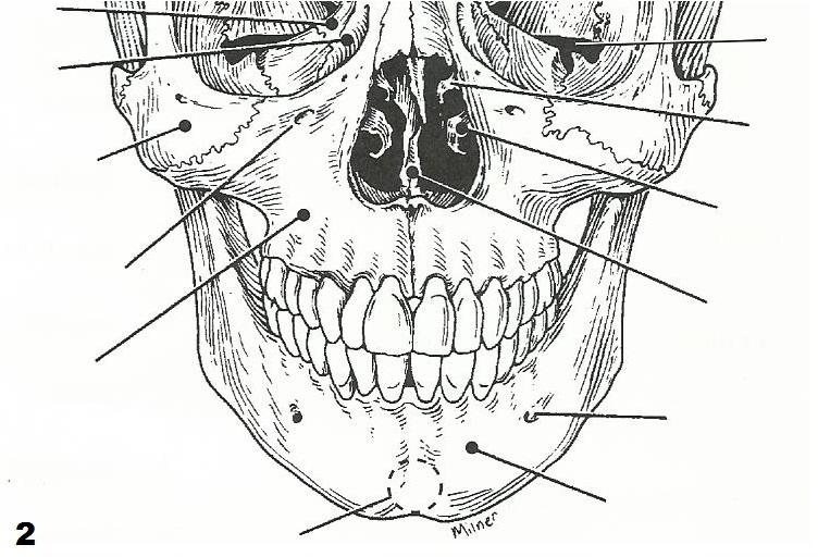 | back 26  |
front 27 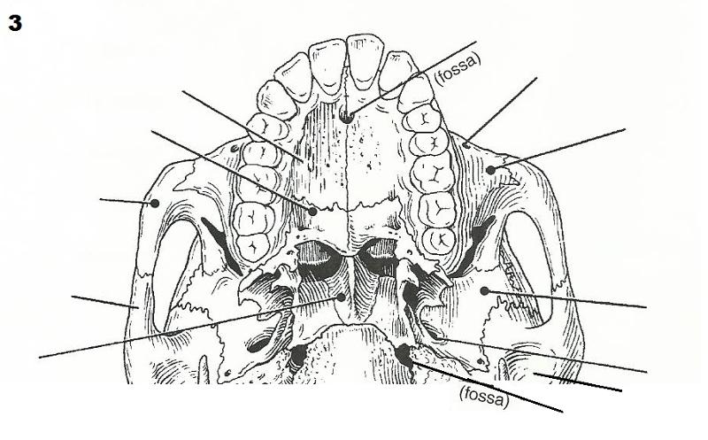 | back 27 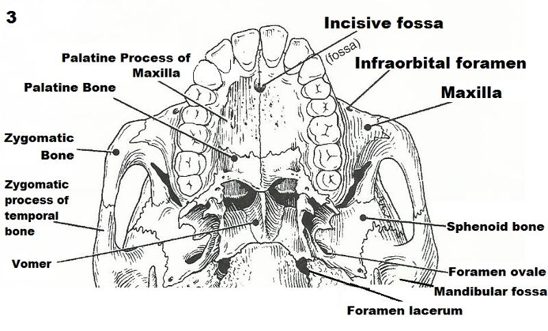 |
front 28 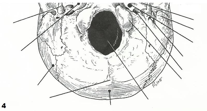 | back 28 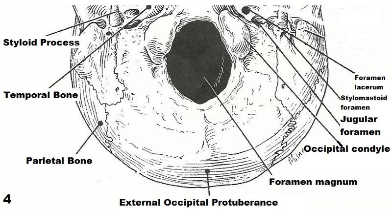 |
front 29 Define suture | back 29 All but one of the bones of the skull are joined by interlocking joints. |
front 30 With one exception, the skull bones are joined by sutures. Name the exception. | back 30 With the exception of 2 paired bones (the parietal and temporal), are all single bones. |
front 31 What bones are connected by the lambdoid suture? | back 31 connects the parietal and temporal bones with the occipital bone |
front 32 What bones are connected by the squamous suture? | back 32 temporal and parietal bones on each side of the skull. |
front 33 Name the eight bones composing the cranium. | back 33 frontal bone, 2 parietal bones, 2 temporal bones, occipital bone, sphenoid, ethmoid |
front 34 Give two possible functions of the sinuses: | back 34 They lighten the facial bones and act as resonance chambers for speech. |
front 35 What is the orbit? | back 35 Eye Socket |
front 36 What bones contribute to the formation of the orbit? | back 36 Frontal bone, maxilla, lacrimal, ethnoid, sphenoid, palatine, zygomatic. |
front 37 Why can the sphenoid bone be called the keystone of the cranial floor? | back 37 Since it is in contact with all of the other cranial bones. |
front 38 cervical vertebra - typical | back 38 vertebral type containing foramina in the transverse processes, through which the vertebral arteries ascend to reach the brain |
front 39 axis | back 39 dens here provides a pivot for rotation of the first cervical vertebra (C1) |
front 40 thoracic vertebra | back 40 transverse processes faceted for articulation with ribs, spinous process pointing sharply downward |
front 41 sacrum | back 41 composite bone, articulates with the hip bone laterally |
front 42 lumbar vertebra | back 42 massive vertebrae, weight sustaining |
front 43 coccyx | back 43 "tail bone:; vestigial fused vertebrae |
front 44 atlas | back 44 supports the head; allows a rocking motion in the conjunction with the occipital condyles |
front 45 vertebral foramen | back 45 cavity enclosing the spinal cord |
front 46 body | back 46 weight bearing portion of the vertebra |
front 47 spinous process & transverse process | back 47 provide levers against which muscles pull |
front 48 body & transverse process | back 48 provides an articulation point for the ribs |
front 49 intervertebral foramina | back 49 openings providing for exit of spinal nerves |
front 50 body & vertebral arch | back 50 structures that form an enclosure for the spinal cord |
front 51 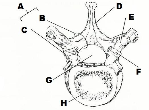 | back 51 A. INTERVERTEBRAL FORAMINA
|
front 52 Describe how a spinal nerve exits from the vertebral column. | back 52 Spinal nerves ( motor axons) exit the vertebral column via the ventral root (where they synapse on motor neuron ganglia ) then the ventral horn. Sensory nerves enter the spinal cord via the dorsal horn, synapse on the dorsal ganglia and enter the spinal cord. |
front 53 name two factors/structures that permit flexibility of the vertebral column | back 53 discs and the S-shaped of the vertebral column prevent shock to the head in walking and running and provide flexibility to the body trunk |
front 54 What kind of tissue compose the intervertebral discs? | back 54 fibrocartilage |
front 55 What is a herniated disc? What problems might it cause? | back 55 a disc in which the nucleus puposus herniates through the annulus;
|
front 56 Which two spinal curvatures are observed at birth? | back 56 The two primary curvatures that we're born with are the concave forward curvatures in the thoracic and sacral spines. |
front 57 Under what conditions do the secondary curvatures develop? | back 57 The "secondary" curvatures, the compensatory curvatures, occur with normal development. (Normal development is the condition under which they occur) These are the cervical curvature, which develops first with infant head lifting and the lumbar curvature, which develops next sitting up. These curvatures prepare the spine for ambulation. |
front 58 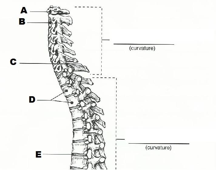 | back 58 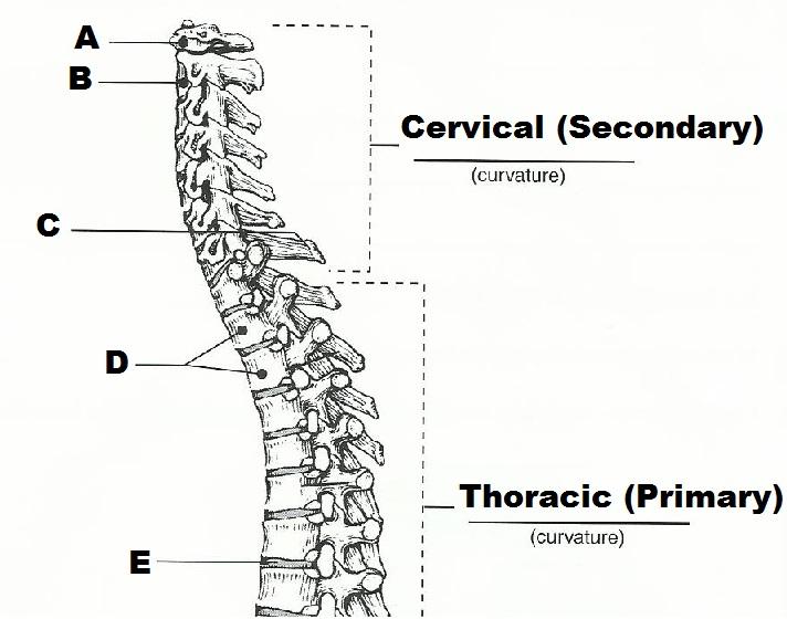 A. ATLAS
|
front 59 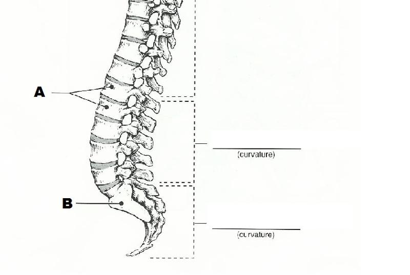 | back 59 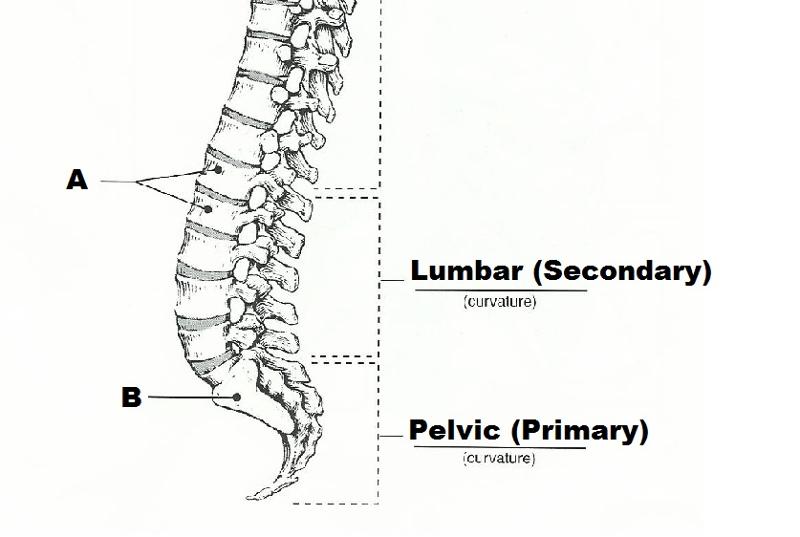 A. TWO LUMBAR VERTEBRAE
|
front 60 The major bony components of the thorax (excluding the vertebral column) are the ___________ and the _______________. | back 60 ribs and sternum |
front 61 Differentiate between a true rib and a false rib. | back 61 a true rib is attached to cartilage that directly articulates with the sternum |
front 62 Is a floating rib a true or false rib? | back 62 A free floating rib is neither a true or false rib. Ribs 1-7 are considered "true" ribs because they are directly attached to the sternum by individual coastal cartilages. Ribs 8-10 are considered "false" ribs because they are indirectly attached to the sternum by a common coastal cartilage. Ribs 11 and 12 are considered "free floating" because they are neither directly or indirectly attached to the sternum. Instead, they end in posterior abdominal musculature. They are still capped with cartilage though |
front 63 What is the general shape of the thoracic cage? | back 63 cone-shaped |
front 64 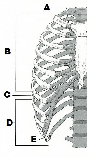 | back 64 A. COSTAL CARTILAGE
|
front 65 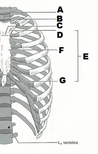 | back 65 A. JUGULAR NOTCH
|