Instructions for Side by Side Printing
- Print the notecards
- Fold each page in half along the solid vertical line
- Cut out the notecards by cutting along each horizontal dotted line
- Optional: Glue, tape or staple the ends of each notecard together
A&P CH23 Digestive System
front 1 digestion | back 1 The process by which food is converted into nutrient molecules which can be absorbed and assimilated by the body; it is accomplished in the digestive system by the mechanical and enzymatic breakdown of foods into simpler chemical compounds. |
front 2 absorption | back 2 The process by which the molecules of growth and nutrition are taken in across a membrane and conveyed to cells, tissues and organs; the movement is accomplished by a variety of molecular processes including diffusion, osmosis, passive and active transport. [Note: From the perspective of chemistry: a process in which one substance permeates another; a fluid permeates or is dissolved by a liquid or solid. This is a part of physiological absorption.] |
front 3 digestive system | back 3 The gastrointestinal tract = alimentary canal (mouth pharynx, esophagus, stomach, small intestine, large intestine) and the accessory digestive structures (lips, cheeks, tongue, teeth) and glands (salivary glands, pancreas, liver) regarded as an integrated system responsible for the ingestion, mechanical and chemical breakdown, and absorption of foods. |
front 4 gastrointestinal tract = GI tract = alimentary canal | back 4 The mucous membrane-and smooth muscle- lined tube of the digestive system through which food passes, in which digestion takes place, and from which wastes are eliminated; it extends from the mouth to the anus and includes the pharynx, esophagus, stomach, and small and large intestines. aka - digestive tract, gut tube. |
front 5 accessory structures | back 5 The additional connected parts which contribute to the main function of a major organ or system; in the gastrointestinal tract, the mouth (lips, cheeks, salivary glands, tongue, teeth) and the small intestine (pancreas, liver) are associated with such collateral components. |
front 6 ingestion | back 6 The voluntary process of taking foods or liquids into the body by means of the mouth for digestion or absorption. aka - eating. |
front 7 secretion | back 7 The physiological process of synthesizing and releasing some functionally specialized substance (especially one which is not a waste) from a gland or cell; at the molecular level, a variety of processes are involved including diffusion, osmosis, passive and active transport mechanisms or exocytosis. |
front 8 mixing | back 8 The mechanical physiological process of combining or blending diverse ingredients or components into one mass or mixture so that constituent parts or elements are diffused among each other and coalesced. |
front 9 propulsion | back 9 The process of driving or propelling something forward or away; in the gastrointestinal tract, food is moved by deglutition (swallowing) and peristalsis. |
front 10 motility | back 10 Having the power to move; in the gastrointestinal tract, propulsive movement is powered by muscular contractions (deglutition = swallowing and peristalsis). |
front 11 mechanical digestion | back 11 The muscular process by which solid food is broken down into smaller particles by the chewing action of the jaws, teeth and tongue in the oral cavity and by the mixing waves created in the stomach; at the same time the food is softened and moistened by the digestive juices (saliva in the mouth, gastric juice in the stomach); reduction in the size of ingested food particles increases the surface area of the particles available to be acted upon by digestive enzymes; it does not alter the molecular structure of the nutrient molecules. |
front 12 chemical digestion | back 12 The physiological process by which food = nutrient molecules have their molecular structure modified by interacting with substances secreted by various digestive glands and tissues; in general, nutrient molecules are broken down into smaller constituent molecules which will be more easily absorbed in the intestines. [Note: There is a limited amount of indiscriminant acid hydrolysis of nutrients in the stomach due to the action of gastric HCl; carbohydrates are split into di- and mono-saccharides by salivary amylase in the mouth and pancreatic amylase in the duodenum and disaccharides are split into monosaccharides by disaccharidases (brush border enzymes) in the duodenum; proteins are split into smaller peptides and ultimately into individual amino acids by pepsin in the stomach and pancreatic proteases (trypsin, chymotrypsin, carboxypeptidase) in the duodenum; lipids are split into glycerol and fatty acids by lingual lipase in the mouth and pancreatic lipase and phospholipase in the duodenum; nucleic acids are split into nucleotides by pancreatic nucleases and nucleotides are split into smaller constituents by nucleotidases (brush border enzymes) in the duodenum.] |
front 13 defecation | back 13 The mechanical muscular process of voiding or eliminating feces from the bowels; a complex process involving the smooth muscles of the large intestine, especially those of the sigmoid colon, rectum and anal canal; it is usually initiated consciously but coordinated by the autonomic nervous system. |
front 14 List: the six processes of Digestion. | back 14 (1) Ingestion (2) Propulsion {peristalsis} (3) Mechanical Digestion (4) Chemical Digestion (5) Absorption (6) Elimination {defecation}
|
front 15 List: the organs of the GI tract in the sequence in which they occur starting with the mouth and ending at the anus (include the regions of each organ, ex: oropharynx and laryngopharynx of pharynx). | back 15 1.Mouth vestibule - buccal cavity - oral cavity
|
front 16 List:the hormones, digestive enzymes, and other chemicals related to digestion secreted by the salivary glands, stomach, small intestine, liver/gall bladder, and pancreas. | back 16 1.Salivary Glands none salivary amylase bicarbonate ions, mucin
|
front 17 List:several physical or mechanical processes of digestion. | back 17 mastication (chewing) - bolus formation - deglutition (swallowing) - peristalsis (propulsion) - gastric churning - intestinal segmentation - haustral churning (compaction) - mass peristaslis - defecation |
front 18 List:the sphincters of the GI tract in the sequence they occur, starting with the mouth and ending at the anus. | back 18 upper esophageal sphincter → lower esophageal sphincter = cardiac sphincter → pyloric sphincter → ileo-cecal valve → internal anal sphincter →
|
front 19 Describe: carbohydrates (starch), proteins, nucleic acids, and lipids. (Hint: You may use summary chemical equations to do this). | back 19 1.carbohydrates (starch) salivary amylase begins starch hydrolysis in the mouth; pancreatic amylase continues starch hydrolysis in the duodenum; intestinal brush border dextrinase and disaccharidases complete the reduction of starch to monosaccharides which can be absorbed
|
front 20 Describe:the secretion and reabsorption of the water involved in digestion. Indicate the specific gastrointestinal organs involved in the secretion or reabsorption of the water involved in digestion. | back 20 *Secretion of The Water Involved In Digestion*
|
front 21 Describe:the type of movement associated with each organ of the digestive (gut) tube. | back 21 1.Mouth mastication (chewing) - bolus formation - deglutition (swallowing)
|
front 22 periodontal disease | back 22 Any inflammatory disease which attacks the gingiva (gums) and the alveolar bone, especially around the tooth sockets (alveoli); it is usually caused by bacteria growing at the base of the teeth; mild inflammation is termed gingivitis while more severe disease with alveolar bone loss, loosening of teeth and pronounced inflammation inflammation is termed periodontitis; it is more often a disease of adults after the period of greatest risk for tooth decay which is during the first two decades of life. |
front 23 root canal | back 23 1.A pulp-filled channel within the dentine of a tooth which is the passage in the root of a tooth through which its nerve and blood vessels enter the pulp cavity.
|
front 24 orthodontics | back 24 The dental specialty and practice of preventing and correcting irregularities of the teeth, e.g., by the use of removable appliances or fixed adjustable braces. |
front 25 mumps | back 25 An acute, inflammatory, contagious disease caused by a paramyxovirus and characterized by swelling of the salivary glands, especially the parotids, and sometimes of the pancreas, ovaries, or testes; this disease, mainly affecting children, can be prevented by vaccination (MMR). Mumps can affect one or both sides of the body. |
front 26 Image: mumps | back 26 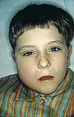 |
front 27 Image: mumps | back 27  |
front 28 gastroenterology | back 28 The branch of medicine dealing with the study, diagnosis and treatment of disorders affecting the stomach, intestines, and accessory organs (liver, gall bladder, pancreas). |
front 29 proctology | back 29 The branch of medicine dealing with the diagnosis and treatment of disorders affecting the colon, rectum, and anus. |
front 30 esophageal hiatus | back 30 The snug opening in the mid-dorsal edge of the diaphragm which is the passage for the esophagus as it passes from the thoracic to the abdominal cavity. |
front 31 hiatal hernia | back 31 A protrusion in which an anatomical part (e.g., the stomach) protrudes through the esophageal hiatus of the diaphragm. |
front 32 Diagram: hiatal hernia | back 32 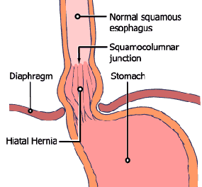 |
front 33 achalasia | back 33 The failure of a ring of smooth muscle fibers, e.g., the lower esophageal = cardiac sphincter of the esophagus, to relax; symptoms include difficulty in swallowing, chest pain, vomiting and heart burn; treatments include surgical dilation and various medications. |
front 34 heartburn | back 34 Indigestion and a hot, fiery sensation, usually centered in the middle of the chest near the sternum, caused by the reflux of acidic stomach fluids which enter, and can potentially erode, the lower end of the esophagus; often associated with nausea and vomiting. aka - acid reflux, cardialgia, pyrosis |
front 35 vomiting | back 35 The act or an instance of ejecting part or all of the contents of the stomach through the mouth, usually in a series of involuntary spasmic movements; regulated by the vomiting center in the medulla oblongata. aka - regurgitation, emesis |
front 36 Diagram: Vomitting | back 36 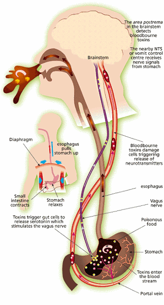 |
front 37 ascites | back 37 An abnormal accumulation of serous, peritoneal fluid in the abdominal cavity, usually caused by liver disease (~80% of cases) and exhibiting reduced synthesis of albumin and other oncotic plasma proteins (proteins which contribute to the osmotic = oncotic pressure of the blood), and less often caused by heart or kidney diseases or pancreatitis. |
front 38 Image: ascites | back 38 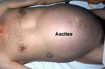 |
front 39 Image: ascites | back 39 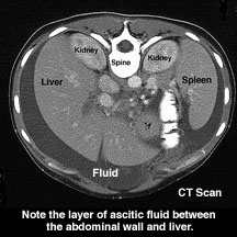 Cirrhosis of the liver
|
front 40 peritonitis | back 40 inflammation of the peritoneum and abdominal cavity; the peritoneal fluid may become mixed with serum, fibrin, inflammatory cells, microorganisms and pus; symptoms include abdominal pain, tenderness, constipation, vomiting and moderate fever. aka - acute abdomen |
front 41 Diagram:sectional view of layers of GI tract | back 41 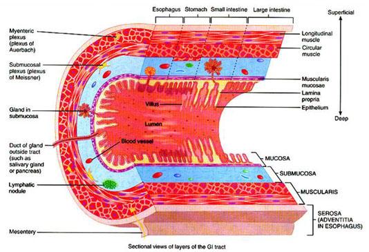 |
front 42 Image: miscoscopic view of GI histology | back 42 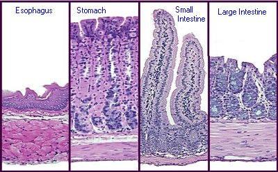 |
front 43 mucous membranes = mucosae | back 43 The specialized epithelial lining which produces a lubricating fluid containing the sticky protein mucin which traps microbes and dirt particles and protects any body structure which is continuous with the external environment except for the skin itself. |
front 44 epithelium | back 44 Tissues with a high degree of cellularity and limited extracellular material, connected with specialized contact structures such as desmsomes, often organized in layers, which have no direct blood supply and which are derived from embryonic ectoderm and endoderm. |
front 45 lamina propria | back 45 The supportive areolar = loose fibrous connective tissue layer found immediately beneath the mucosal epithelium and its basement membrane and the muscularis mucosae. |
front 46 muscularis mucosae | back 46 The thin layer of smooth muscle which forms the boundary between the lamina propria of the mucosa and the submucosa in the walls of the gastrointestinal tract; contraction of this layer of smooth muscle pushes the mucosal membrane into small folds which increase the surface area for digestion and absorption of nutrients. |
front 47 enteroendocrine cell | back 47 Any of a group of diffuse neuroendocrine cells,.also referred to as APUD cells (amine precursor uptake and decarboxylation cells), scattered individually among the cells of the exocrine glands from the stomach to the colon; their amine hormones diffuse into the blood capillaries, and ultimately influence several digestive system target organs; these cells, along with the nervous system, control and coordinate the muscular and secretory activities of the gastrointestinal tract; enteroendocrine cells typically extend a cytoplasmic process into the digestive tract lumen forming an important communication site to monitor the luminal contents, of which dietary amino acids are the most important stimulants; there are more than a dozen different types of enteroendocrine cell and a given enteroendocrine cell will only secrete one type of hormone, or hormone-like substance; these include gastrin, histamine, endorphins, serotonin, cholecystokinin and somatostatin. |
front 48 MALT = mucosa-associated lymphatic tissue | back 48 The general term used for the various aggregations of lymphoid tissue found associated with the mucous membranes of the respiratory and digestive tracts; the consist of lymphatic nodules within the connective tissues walls (in the mucosa and submucosa) of these tubular organs; these lymphocytes are involved in immune defenses. |
front 49 submucosa | back 49 The supportive loose fibrous connective tissue layer found immediately beneath the mucosal epithelium and its basement membrane; in the gastrointestinal tract it lies between the inner muscularis mucosae and the outer muscularis layer; it is the location for networks of nerves, blood vessels, lymphatic vessels, and lymphatic tissue and may be the location for mucous secreting glands. |
front 50 neural plexus | back 50 The interlacing networks of nerve fibers which originate from certain cranial nerves, e.g., the Vagus (X) and the ventral rami of spinal nerves in which individual autonomic nerve processes from the brain stem and individual spinal segments become redistributed to that (1) each resulting branch of the plexus contains fibers from several cranial and spinal nerves and (2) fibers from each ventral ramus travel to the peripheral tissues of the body by several routes; this arrangement permits portion of the digestive tube to be innervated by autonomic motor (afferent) fibers from more than one spinal segment -- as a result, an injury to a single spinal segment or spinal motor (ventral) root cannot completely paralyze any organ. |
front 51 submucosal plexus | back 51 The interlacing networks of unmyelinated nerve fibers derived chiefly from the superior mesenteric plexus which is located within the submucosa of the gastrointestinal tract, especially in the intestines; it consists chiefly of visceral sensory fibers and postganglionic autonomic motor fibers; it is concerned with the control of functions within the inner walls of each gut segment, i.e., local absorption, secretion, and contraction. |
front 52 myenteric plexus | back 52 The interlacing networks of unmyelinated nerve fibers derived chiefly from the superior mesenteric plexus which is located within the muscularis of the gastrointestinal tract (between the inner circular and outer longitudinal layers); it consists chiefly of postganglionic autonomic motor fibers; it is concerned with the control of smooth muscle tone, peristalsis, and the relaxation of the pyloric and ileocecal sphincters. |
front 53 muscularis | back 53 The muscular coat or tunic of any hollow organ or tubular structure; its contractions propel the contents of the organ elsewhere; it lies between the inner submucosa and the outer adventitia or serosa; in the gastrointestinal tract, it typically consists of an inner circular layer and an outer longitudinal layer of smooth muscle, but, in the stomach, it also contains a middle oblique layer; it is the location for plexuses of postganglionic autonomic motor fibers concerned with the control of enteric smooth muscle tone, mixing and peristalsis. |
front 54 serous membrane = serosa | back 54 A simple squamous lining found in the various ventral body cavities which lines the walls of the cavities and the organs contained therein and produces a watery serous fluid which lubricates the lining surfaces. |
front 55 List:the type of epithelium lining each part of the digestive (gut) tube and explain why this epithelium promotes the functions of its respective part. | back 55 1.Esophagus nonkeratinized stratified squamous epithelium protective lining to limit abrasion during deglutition (swallowing)
|
front 56 Diagram and Label: a cross-section of the GI tract (= alimentary canal) showing the main tissue layers and their sublayers. List unique features of the histology of each organ (or region of each organ) of the GI tract. | back 56  1.Esophagus nonkeratinized stratified squamous epithelium; no mucosal glands, adventitia
|
front 57 peritoneal cavity = abdominal cavity | back 57 the interior of the peritoneum; a potential space between layers of the peritoneum bounded by the diaphragm above; it is lined by the parietal layer of the serous peritoneum and contains the abdominal organs (stomach, intestines, spleen, liver, pancreas, adrenal glands -- each covered by its portion of the visceral peritoneum) and the various abdominal mesenteries; it also contains a small amount of serous fluid = peritoneal fliuid. |
front 58 visceral peritoneum | back 58 The serous membrane which covers the various abdominal organs (stomach, intestines, pancreas, liver, spleen, etc.) in the abdominal cavity and produces the peritoneal fluid. |
front 59 parietal peritoneum | back 59 The serous membrane which lines the abdominal cavity which houses the various abdominal organs and produces the peritoneal fluid. |
front 60 retroperitoneal | back 60 A directional term used to describe the location of certain organs as behind or beneath the parietal peritoneum which lines the peritoneal cavity; e.g., the kidneys, ureters, urinary bladder, portions of the ascending and descending colon, etc. |
front 61 mesentery | back 61 Any of several folds of the peritoneum (fibrous connective tissue covered by the visceral peritoneum serosal membrane) which support and connect the intestines (or other abdominal organs) to the dorsal abdominal wall, especially the fold which envelops the jejunum and ileum ("the mesentery proper"); the other mesenteries being called mesocecum, mesocolon, mesorectum, etc.); the interior of a mesentery contains a rich blood supply, lymphatics and lymph nodes, and nerves. |
front 62 Diagram: mesenteries | back 62  |
front 63 mesocolon | back 63 The fold of the peritoneum (mesentery -- fibrous connective tissue covered by the visceral peritoneum serosal membrane) which supports and connects the the lower colon to the dorsal abdominal wall; the interior contains a rich blood supply, lymphatics and lymph nodes, and nerves. |
front 64 falciform ligament | back 64 The anteroposterior fold of the parietal peritoneum (fibrous connective tissue covered by the visceral peritoneum serosal membrane) attached to the under surface of the diaphragm and the sheath of the rectus muscle and along a line on the anterior and upper surfaces of the liver extending back from the notch on the anterior margin of the liver; it is a main support for the liver in the abdominal cavity. |
front 65 Image: falciform ligament | back 65 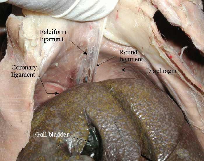 |
front 66 lesser omentum | back 66 The fold of the peritoneum (mesentery -- fibrous connective tissue covered by the visceral peritoneum serosal membrane) which supports and connects parts of the stomach and duodenum to the liver and and supports the hepatic vessels; the interior contains a rich blood supply, lymphatics and lymph nodes, and nerves. |
front 67 greater omentum | back 67 The large fold of the peritoneum (mesentery -- fibrous connective tissue covered by the visceral peritoneum serosal membrane) which supports and connects the stomach to the transverse colon; the fold is large enough to entirely cover the intestines anteriorly; the interior contains a rich blood supply, lymphatics and lymph nodes, and nerves. |
front 68 Image: lesser omentum | back 68 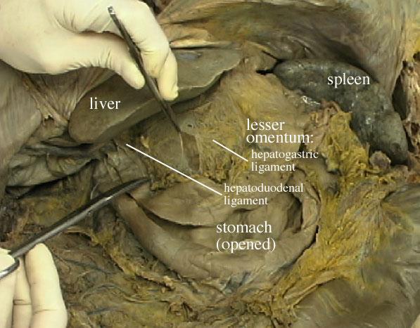 |
front 69 Image: greater omentum | back 69 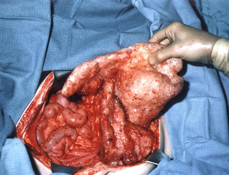 The greater omentum has been elevated away from the small bowel during a surgery to remove cancer. The surgeon's hand is holding the transverse colon. The small intestine appears most clearly to the left. [A large volume of mucinous tumors heavily coats the greater omentum and transverse colon. Each tumor appears as a small glistening raised nodule.] You can also see the three layers of abdominal muscle in the cut abdominal wall. |
front 70 Diagram: Oral Cavity | back 70 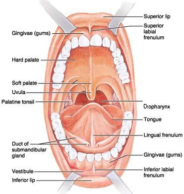 |
front 71 oral cavity | back 71 The first portion of the gastrointestinal tract, the opening through which food is taken in and vocalizations emerge, it is bounded by the lips, cheeks, palate, and tongue, and ends where it merges with the oropharynx; it is lined by a wet stratified squamous epithelium and has as accessory structures, the teeth and salivary glands. nickname - mouth. |
front 72 buccal cavity | back 72 The external portion of the oral cavity located between the jaws and teeth and the cheeks; it serves as a space to trap food while it is masticated by the chewing action of the jaws and teeth; it is lined by a wet stratified squamous epithelium. |
front 73 labia | back 73 The folds (superior and inferior) of the integument at the opening of the oral cavity and of the vulva; in the oral cavity they are identified with the red margin = vermilion border (extraoral labial cutaneous junction), where ordinary skin with hair follicles is replaced by a thin hairless keratinised stratified squamous epithelium; the help process and trap food while it is masticated by the chewing action of the jaws and teeth and assist in the articulation of speech. aka - lips. |
front 74 red margin = vermilion border | back 74 The red margin of the upper and lower lips which commences at the exterior edge of the intraoral labial mucosa ("moist line") and extends outward, terminating at the extraoral labial cutaneous junction; a keratinised stratified squamous epithelium deeply penetrated by well-vascularised dermal papillae which show through the translucent epidermis to impart the typical red appearance of the lips; some individuals have considerable melanin deposition in this region as well. |
front 75 labial frenulum | back 75 The thin band or fold of mucosal tissue (superior and inferior) which attaches the buccal surface of the labia to the midline at the juncture of the labia and the gingivae in the buccal cavity. |
front 76 cheeks | back 76 The fleshy part of either side of the face below the eye and between the nose and ear which form the lateral walls of the oral (and buccal) cavity; they serve as a barrier to trap food in the buccal cavity while it is masticated by the chewing action of the jaws and teeth; they are lined by a wet stratified squamous epithelium; they contain facial muscles and the ducts of the parotid salivary glands. |
front 77 (oral) vestibule | back 77 In general, a cavity, chamber, or channel which leads to or is an entrance to another cavity; in the oral cavity, it is the medial continuation of the buccal cavity, located between the jaws and teeth and the labia; it serves as a space to trap food while it is masticated by the chewing action of the jaws and teeth; it is lined by a wet stratified squamous epithelium. |
front 78 oral cavity proper | back 78 The larger inner portion of the oral cavity, the space where food is chewed and moistened by saliva and where some articulations of speech sounds occur, it is bounded by the alveolar processes of the jaws and their teeth, palate, and tongue, and ends where it merges with the oropharynx; it is lined by a wet stratified squamous epithelium. |
front 79 fauces | back 79 The distal inner portion of the oral cavity proper, the passageway for food from the back of the oral cavity to the oropharynx; it is bounded by the soft palate, the base of the tongue, and the palatine arches; on either side of the passage two membranous folds, called the pillars of the fauces, enclose the tonsils. |
front 80 hard palate | back 80 The relatively hard, vaulted anterior portion of the palate forming the anterior portion of the roof of the mouth, beneath the mucosa lie the palatine processes of the maxillae and the horizontal plates of the palatine bones; it is lined by a wet stratified squamous epithelium. |
front 81 soft palate | back 81 The movable fold, consisting of skeletal muscular fibers enclosed in a mucous membrane, which is suspended from the rear of the hard palate and closes off the nasal cavity from the oral cavity during swallowing or sucking; the uvula depends from it; it is lined by a wet stratified squamous epithelium. |
front 82 uvula | back 82 The small, conical, fleshy mass of mucosal tissue suspended from the center of the soft palate; it assists in completing the closure of the nasal cavity by the soft palate during swallowing or sucking; it is lined by a wet stratified squamous epithelium. |
front 83 lingual frenulum | back 83 The thin band or fold of mucosal tissue which attaches the bottom of the tongue to the floor of the mouth and limits the extension of the tongue from the oral cavity. |
front 84 papillae | back 84 1.A small nipplelike projection, such as a protuberance on the skin, at the root of a hair or feather, or at the base of a developing tooth.
|
front 85 filiform papillae | back 85 The most common of the small, thin, pointed, cone-shaped protuberances on the anterior dorsal surface of the tongue which contain taste buds; they are lined by a wet stratified squamous epithelium. |
front 86 fungiform papillae | back 86 The second most common of the small, rounded eminences scattered on the anterior dorsal surface of the tongue which contain taste buds; in cross-section they appear to have the outline of a mushroom; they are lined by a wet stratified squamous epithelium. |
front 87 circumvallate papillae | back 87 Eight or ten larger projections from the dorsum of the tongue forming a row anterior to and parallel with the sulcus terminalis, which separates the anterior from the posterior dorsal surface of the tongue; each one is surrounded by a circular trench (fossa) having a slightly raised outer wall ("vallum"); on their sides and the opposed margin of the vallum are numerous taste buds; they are lined by a wet stratified squamous epithelium. |
front 88 lingual lipase - | back 88 An enzyme, produced by special cells on the tongue, which catalyses the hydrolysis of fats (monoglycerides, diglycerides and triglycerides) to glycerol and fatty acids, it works in the oral cavity and in the stomach to some degree. |
front 89 List: structures which increase the surface area of the tongue. | back 89 The various papillae on the dorsal surface (filiform papillae, fungiform papillae, circumvallate papillae) - the filiform papillae are the most important for increasing the surface area of the tongue because they are the most numerous. |
front 90 List:the functions of the tongue. | back 90 assists in: ingestion, perception of touch and temperature, perception of taste, mastication (chewing), bolus formation, chemical digestion of lipids (lingual lipase), articulation of speech, oral hygiene, deglutition (swallowing), closing the epiglottis during deglutition (swallowing), immune defense of the oral cavity (lingual tonsils) |
front 91 gingivae | back 91 The firm, fleshy mucosal tissue covering the alveolar parts of either jaw (maxillae and mandible) and enveloping the necks of the teeth; they are lined by a wet stratified squamous epithelium. nickname - gums. |
front 92 periodontal ligament | back 92 The dense fibrous connective tissue surrounding the root of a tooth which anchors it into the socket = alveolus of the alveolar process of either jaw (maxillae and mandible); it forms a type of immovable joint (synarthrosis) termed a gomphosis. |
front 93 dentin | back 93 The main, mineralized (calcium salts, e.g., hydroxyapatite) part of a tooth, beneath the enamel and surrounding the pulp chamber and root canal(s); it is a living tissue capable of limited growth and repair; it is intermediate in density and hardness between bone and enamel. |
front 94 pulp cavity | back 94 The central cavity of a tooth, within the dentin, containing the pulp (including the root canal). |
front 95 pulp | back 95 The loose fibrous connective tissue forming the inner structure of a tooth and containing nerves, blood vessels and lymphatic vessels. |
front 96 enamel | back 96 The external mineralized (calcium salts, e.g., hydroxyapatite) white part of a tooth, covering the dentin of the crown of the tooth; it is a not living tissue and is incapable of repair; it is more dense and harder than bone and dentin. |
front 97 dentitions | back 97 The sets of teeth which follow one another in a developmental sequence and differ in type, form, number, and arrangement; humans have two such sets of teeth, the primary teeth = deciduous teeth and the permanent teeth. |
front 98 primary teeth = deciduous teeth | back 98 The first set of teeth, comprising a total of 20, which erupt between the mean ages of 6 and 28 months of age; they are shed and replaced by corresponding permanent teeth over a period of a decade or more; they participate in the mechanical digestion of food and in the articulation of speech. nicknames - milk teeth, baby teeth. |
front 99 permanent teeth | back 99 The second set of teeth which erupt following the eruption of the primary teeth, and typically persist into old age; there are 32 in all, including 4 incisors, 2 canines, and 4 premolars and 6 molars in each jaw; the 12 molars in the permanent dentition do not replace primary teeth; they participate in the mechanical digestion of food and in the articulation of speech. |
front 100 mastication | back 100 The first phase of mechanical digestion of food which occurs in the mouth where the jaws and teeth, assisted by the tongue, bite and grind the food and mix it with saliva, softening the food enough to form a bolus for swallowing. nickname - chewing. |
front 101 bolus | back 101 A soft moist mass of chewed food within the mouth or esophagus. |
front 102 salivary glands | back 102 Any of the various exocrine glands which discharge into the oral cavity the fluid secretions which form saliva; the main glands in humans are the parotid glands, the sublingual glands, and the submandibular glands; they also produce the digestive enzyme, salivary amylase, which breaks down carbohydrates. |
front 103 buccal glands | back 103 Any of the small mucous glands found in the mucous membrane lining the cheeks; they make a minor contribution to the composition of saliva. |
front 104 parotid glands | back 104 The largest pair of salivary glands, situated on each side of the face below and in front of the ear, composed primarily of serous acini; their serous secretions make a major contribution to the composition of saliva; their secretions are delivered to the buccal cavity by the parotid ducts. |
front 105 submandibular glands | back 105 The second largest pair of salivary glands, situated inside of and near the lower edge of each side of the mandible, composed of both serous and mucous acini; their secretions make a major contribution to the composition of saliva; their secretions are delivered to the floor of the mouth under the tongue by large ducts. |
front 106 sublingual glands | back 106 The small pair of salivary glands situated on each side of the mouth lying beneath the mucous membrane in a fossa in the mandible near the symphysis and under the tongue, composed of both serous and mucous acini; their secretions make a minor contribution to the composition of saliva; their secretions are delivered to the floor of the mouth under the tongue by a series of small ducts. |
front 107 saliva | back 107 The slightly alkaline secretion of the various salivary glands with contributions from some specialized cells of the lining epithelium of the oral cavity, consisting of water, mucin, other protective proteins, salts, and two digestive enzymes, salivary amylase -- splits starch, and lingual lipase -- splits lipids; this fluid moistens, lubricates and softens ingested food, and begins the chemical digestion of starches and lipids. |
front 108 salivation | back 108 The process of secreting saliva from the various salivary glands and small mucous glands of the oral cavity; it may be stimulated by the thought, taste or smell of foods, and by the actual masticating of food; it is coordinated by parasympathetic impulses of the ANS and inhibited by sympathetic impulses from the ANS during "fight-or-flight" emergencies. |
front 109 salivary amylase | back 109 The enzyme, produced by the salivary glands, which is present in saliva and catalyzes the hydrolysis of starch to sugar, beginning the chemical digestion of complex carbohydrates; it works well in the slightly alkaline oral cavity but becomes denatured and ineffective in the highly acidic environment of the stomach. |
front 110 List: the functions of the salivary glands. | back 110 (1) initiate chemical digestion of starch with salivary amylase
|
front 111 esophagus | back 111 The muscular membranous tube, which lies posterior to the trachea, which creates peristaltic waves for the passage of each bolus of food from the pharynx to the stomach; its wall consists of an inner stratified squamous epithelium, a lamina propria of loose fibrous connective tissue, a muscularis, and an adventitia. |
front 112 adventitia | back 112 A loose fibrous connective tissue outer covering of an organ or a blood vessel which is not within a body cavity and therefore is not covered by a serosal membrane; this outer covering simply extends outward until the next organ or structure is encountered. |
front 113 deglutition | back 113 The act or process of swallowing food or liquid; it is a complex process which may be under voluntary or involuntary control; it requires the coordinated contraction of skeletal muscles including the tongue, those in the soft palate, pharynx and throat, and the skeletal and smooth muscle of the esophagus; during swallowing the larynx must shift superiorly and anteriorly in order to prevent food or liquid from accidentally entering the respiratory tree. |
front 114 peristalsis | back 114 The wavelike contractions of the smooth muscles of the alimentary canal = gastrointestinal tract or other tubular structures (e.g., the ureters, the uterine tubes) by which contents are forced onward through the tube; to achieve this movement the smooth muscle is arranged in an inner circular and an outer longitudinal layer which act as an antagonistic pair; it is controlled by the ANS and certain local hormones. |
front 115 upper esophageal sphincter | back 115 The ring-like muscle which separates the pharynx from the esophagus; by remaining closed in its resting state, it prevents air from entering the gastrointestinal tract during inspiration, and protects the airway by preventing the reflux of material from the esophagus into the pharynx; it relaxes during swallowing to allow ingested material to enter the esophagus; it consists of at least three groups of skeletal muscles; it is controlled by the ANS. |
front 116 lower esophageal sphincter | back 116 The ring of smooth muscle fibers at the junction of the esophagus and stomach; by remaining closed except when a bolus of food or liquid is being passed to the cardiac portion of the stomach, it protects the esophageal lining from erosion by preventing the acidic stomach contents from being refluxed into the esophagus; it is controlled by the ANS. aka gastroesophageal sphincter, cardiac sphincter |
front 117 DIagram: Stomach | back 117 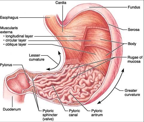 |
front 118 stomach | back 118 The enlarged, muscular sac-like portion of the gastrointestinal tract, located in the upper abdominal cavity, between the esophagus (at the lower esophageal sphincter) and the duodenum of the small intestine (at the pyloric sphincter); where mechanical digestion is completed and chemical digestion, particularly of protein, continues; its glands secrete mucous, HCl, pepsin(ogen), intrinsic factor, and in infants, gastric lipase and rennin; it is roughly divided into four parts (cardia, fundus, body, and pylorus); its inner lining is covered by longitudinal folds of the mucosa, rugae, and its wall consists of an inner simple columnar epithelium with some goblet cells, a lamina propria of loose fibrous connective tissue, a muscularis unusual in having a third layer of oblique fibers, and an outer serosa. |
front 119 cardia | back 119 The upper tapered portion of the stomach which extends a short distance from the opening of the esophagus and where the gastric glands lack parietal and chief cells; the part of the stomach usually involved when hiatal hernia occurs. |
front 120 fundus | back 120 The rounded base of the stomach, that portion farthest from the pyloric sphincter. |
front 121 pylorus | back 121 The long, tapering passage at the lower, distal end of the stomach which opens into the duodenum at the pyloric sphincter; the part of the stomach responsible for gastric emptying which is regulated by GIP, CCK and secretin. |
front 122 rugae | back 122 The longitudinal folds or creases on the inner lining of the stomach, composed primarily of the epithelial and mucosal layers, which contribute to effective mixing of chyme in the stomach; they increase the surface area for gastric secretion and also contribute to gastric mixing, acting as modest mixing "blades"/"spatulas". |
front 123 Image: rugae microscopic | back 123 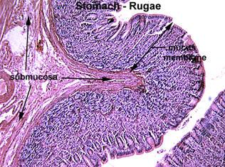 |
front 124 pyloric sphincter | back 124 The ring of smooth muscle fibers around the distal opening of the pylorus of the stomach into the duodenum; it regulates gastric emptying by remaining closed except when a small amount of liquified, partially digested food, chyme, or liquid is being passed to the duodenum; it protects the duodenal lining from erosion by the acidic stomach contents; its opening is regulated by the content of the meal, by hyperglycemia, by digestive hormone (gastrin, cholecystokinin, vasoactive intestinal polypeptide and gastric inhibitory peptide) and regulatory impulses from the ANS. |
front 125 lesser curvature | back 125 The boundary of the stomach which forms a relatively short concave curve on the right, medial, side of the stomach, from the opening for the esophagus to the opening into the duodenum. |
front 126 greater curvature | back 126 The boundary of the stomach which forms a relatively long convex curve on the left and inferior sides of the stomach, from the opening for the esophagus to the opening into the duodenum. |
front 127 mixing waves | back 127 - Slow segmenting movements, every 15 to 25 seconds, within the stomach involving complex patterns of contraction of the three layers of gastric smooth muscle (circular, longitudinal, and oblique) which complete the mechanical digestion of food while combining the bolus of swallowed food with the gastric secretions to continue to chemical digestion of food. |
front 128 chyme | back 128 The thick, semifluid mass of partly digested food which has been acted upon by gastric secretions and is transferred slowly, in small quantites, by the stomach into the duodenum. |
front 129 List: structures which increase the surface area of the stomach. | back 129 rugae |
front 130 List: the functions of the stomach. | back 130 (1) serve as a storage chamber for ingested foodstuffs
|
front 131 Diagram: stomach histology | back 131 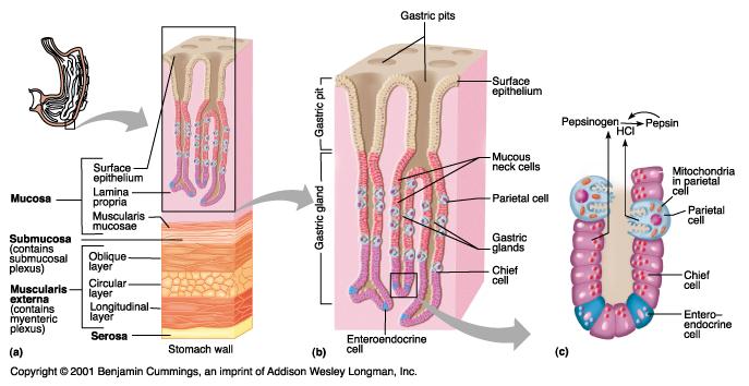 |
front 132 gastric pits | back 132 The numerous small indentations in the mucous membrane of the stomach which are the mouths of the gastric glands. |
front 133 gastric glands | back 133 Branched tubular glands located in the mucosa of the stomach wall; they contain parietal cells which secrete HCL and intrinsic factor, chief cells which secrete pepsin(ogen) and mucous neck cells which secrete mucin; their secretion is regulated by gastrin release from G cells in the stomach and by gastric inhibitory peptide = GIP secreted by enteroendocrine cells from the mucosal epithelial cells of the duodenum and by parasympathetic autonomic motor impulses. |
front 134 mucous neck cells | back 134 The secretory cells which produce an acidic solution containing the protein mucin; they are located primarily in the upper, more superficial portions of the gastric glands; they are believed to be the stem cells for all the other cells in the mucosal glands of the stomach. |
front 135 chief cells | back 135 One of the two glandular epithelial cell types located in the basal half of gastric glands; they synthesize and secrete pepsinogen, the inactive precursor to the proteolytic enzyme pepsin; they appear smaller and with a more darkly staining granular cytoplasm than the neighboring parietal cells. |
front 136 parietal cells | back 136 One of the two glandular epithelial cell types located in the basal half of gastric glands; they synthesize and secrete HCl and intrinsic factor, which is necessary for vitamin B12 absorption in the small intestine; they appear larger and with a pale staining cytoplasm than the neighboring chief cells. |
front 137 Image: Gastric parietal cells and chief cells | back 137 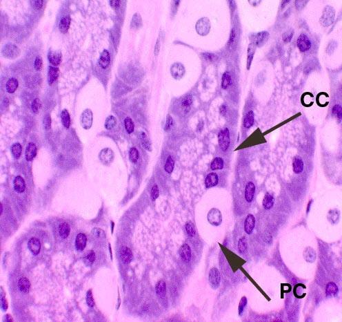 |
front 138 gastric juice | back 138 The clear, viscous, strongly acid secretion of the gastric glands, consisting of enzymes (pepsin(ogen) in adults, and, in neonates, additionally, rennin and gastric lipase) produced by the chief cells, HCL and intrinsic factor produced by the parietal cells, and an acidic mucus fluid secreted by the mucous neck cells; it continues the chemical digestion of food already acted on by saliva. |
front 139 pepsinogen | back 139 The inactive precursor to the proteolytic enzyme pepsin, synthesized and secreted by the chief cells of the gastric glands of the stomach; it is converted to pepsin in the lumen of the stomach by autocatalysis in the presence of gastric hydrochloric acid. |
front 140 pepsin | back 140 The active proteolytic enzyme, synthesized as an inactive precursor precursor, pepsinogen, and secreted by the chief cells of the gastric glands of the stomach; it is activated in the lumen of the stomach by autocatalysis in the presence of gastric hydrochloric acid; it catalyzes the hydrolysis of most large proteins into somewhat smaller units (peptones). |
front 141 gastric lipase | back 141 The enzyme present in gastric juice which catalyzes the hydrolysis of neutral fats (mono-, di-, and triglycerides) into glycerol and free fatty acids in the stomach; it is synthesized and secreted by the chief cells of the gastric glands of the stomach of neonates to contribute to the digestion of milk's butterfat; synthesis is regulated off soon after the infant is weaned. |
front 142 mucin | back 142 mucin |
front 143 hydrochloric acid = HCl | back 143 A solution of hydrogen chloride gas in water; it is a highly corrosive, strong mineral (inorganic) acid which is secreted by the parietal cells of gastric glands; it creates a low pH environment in the stomach which serves a protective function by destroying most microorgaisms, it contributes in a minor way to chemical digestion of foods with a general hydrolysis of chemical bonds, and it activates the conversion of pepsinogen to pepsin. |
front 144 intrinsic factor | back 144 A transport glycoprotein which is secreted by the parietal cells of the gastric glands of the mucous membrane of the stomach and is essential for the absorption of vitamin B12 in the intestines; hereditary failure to secrete it is a major cause of pernicious anemia. |
front 145 rennin | back 145 enzyme, synthesized and secreted by the chief cells of the gastric glands of the stomach of neonates which catalyzes the coagulation of milk; synthesis is regulated off soon after the infant is weaned. [Note: it is also obtained from the gastric juice of the fourth stomach of young ruminants and used in making cheeses and junkets (where it is called rennet); a similar microbial rennin enzyme is also used in cheese production.] |
front 146 G cells | back 146 Enteroendocrine cells, located in the gastric glands in the mucous membrane of the stomach, which synthesize and secrete the digestive hormone gastrin, a peptide hormone; they secrete gastrin in response to gastric distension or to a rise in gastric pH of the stomach, which then stimulates the production of HCl by parietal cells; it also promotes gastric mucosal growth. |
front 147 Image: gastic cief calles and G cells | back 147 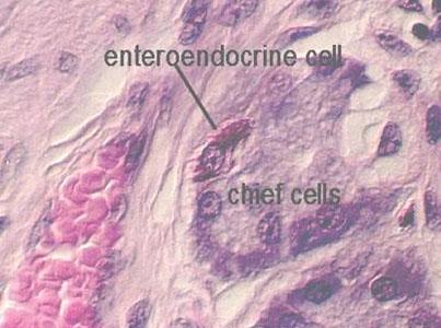 G cell (enteroendocrine cell) stained dark red under very high power |
front 148 cephalic phase | back 148 The first stage of regulation of digestive system activity, particularly in terms of the activities of the stomach, duodenum, pancreas, and gall bladder; sensory information about food (visual, olfactory, gustatory = taste) create conditioned reflexes that trigger some parasympathetic output to the stomach; other stimuli include the actions of chewing and swallowing (mastication and deglutition) and the recognition of hypoglycemia by the hypothalamus; digestive system responses occur within minutes but are temporary if no food actually enters the stomach; during this period small amounts of gastric secretions are produced, particularly H+ from the parietal cells; this stage is mediated by parasympathetic autonomic motor impulses and may involve some secretion of gastrin from G cells. |
front 149 Diagram: cephalic phase | back 149 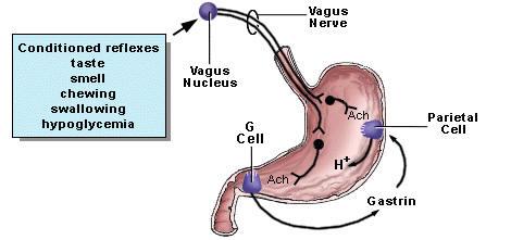 |
front 150 gastric phase | back 150 The second stage of regulation of digestive system activity, particularly in terms of the activities of the stomach, duodenum, pancreas, and gall bladder; visceral sensory stimulation of mechanoreceptors caused by distension of the stomach and detection of protein digestion in the bolus (the presence of peptones, peptides, and amino acids); during this period large amounts of gastric secretions are produced; it is mediated by parasympathetic autonomic motor impulses and by gastrin release from G cells and secretin release from enteroendocrine cells of the mucosal epithelium cells of the duodenum. |
front 151 Digram: gastric phase | back 151 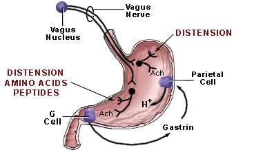 |
front 152 intestinal phase | back 152 The third stage of regulation of digestive system activity, particularly in terms of the activities of the stomach, duodenum, pancreas, and gall bladder; this period is directly related to control of gastric emptying; during this period negative feedback inhibition of further gastric secretions occur; the presence of chyme in the duodenum brings about neural and endocrine responses which first stimulate and later inhibit gastric acid secretion by the stomach; when gastric chyme is at a pH above 3, during early gastric emptying, gastric acid is stimulated; later, when the buffering capacity of gastric chyme is exhausted and the pH of chyme falls below a pH of 2, gastric secretion is inhibited; during this third stage, gastric secretion is first stimulated by the presence of peptides and amino acids in the distended duodenum; G cells in the duodenum and upper jejunum release gastrin in response to peptides and amino acids; this gastrin is released into the blood and can stimulate parietal cells of the stomach to secrete gastric acid; later in this third stage, the enterogastric reflex takes effect, during which, sensory signals from distension of the small intestine and the detection of gastric HCl in the duodenal chyme inhibit gastric motility and gastric secretion; it is mediated by parasympathetic autonomic motor impulses and by gastrin release from G cells and by gastric inhibitory peptide = GIP and secretin release from enteroendocrine cells of the mucosal epithelium cells |
front 153 Diagram: Intestional phase | back 153 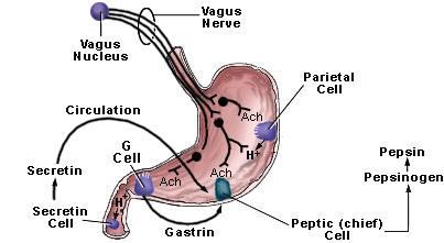 |
front 154 List:the three phases of digestion in the stomach (Can also be diagramed). Describe the neural and hormonal mechanisms involved in each stage, including the stimuli for each. | back 154 1.cephalic phase Neuronal: Sensations of smell and taste or even thoughts or memories of food stimulate cortical centers which activate parasympathetic impulses delivered by the Vagus nerve (X) to initiate gastric secretion.
|
front 155 Diagram: stimulatory and inhibitory factors of the three phases of digestion | back 155 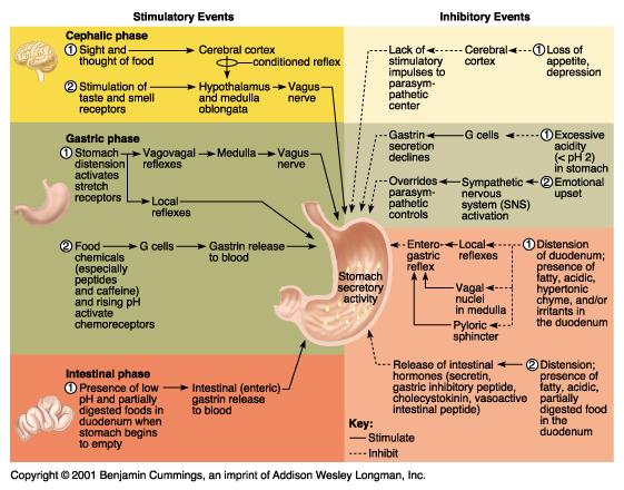 |
front 156 endoscopy | back 156 The visual examination of the interior of a body canal or a hollow body organ by use of an endoscope (an instrument for examining visually the interior of a body canal or a hollow organ), e.g., the colon, bladder, or stomach, etc |
front 157 diagram: endoscope use | back 157 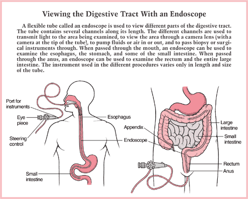 |
front 158 gastroscopy | back 158 The visual examination of the interior of the stomach by use of an endoscope = gastroscope. |
front 159 pylorospasm | back 159 Spasmodic contractions of the smooth muscle in the pylorus of the stomach which prevents chyme from passing to the duodenum; it is a cause of vomiting in neonates, especially those with cerebral birth injuries; it must be distinguised from pyloric stenosis. |
front 160 pyloric stenosis | back 160 A condition where the pylorus of the stomach has a constriction or narrowing, and will thus not allow the passage of food; the main presenting symptom is projectile vomiting. |
front 161 peptic ulcer disease = PUD | back 161 A spectrum of diseases in which the lining of the stomach, pylorus, or duodenum develops a cratering necrotic lesion (ulcer) of the mucosal membrane which becomes eroded by the stomach acid and proteolytic enzyme (pepsin) attack; symptoms may be absent, but usually include indigestion, heartburn, abdominal or chest pain, bleeding, vomiting blood; risk factors include heredity, smoking, heavy drinking, use of aspirin an other anti-inflammatory NSAIDs, and infection by Helicobacter pylori bacteria. |
front 162 living-donor liver transplant | back 162 The transplantation of a portion of a liver from a living donor, who must be a healthy adult; the donor has to be a family member or emotionally related friend of the family; the selection criteria are very strict and potential donors have to undergo an extensive series of tests in order to decide if they are suitable. [Donor Inclusion Criteria: age 18-55; no current or prior history of heart, lung, or other medical problems; no history of liver disease; no history of malignancy; ABO compatibility with the recipient; if female and pre-menopausal, a negative pregnancy test; a stable relationship with recipient. |
front 163 pancreatitis | back 163 Any acute or chronic inflammation of the pancreas; it is difficult to diagnosis; patients may be asymptomatic, but often present with abdominal pain, nausea and vomiting; it is most often caused by alcoholism or biliary tract disease, but may be associated with hyperlipidemia, hyperparathyroidism, trauma, vascular or kidney disease. |
front 164 jaundice | back 164 discoloration of the whites of the eyes, skin, and mucous membranes caused by deposition of bilirubin in these tissues; it occurs as a symptom of various diseases, e.g., internal hemorrhage, hepatitis, or blockages of the biliary tree, which affect the processing or secretion of bile into the duedenum. aka - icterus |
front 165 obstructive (= posthepatic or extrahepatic) jaundice | back 165 Yellowish discoloration of the whites of the eyes, skin, and mucous membranes caused by deposition of bilirubin in these tissues; this form occurs when there has been no internal hemorrhage or hepatic injury or disease, and is due to some blockage of the biliary tree, e.g., from gall bladder stones, or scarring of the bile ducts, which reduce the secretion of bile into the duodenum. |
front 166 hepatitis | back 166 Inflammation of the liver, caused by infectious* or toxic agents and characterized by jaundice, fever, liver enlargement, and abdominal pain. [*Note: viral pathogens, hepatitis A, hepatitis B, hepatitis C, hepatitis D, and hepatitis E are particularly important causes.] |
front 167 cirrhosis | back 167 A chronic disease of the liver characterized by the replacement of normal parenchymal tissue with dense irregular fibrous (scar) tissue and the loss of functional liver cells and marked reduction in liver function; the cirrhotic liver is usually smaller than normal; it can result from alcohol abuse, nutritional deprivation, or infection, especially by the hepatitis viruses; symptoms include exhaustion, fatigue, loss of appetite, nausea, weakness, weight loss, and abdominal pain; complications include edema and ascites, bruising and bleeding, jaundice, gallstones, CNS effects, sensitivity to medications, portal hypertension, varices (enlarged veins), insulin resistance and type II diabetes, immune dysfunction and possible liver cancer. |
front 168 gall stones | back 168 The crystalized solids which precipitate out of the bile stored in the gall bladder under certain circumstances; the vary in size from similar to a grain of sand to as much as an inch in diameter; they may be rough or smooth; they are either composed of a variety of calcium salts or of cholesterol or a mixture of the two; symptoms include fluctuating jaundice, intermittent or constant upper right quadrant pain, nausea and vomiting. |
front 169 Image: Gallstones | back 169 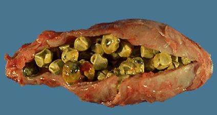 |
front 170 lactose intolerance | back 170 The inability to digest significant amounts of lactose, milk sugar, this inability results from a shortage of the brushborder enzyme lactase, which is normally produced by the epithelial lining cells of the small intestine. |
front 171 appendicitis | back 171 Inflammation of the appendix, a finger-like projection extending from the caecum of the large intestine; symptoms include lower quadrant abdominal pain, fever, and loss of appetite. |
front 172 hemorrhoids | back 172 An itching or painful swollen mass of dilated or varicose veins in the anal mucosal tissue; they may be internal or external or shift between the two locations; they result from a persistent increase in local venous pressure; the may become abraded and eroded and bleed. |
front 173 diverticulitis | back 173 Inflammation of a diverticulum/diverticula (abnormal pouch-like outpockets in the wall of the large intestine, most commonly found in the regions of the sigmoid colon) in the intestinal tract, causing fecal stagnation and pain; they may perforate or form abscesses. |
front 174 diarrhea | back 174 Any excessive and frequent evacuation of loose or watery feces, usually indicating gastrointestinal distress or disorder. |
front 175 constipation | back 175 Difficult, incomplete, or infrequent evacuation of dry hardened feces from the bowels; it can be a symptom of intestinal obstruction or diverticulitis. |
front 176 anorexia nervosa | back 176 A psycho-physiological condition, occurring especially in girls and young women, which is characterized by the inability or refusal to eat, an abnormal fear of becoming obese, and a distorted self-image (somatic delusions that you are too fat despite being emaciated); the main clinical features of the condition are a reduction in body weight, with a body mass index below 17.5, an intense desire to remain thin, self-induced vomiting, excessive exercise, malnutrition, amenorrhea in females, and other physiological changes; it is a condition of Western Societies. [The prevalence rate of this condition is estimated at 1 to 2% of schoolgirls and female students in higher education; the male prevalence rate is much lower.] |
front 177 bulimia | back 177 A psycho-physiological condition characterized by binge-eating with associated feelings of loss of control, intense guilt, periods of dietary restraint; it may include starvation, self-induced vomiting and laxative abuse; these activities may relieve feelings of guilt; it is not usually associated with severe weight loss; it is a condition of Western Societies. [The prevalence rate of this condition is estimated at 1 to 2% of the Western Caucasian female population, with an increased incidence in the higher social classes; the male prevalence rate is much lower.] aka - bulimia nervosa |
front 178 dysentery | back 178 An inflammatory disorder of the lower intestinal tract, usually caused by a bacterial, parasitic worm, or protozoan infection or certain chemical irritants, and resulting in pain, fever, a constant desire to evacuate the bowels, and severe diarrhea, often accompanied by the ulceration of the colon and rectum, passage of blood and mucus. |
front 179 fecal impaction | back 179 A severe form of constipation in which a large mass of stool cannot be passed; it occurs most commonly in older or bedridden people; symptoms include swelling of the abdomen, nausea and vomiting, thin watery discharge from the rectum, abdominal distress or cramping, and fever. |
front 180 Crohn's disease | back 180 A serious, chronic, progressive inflammatory disorder of the lower intestinal tract, usually involving the terminal portion of the ileum and characterized by nausea, fever, weight loss, frequent bouts of diarrhea, abdominal pain, ulceration, and fibrous tissue buildup. |
front 181 ulcerative colitis | back 181 A serious chronic inflammatory disease of the large intestine and rectum characterized by recurrent episodes of fatigue, loss of appetite, abdominal pain, fever and chills, and profuse diarrhea, weight loss and possible rectal bleeding. |
front 182 intestinal polyps | back 182 Abnormal tissue growths which projects (usually on a stalk) from the mucosal lining of the colon or rectum; they are usually benign and asymptomatic, but may cause painless rectal bleeding; certain types of polyps, called adenomatous polyps, can develop into cancer; risk factors include advancing age, cigarette smoking, high fat or low fiber diet, and family history. aka - colon polyps |
front 183 colon cancer | back 183 A tumor of the colon or rectum, frequently malignant; an early symptom is bloody stools; the second-leading cause of cancer-related deaths in the United States. |
front 184 gastrin - | back 184 A peptide hormone, secreted in response to gastric distension or a rise in gastric pH by enteroendocrine cells (G cells) within the glandular pits in the mucous membrane of the stomach, which stimulates the production of HCl by parietal cells; an important regulator of stomach digestive processes during the middle gastric phase; it also promotes gastric mucosal growth. |
front 185 gastric inhibitory peptide = GIP | back 185 A peptide hormone, secreted in response to duodenal distension or the detection of glucose and fatty acids in the chyme arriving from the stomach; it is secreted by enteroendocrine cells beneath the mucosal epithelial cells of the duodenum; it stimulates the release on insulin from beta cells of the pancreatic islets and inhibits the release of gastric HCl from the parietal cells and pepsin from the chief cells of the gastric mucosa. |
front 186 secretin | back 186 A peptide hormone, secreted in response to the detection of gastric HCl in the chyme arriving from the stomach; it is secreted by enteroendocrine cells from the mucosal epithelium cells of the duodenum; it stimulates the production of bicarbonate-rich alkaline secretion from pancreatic ductal cells, and, to a lesser degree, from bile duct cells. |
front 187 cholecystokinin = CCK | back 187 A peptide hormone produced principally by the enteroendocrine cells from the mucosal epithelium of the duodenum in response to the presence of fatty acids and proteins in the chyme arriving from the stomach; it stimulates the contraction of the gallbladder, release of bile, and secretion of pancreatic digestive enzymes from the pancreatic acinar cells. aka - pancreozymin (older terminology). [Note: It is also found in the CNS and may be related to the control of feelings of satiety (fullness, lack of hunger).] |
front 188 enterogastric reflex | back 188 One of the visceral = autonomic reflexes involved in the regulation of digestion in which sensory signals from distension of the small intestine and the detection of gastric HCl in the chyme inhibit gastric motility and gastric secretion; it also encourages secretion of bile from the gall bladder and pancreatic juice from the pancreas; it is mediated by parasympathetic autonomic motor impulses. |
front 189 gastric emptying | back 189 The passage of the contents of the stomach into the small intestine; it is influenced by the content of the meal, by hyperglycemia, by digestive hormone (gastrin, cholecystokinin, vasoactive intestinal polypeptide and gastric inhibitory peptide) levels, and regulatory impulses from the ANS; solid foods generally require approximately 90 minutes/half-time while liquids move faster. |
front 190 distension | back 190 The act or process of expanding or stretching a structure by pressure from within, e.g., by filling the structure with material. |
front 191 Describe:the hormones secreted by the duodenum and how they regulate secretion by the stomach (gastric), liver (hepatic), gall bladder (cystic), and pancreas (pancreatic). | back 191 *a.Duodenal Hormone b.Stomach Secretion c.Liver Secretion d.Gall Bladder Secretion e.Pancreas Secretion
|
front 192 Describe: how gastric emptying is regulated. | back 192 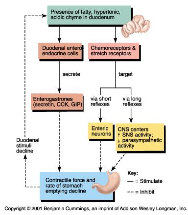 Autonomic NS Regulation: Increased sympathetic impulses and decreased parasympathetic impulses to gastric smooth muscle stimulate emptying. Duodenal proprioceptive stretch recepters provide negative feedback information to inhibit emptying if the duodenum becomes overfilled.
|
front 193 pancreas | back 193 A long, flattened, irregularly shaped, fragile accessory digestive gland, lying behind the stomach and between the duodunum and the spleen; its parenchyma consists primarily of exocrine acinar cells drained by the pancreatic duct system, but interspersed among them are small round collections of endocrine cells (alpha, beta, and delta cells) termed islets; the acinar cells secrete an enzyme-rich alkaline pancreatic juice into the duodenum while the islet cells secrete insulin (alpha cells), glucagon (beta cells), and somatostatin (delta cells) into the bloodstream. |
front 194 DIagram: Pancreas | back 194  |
front 195 pancreatic duct | back 195 The major excretory duct of the pancreas, composed of a simple cuboidal epithelium, by which pancreatic juice is secreted into the duodenum at the hepatopancreatic ampulla. |
front 196 hepatopancreatic ampulla | back 196 The goblet-shaped dilatation (expansion) of the ducts coming from the liver and gall bladder (common bile duct) and the pancreas (pancreatic duct) at the point (major duodenal papilla) where they enter the duodenum. |
front 197 major duodenal papilla | back 197 A nodular protuberance on the medial wall of the duodenum through which the common bile duct and pancreatic duct expel bile and pancreatic juice respectively into the duodenum by way of the hepatopancreatic ampulla to aid in the processes of digestion. |
front 198 accessory duct | back 198 A variable duct of the pancreas, composed of a simple cuboidal epithelium, which branches from the main pancreatic duct and opens into the duodenum above it at the minor duodenal papilla |
front 199 pancreatic acini | back 199 The majority of pancreatic tissue which is exocrine tissue collected into a complex system of secretory cell clusters (acini) attached to the pancreatic duct system; the acinar cells secrete a variety of digestive enzymes into the pancreatic juice which is transported to the duodenum. |
front 200 pancreatic juice | back 200 The clear alkaline secretion of pancreas, consisting of enzymes (e.g., pancreatic amylase, trypsin, chymotrypsin, carboxypeptidase, elastase, pancreatic lipase, ribonuclease, deoxyribonuclease, etc.) produced by the acinar cells and an alkaline mucus fluid secreted by the pancreatic ductal cells which flows via the pancreatic duct system to the duodenum and continues the chemical digestion of food already acted on by the gastric juice and saliva. |
front 201 pancreatic islets = Islets of Langerhans | back 201 Any of several masses of endocrine cells in the pancreas including alpha cells which secrete the protein hormone glucagon, beta cells which secrete the protein hormone insulin, and delta cells which secrete the protein hormone somatostatin. |
front 202 pancreatic amylase | back 202 - The enzyme present in pancreatic juice which catalyzes the hydrolysis of starch to sugar to produce carbohydrate derivatives in the duodenum; elevated serum pancreatic amylase levels are associated with pancreatitis and many other abdominal disorders. |
front 203 trypsinogen | back 203 The inactive precursor of trypsin, produced by the pancreatic acinar cells and converted to trypsin in the small intestine by the intestinal brush border enzyme enterokinase. |
front 204 trypsin | back 204 The pancreatic proteolytic enzyme which catalyzes the hydrolysis of proteins and large peptides into smaller oligopeptide units in the small intestine; it acts on the breakdown products of the gastric digestion of proteins by pepsin; it is secreted by the acinar cells in the form of an inactive precursor, trypsinogen. |
front 205 chymotrypsin | back 205 The pancreatic proteolytic enzyme which catalyzes the hydrolysis of peptides into smaller oligopeptides into small peptides and amino acids in the small intestine; it is secreted by the acinar cells in the form of an inactive precursor, chymotrypsinogen. |
front 206 carboxypeptidase | back 206 The pancreatic proteolytic enzyme which catalyzes the hydrolysis of the terminal amino acid of a polypeptide or peptide from the C-terminal end (which contains a free carboxylic acid group) in the small intestine; two forms exist, A and B; they are secreted by the acinar cells in the form of inactive precursors, procarboxypeptidase. |
front 207 elastase | back 207 The pancreatic proteolytic enzyme which catalyzes the hydrolysis of elastin (and collagen) in the small intestine. |
front 208 pancreatic lipase | back 208 The enzyme present in pancreatic juice which catalyzes the hydrolysis of neutral fats (mono-, di-, and triglycerides) into glycerol and free fatty acids in the duodenum; elevated serum pancreatic lipase levels are associated with pancreatitis and some other abdominal disorders. |
front 209 ribonuclease | back 209 The enzyme present in pancreatic juice which catalyzes the hydrolysis of RNA (ribonucleic acids) into constituent ribonucleotides in the small intestine. aka - RNase. |
front 210 deoxyribonuclease | back 210 The enzyme present in pancreatic juice which catalyzes the hydrolysis of DNA (deoxy-ribonucleic acids) into constituent ribonucleotides in the small intestine. aka - DNase. |
front 211 List: the functions of the pancreas. | back 211 (1) provides some bicarbonate ions to assist in buffering acidic chyme transferred from the stomach to the duodenum
|
front 212 Describe: how pancreatic secretion (endocrine) is regulated. | back 212 Pancreatic islet cells respond to autoregulation. Pancreatic islet cells monitor plasma glucose levels. If plasma glucose levels rise, e.g., after the absorption of a meal, then beta cells will release more insulin into the blood stream; if plasma glucose levels fall, then alpha cells will release more glucagon into the blood stream. |
front 213 Describe: how pancreatic secretion (exocrine) is regulated. | back 213 The stimuli that trigger increased pancreatic exocrine secretion are (1) the presence of an acidic chyme in the duodenum and (2) the presence of fatty acids and amino acids in the chyme in the duodenum. These stimuli produce three different regulatory feedback actions to stimulate increased pancreatic exocrine secretion: (a) autoregulation involving local hormones within the head of the pancreas; (b) release of CCK (cholecystokinin) and secretin from enteroendocrine cells in the walls of the duodenum [Note: CCK stimulates digestive enzyme secretion by pancreatic acinar cells while secretin stimulates bicarbonate ions and mucous secretions from the pancreatic duct cells.]; and (c) autonomic parasympathetic impulses, delivered via the vagus nerve (X), which increase pancreatic secretions. |
front 214 liver | back 214 The large, reddish-brown, glandular digestive organ located in the upper right quadrant of the abdominal cavity beneath the diaphragm which is divided into three lobes, the right, left, and caudate; it synthesizes and secretes bile, is active in the formation of certain blood proteins and lipoproteins; plays a major role in the metabolism of carbohydrates, fats, and proteins; stores or detoxifies toxins; it receives blood from both the hepatic artery and the hepatic portal system. |
front 215 liver lobes | back 215 liver lobes |
front 216 falciform ligament | back 216 The anteroposterior fold of the parietal peritoneum attached to the under surface of the diaphragm and the sheath of the rectus muscle and along a line on the anterior and upper surfaces of the liver extending back from the notch on the anterior margin of the liver; it is a main support for the liver in the abdominal cavity. |
front 217 ligamentum teres | back 217 A dense fibrous connective tissue cord resulting from the obliteration of the umbilical vein of the fetus and passing from the navel to the notch in the anterior border of the liver and along the undersurface of that organ. |
front 218 coronary ligaments | back 218 Several folds of the parietal peritoneum attached to the under surface of the diaphragm and to the liver; they are supports for the liver in the abdominal cavity. |
front 219 lobules | back 219 Small sections or subdivisions of the lobe of an organ; in the liver, the structural subunit which consists of roughly hexagonal arrangements of plates or cords of hepatocytes radiating outward from a central vein and separated from each other by radiating capillary sinusoids; each lobule is surrounded by a thin layer of dense fibrous connective tissue; the liver contains ~1,000,000 lobules. |
front 220 central vein | back 220 The single large blood vessel in the middle of each lobule of the liver which runs from the apex to the base of the lobule, receives blood from the sinusoids, and empties into the sublobular veins from where the blood will eventually return to the inferior vena cava. |
front 221 hepatocytes | back 221 The parenchymal (chief functional) cells of the liver organized into plates or cords radiating outward from the central vein of each liver lobule and separated from each other by radiating capillary sinusoids; they are polygonal in shape with a large oval nucleus and a granular cytoplasm; they carry out a wide variety of metabolic, endocrine, and secretory (exocrine) functions; they synthesize and secrete bile, synthesize and secrete certain blood proteins and lipoproteins; play a major role in the metabolism of carbohydrates, fats, and proteins; store or detoxify toxins, etc. |
front 222 portal triad | back 222 A characteristic histological feature of the borders of liver lobules, consisting of one or more of each of the following structures, a branch of the hepatic artery bringing oxygenated blood to the hepatocytes, a branch of the hepatic portal vein bringing nutrient laden blood from the capillary beds of the stomach, small and large intestines, spleen, and pancreas, and an interlobar branch of the bile duct system. |
front 223 sinusoid (capillary) | back 223 The category of capillaries, primarily the capillaries in the liver, bone marrow, some endocrine glands and in lymphoid tissue, which lack pores = fenestrations but in which adjacent endothelial cells exhibit some wider intercellular gaps which permit some fluid exchange between the plasma and the tissue fluid by transfer between endothelial cells; they often have an irregular cross-section. |
front 224 stellate reticuloendothelial cells | back 224 The fixed macrophages of the walls of the liver sinusoids which have long cytoplasmic projections, a large oval nucleus and their cytoplasm is commonly packed with fragments resulting from phagocytic action; they play a major role, along with the spleen, in clearing the blood of damaged RBCs and in removing immune (Ab-Ag) complexes from the blood. aka - Kupffer cells |
front 225 hepatic portal* circulation = hepatic portal* system | back 225 The separate pathway for a portion of the blood returned to the heart from certain the abdominal organs; the vessels involved are the hepatic portal vein and its branches (splenic vein, superior and inferior mesenteric veins and gastric vein and their smaller branches); venous blood from the capillary beds of the spleen, pancreas, stomach and intestines (large and small) is routed to the capillary beds of the liver where various compounds in the blood are processed or stored by the liver; after leaving the liver this blood is returned to the general venous return of the inferior vena cava. [Note: A portal system is a vascular arrangement in which blood from the capillaries of one organ is transported to the capillaries of another organ by a connecting vein or veins without returning to the heart.] |
front 226 hepatic portal vein | back 226 The large blood vessel carrying blood from the capillary beds of the spleen, pancreas, stomach and intestines (large and small) to the liver where the nutrients carried by the blood are processed by the hepatocytes before passing into the systemic circulation. |
front 227 enterohepatic circulation | back 227 circular pathway in which molecules, particularly bile salts and bile acids, are recycled; the path is from the liver to the small intestine via the biliary tree, reabsorption from the chyme in the intestines into the hepatic portal circulation, and back to the liver. |
front 228 List: the five organs whose venous blood is routed by the hepatic portal system to the liver and a reason why each of these organs has its venous blood routed to the liver. | back 228 1. Stomach (1) absorbed nutrients (minimal) will be processed and stored by the liver
|
front 229 Diagram and Label: the structure of a liver lobule illustrating the general pattern of blood and bile flow. Identify the factors that increase bile secretion by the liver. | back 229 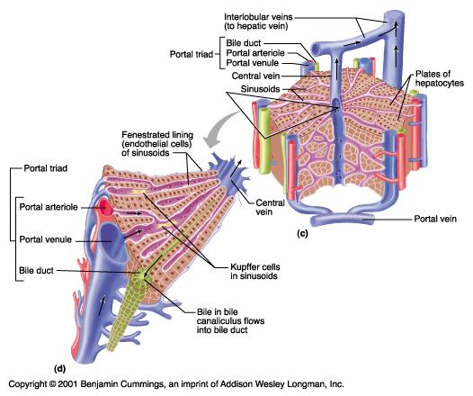 Blood Flow: Oxygenated blood from the systemic circulation via the abdominal aorta and hepatic artery and deoxygenated blood from the stomach, small intestines, large intestine, pancreas, and spleen are delivered to the periphery of each liver lobule (at hepatic = portal triads); mix in the capillary sinusoids of the liver lobule and drain to the central vein which carries the hepatic venous blood out of the liver via the hepatic veins, then to the inferior vena cava and from there back to the right atrium of the heart.
|
front 230 bile canaliculi | back 230 The minute channels which run between hepatocytes, and into which they secrete bile; they form the smallest and most distal branches of the biliary tree and carry bile toward the periphery of the lobules of the liver where it is collected into interlobar bile ducts at the "hepatic triad." aka - bile capillaries |
front 231 DIagram: biliary tree | back 231 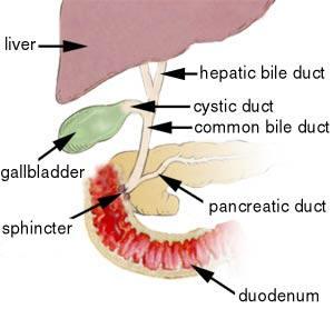 |
front 232 hepatic ducts | back 232 Any portion of the biliary tree; especially, the larger duct(s) which carry the bile from the lobes of the liver to the cystic and common bile ducts; they are lined by columnar epithelium and a delicate layer of smooth muscle. |
front 233 cystic duct | back 233 The portion of the biliary tree which conveys bile to the gall bladder from the liver via the hepatic ducts between meals and from the gallbladder to the common bile duct (and on to the duodenum) during the intestinal phase of the digestion of a meal; it is lined by columnar epithelium and a delicate layer of smooth muscle. |
front 234 common bile duct | back 234 The portion of the biliary tree formed by the union of the cystic duct and the hepatic duct which carries bile from the liver and the gallbladder to the duodenum during the intestinal phase of the digestion of a meal; it is lined by columnar epithelium and a delicate layer of smooth muscle. |
front 235 gallbladder | back 235 The small, pear-shaped sac, located under the right lobe of the liver, in which bile secreted by the liver is stored until needed by the body for digestion of a fatty meal; it is lined by columnar epithelium and a layer of smooth muscle innervated by the Vagus Nerve and responsive to CCK. |
front 236 sphincter of hepatopancreatic ampulla | back 236 The ring of smooth muscle which guards the opening of the merged common bile duct and pancreatic duct located in the wall of the duodenum; it is innervated by the Vagus Nerve and responsive to CCK. |
front 237 bile | back 237 A bitter, alkaline, viscous, brownish-yellow or greenish-yellow mucous fluid which is secreted by the liver hepatocytes, collected into bile canaliculi and delivered by the biliary tree to be stored in the gallbladder, and discharged into the duodenum; it contains bile salts and bile acids which aid in the emulsification of ingested fats to improve the efficiency of lipid digestion and absorption; it also contains a variety of wastes such as bilirubin from the breakdown of the heme in hemoglobin and various bile pigments; adult humans produce 0.4 - 0.8 L daily. |
front 238 bile salts and bile acids | back 238 A group of amphipathic (both lipid and water soluble) metabolic waste products of the hepatic catabolism of cholesterol and steroid hormones; they are produced by hepatocytes and added to bile; in bile they function as a surfactant to emulsify ingested fats to improve the efficiency of lipid digestion and absorption in the duodenum; they are reabsorbed into the bloodstream from the small intestine and returned to the liver by the hepatic portal circulation ("enterohepatic circulation") where they are recycled so that each molecule is used approximately 20 times; they also play a negative-feedback regulatory role in the control of hepatic synthesis of cholesterol. |
front 239 emulsification | back 239 To make a preparation of one liquid (discontinuous phase) distributed in small globules or droplets throughout the volume of a second liquid (continuous phase); when water is the continuous phase, the preparation is termed a water emulsion, when a nonpolar liquid (oils, lipids, etc.) is the continuous phase, the preparation is termed an oil or lipid emulsion; bile, especially the bile salts and bile acids acting as a surfactant coating, contribute to the emulsification of ingested fats to improve the efficiency of lipid digestion and absorption in the duodenum. |
front 240 bilirubin | back 240 An orange-yellow lipid pigment excreted by the liver into the bile which forms as a by-product of hemoglobin breakdown and recycyling; excess amounts in the blood or deposited in tissues produce the yellow appearance observed in jaundice. |
front 241 List: the functions of bile. | back 241 (1) provides some bicarbonate ions to assist in buffering acidic chyme transferred from the stomach to the duodenum
|
front 242 carbohydrate metabolism | back 242 The diverse set of biochemical reactions which occur within cells that (1) convert ingested sugars into larger carbohydrates (glycogenesis = synthesis of starch) or (2) add sugars to other large polymers such as lipids and proteins (glycosylation) or (3) create new sugar molecules from non-carbohydrate precursors (gluconeogenesis) or (4) breakdown starch (glycogenolysis) into glucose molecules or (5) breakdown glucose and other simple sugars in oxidative pathways (glycolysis, the citric acid cycle, oxidative phosphorylation) to produce useful chemical energy (ATP) and waste products (CO2 and H2O) or (6) in fermentation pathways (lactic acid production) to produce useful chemical energy (ATP); processes 1, 2 and 3 above are anabolic while processes 4, 5, and 6 are catabolic. |
front 243 lipid metabolism | back 243 The diverse set of biochemical reactions which occur within cells that (1) convert ingested lipid subunits (glycerol, fatty acids, cholesterol, etc.) into larger lipids (lipogenesis = synthesis of neutral fats, di- and tri-glycerides) or (2) add lipid subunits to other large polymers such as carbohydrates and proteins or (3) create new lipid molecules from non-lipid precursors or (4) breakdown lipids (lipolysis) into lipid subunits (glycerol, fatty acids, cholesterol, etc.) or (5) breakdown lipid subunits (glycerol, fatty acids, cholesterol, etc.) in oxidative pathways (beta oxidation, glycolysis, the citric acid cycle, oxidative phosphorylation) to produce useful chemical energy (ATP) and waste products (CO2 and H2O); processes 1, 2 and 3 above are anabolic while processes 4, and 5 are catabolic. |
front 244 beta oxidation | back 244 The catabolic process by which fatty acids are degraded, involving oxidation of the beta carbons and removal of successive two-carbon fragments from the fatty acid to form molecules of acetyl co-enzyme A; acetyl co-enzyme A can transfer the two-carbon acetyl groups to the the citric acid cycle in mitochondria for further oxidation to produce useful chemical energy (ATP) and waste products (CO2 and H2O). |
front 245 protein metabolism | back 245 The diverse set of biochemical reactions which occur within cells that (1) convert ingested protein subunits (amino acids) into larger proteins (proteine synthesis = transcription + translation) or (2) add protein subunits (amino acids) to other large polymers such as carbohydrates and lipids or (3) create new lipid molecules from non-lipid precursors or (4) breakdown lipids (lipolysis) into protein subunits (amino acids) (transamination) or (5) breakdown protein subunits (amino acids) in oxidative pathways (deamination, glycolysis, the citric acid cycle, oxidative phosphorylation) to produce useful chemical energy (ATP) and waste products (CO2, H2O and ammonia → urea); processes 1, 2 and 3 above are anabolic while processes 4, and 5 are catabolic. |
front 246 transamination | back 246 The enzyme-catalyzed transfer of an amino group (-NH2) from one compound to another; an important function of the liver hepatocytes and carried out in lesser amounts by many other cell types. |
front 247 deamination | back 247 The enzyme-catalyzed removal of an amino group (-NH2) from an organic compound; an important function of the liver hepatocytes and carried out in lesser amounts by renal tubular cells and a few other cell types. |
front 248 ammonia | back 248 A colorless, pungent, strongly basic, irritating volatile gas, NH3, extensively used to manufacture fertilizers and a wide variety of nitrogen-containing organic and inorganic chemicals; it is very soluble in water; it is a by-product of protein catabolism in cells, but is highly toxic and must be rapidly excreted or else converted into less toxic nitrogenous wastes, e.g., urea |
front 249 urea | back 249 A highly water-soluble, non-toxic compound, CO(NH2)2, formed from the addition of two amino groups (-NH2) to a molecule of carbon dioxide (CO2) in the liver, which is the major nitrogenous waste product of protein catabolism and is the chief nitrogenous waste component of the urine. |
front 250 List:the functions of the liver. | back 250 (1) carbohydrate metabolism (storage, glycogenesis, glycogenolysis, gluconeogenesis, contribute to regulation of blood glucose levels, etc.)
|
front 251 small intestine | back 251 The narrow, winding, longest part (~22 feet) of the gastrointestinal tract where chemical digestion is completed and where nutrients are absorbed into the blood; it extends from the pyloric sphincter to the ileocecal valve and consists of the duodenum, the jejunum, and the ileum; its wall consists of an inner simple columnar epithelium with some goblet cells, a lamina propria of loose fibrous connective tissue, a muscularis, and an outer serosa. |
front 252 duodenum | back 252 The first or proximal portion of the small intestine, starting at the pyloric sphincter at the lower end of the stomach and extending to the jejunum; it is approximately one foot in length; its wall consists of an inner simple columnar epithelium with some goblet cells, a lamina propria of loose fibrous connective tissue, a muscularis, and an outer serosa; it receives bile from the liver and gall bladder via the common bile duct and pancreatic juice from the pancreatic duct; it is the main site for completion of the chemical digestion of food; it has circular folds (plicae circulares) and only a few Peyer's patches. |
front 253 jejunum | back 253 The middle portion of the small intestine, starting at the duodenum and extending to the ileum; it is ~9-10 feet in length; its wall consists of an inner simple columnar epithelium with goblet cells, a lamina propria of loose fibrous connective tissue, a muscularis, and an outer serosa; it is a main site for absorption of nutrients into the bloodstream; it is larger, thicker-walled, more vascular and has more circular folds (plicae circulares) and fewer Peyer's patches than the ileum. |
front 254 ileum | back 254 The final or distal portion of the small intestine, starting at the jejunum and extending to the ileocecal valve where it joins the large intestine; it is ~12 feet in length; its wall consists of an inner simple columnar epithelium with many goblet cells, a lamina propria of loose fibrous connective tissue, a muscularis, and an outer serosa; it is a site for absorption of nutrients into the bloodstream; it is smaller, thinner-walled, less vascular and has fewer circular folds (plicae circulares) and more Peyer's patches than the jejunum. |
front 255 ileocecal sphincter | back 255 A thickening of the circular layer of smooth muscle at the juncture of the ileum and the large intestine which serves as a valve to control the movement of chyme from the ileum into the cecum; it is controlled by the ANS. aka - ileocecal valve |
front 256 circular folds = plicae circulares | back 256 The numerous permanent crescentic folds or pleats of mucous membrane found in the small intestine, especially in the lower part of the duodenum and the jejunum; they increase the surface area available for absorption. |
front 257 Labeled diagram small intestines | back 257  3 Descending (second) part of duodenum
|
front 258 DIagram small intestines plicae histology | back 258 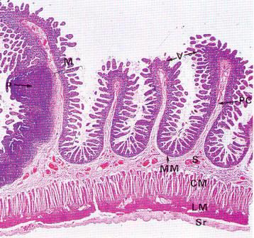 The mucosa M is thrown into transverse folds, the plicae circulares PC, covered with villi V. The muscularis mucosae MM lies immediately beneath the basal crypts and is difficult to see at this magnification. The vascular submucosa S extends into the plicae circularis. Beneath it lie circular CM and longitudinal LM layers of the muscularispropria and the serosa Sr. Peyer's patches P dominate the mucosa at the left of the field. |
front 259 Diagram small intestines villi | back 259 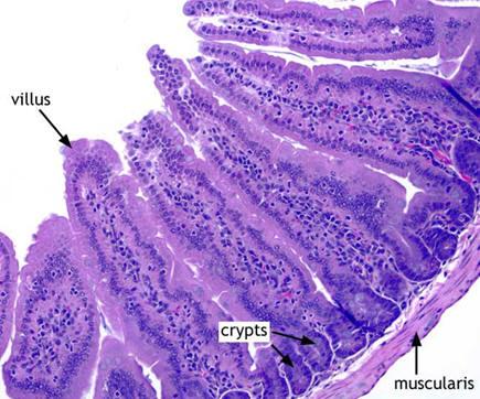 Lacteals convey chyle (lymph containing lipids absorbed from the meal and packaged in chylomicrons) from the intestine to the lymphatic circulation and thereby to the thoracic duct and then to the systemic blood circulation. |
front 260 villi | back 260 A minute finger-like, multicellular projection arising from a mucous membrane which serve to greatly increase the surface area of the mucous membrane; in the gastrointestinal tract, they are found in the small intestine and improve the efficiency of absorption; they are lined by a simple columnar epithelium with goblet cells; the interior contains loose fibrous connective tissue, small blood vessels, a small amount of smooth muscle, small nerve fibers, and a lacteal. |
front 261 segmentation | back 261 A pattern of annular contraction of the smooth muscle layers in the walls of the small intestine which temporarily seems to cut the region affected into individual compartments; this activity serves to mix the chyme within the small intestine; it is controlled by the ANS. |
front 262 List: structures which increase the surface area of the small intestine. | back 262 (1) circular folds = plicae circulares, (2) villi, (3) microvilli |
front 263 List: the functions of the small intestine. | back 263 (1) complete chemical digestion of food with the combination of bile, pancreatic enzymes, and intestinal brush border enzymes
|
front 264 1/3 Diagram and Label: structures of the small intestine that increase its surface area (folding) for digestion and absorption. | back 264 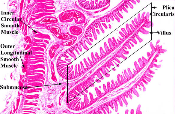 1/3 Plicae Circularies: Lining epithelium covers a series of villi which are supported by a dense fibrous connective tissue support within the plica circularis |
front 265 2/3 Diagram and Label: structures of the small intestine that increase its surface area (folding) for digestion and absorption. | back 265  Intestinal Villus |
front 266 3/3 Diagram and Label: structures of the small intestine that increase its surface area (folding) for digestion and absorption. | back 266 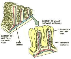 Intestinal Epithelial Lining Cell Microvilli form a Brush Border on the Luminal Surface |
front 267 Diagram and Label: an intestinal villus. | back 267 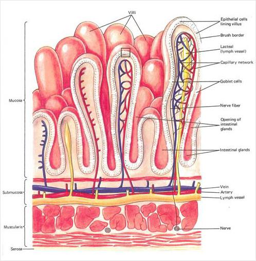 |
front 268 lacteal | back 268 The minute intestinal lymphatic vessel, found in the interior of each intestinal villus, which receives chylomicrons, lipids absorbed from chyle and modified by the simple columnar cells which line the small intestine, these chylomicrons will be transported from the lamina propria of the small intestine to the lymphatic circulation and thereby to the thoracic duct. |
front 269 microvilli | back 269 The minute hair-like or finger-like projections of the outer cell membrane which increase the surface area of the outer cell membrane of certain types of epithelial cells, e.g., the simple columnar cells which line the small intestine, the cubiodal cells of the proximal convoluted tubules of the nephron in the kidney, etc. |
front 270 brush border | back 270 A thin layer of specialized cell membrane, faintly visible in the light microscope, on the apical surface of certain types of epithelial cells; in some cases, it is formed by microvilli, e.g., the simple columnar cells which line the small intestine, the cubiodal cells of the proximal convoluted tubules of the nephron in the kidney, etc.; in other cases, it is formed by cilia, e.g., the pseudostratified columnar epithelium of the respiratory tree, the cells lining the ovarian ducts, etc. |
front 271 intestinal glands | back 271 The simple tubular exocrine secretory structures of the intestinal mucous membrane formed by extension of layer of simple columnar cells which line the small and large intestine; mucus-producing goblet cells, other glandular secretory cells and small numbers of enteroendocrine cells are present. |
front 272 paneth cells | back 272 Specialized granular secretory cells with large acidophilic-staining nuclei found in the depths of intestinal glands in the small intestine which secrete lysozyme and other anti-bacterial compounds. |
front 273 duodenal glands | back 273 The compound tubular exocrine secretory structures of the submucosa of the duodenum; they contain mucus-producing cells which release bicarbonate ions; this alkaline mucus assists in neutralizing the acidic chyme from the stomach (as does the alkaline pancreatic juice). |
front 274 solitary lymphatic nodules | back 274 Isolated minute round to oval collections of lymphoid tissue (T lymphocytes and B lymphocytes, especially those capable of secreting IgA) found in the mucosa of the small and large intestines, containing only a single follicle with germinal center, if any; especially common in the cecum and appendix; an example of MALT = mucosa-associated lymphatic tissue. |
front 275 aggregated lymphatic nodules | back 275 Clusters of minute unencapsulated masses of lymphoid tissue (T lymphocytes and B lymphocytes, especially those capable of secreting IgA) found in the mucosa of the small and large intestines, containing numverous follicles with germinal centers; especially common in the jejunum and ileum; an example of MALT = mucosa-associated lymphatic tissue. aka - Peyer's Patches |
front 276 intestinal juice | back 276 The fluid which is secreted in small quantity in the small intestine, is highly variable in composition, containing mucus and various brush border enzymes (dextrinase, glucoamylase, maltase, lactase, sucrase, carboxypeptidase, dipeptidase, nucleosidases, phosphatases, and enterokinase) and, which, after mixing with bile and pancreatic juice, participates in the final chemical digestion of food. |
front 277 brush border enzymes | back 277 The set of digestive enzymes secreted by the simple columnar cells which line the small intestine, including erepsin, dextrinase, glucoamylase, maltase, lactase, sucrase, dipeptidase, nucleosidases, phosphatases, and enterokinase. |
front 278 dextrinase | back 278 A brush border enzyme secreted by the simple columnar cells which line the small intestine which catalyzes the hydrolysis of dextrins, which are starch fragments containing branching points for glucoses linked in the 1,4 position, into smaller oligosaccharides. |
front 279 maltase | back 279 A brush border enzyme secreted by the simple columnar cells which line the small intestine which catalyzes the hydrolysis of the disaccharide maltose to two glucoses. |
front 280 lactase | back 280 A brush border enzyme secreted by the simple columnar cells which line the small intestine which catalyzes the hydrolysis of the disaccharide lactose, "milk sugar," to one glucose and one galactose. |
front 281 peptidases | back 281 A group of brush border enzymes secreted by the simple columnar cells which line the small intestine which catalyze the hydrolysis of small peptides into tripeptides, dipeptides and individual amino acids. |
front 282 nucleosidases | back 282 A group of brush border enzymes secreted by the simple columnar cells which line the small intestine which catalyze the hydrolysis of nucleotides (a ribose sugar linked to a nitrogenous base) into the constituent ribose sugar and nitrogenous base. |
front 283 phosphatases | back 283 Any of numerous enzymes that catalyze the hydrolysis of esters of phosphoric acid and are important in the absorption and metabolism of carbohydrates, nucleotides, and phospholipids and in the calcification of bone; in the gastrointestinal tract, the group of brush border enzymes secreted by the simple columnar cells which line the small intestine which catalyze the hydrolysis of phosphate ions from phosphorylated nutrient molecules. |
front 284 List: the functions of the brush border enzymes. | back 284 1.dextrinase catalyzes the hydrolysis of dextrins, which are starch fragments containing branching points for glucoses linked in the 1,4 position, into smaller oligosaccharides
|
front 285 carbohydrate digestion | back 285 The process by which carbohydrates are converted into simple sugars which can be absorbed and assimilated by the body; it is accomplished in the digestive system by the mechanical and enzymatic breakdown. [Note: There is a limited amount of indiscriminant acid hydrolysis of carbohydrates in the stomach due to the action of gastric HCl; carbohydrates are split into di- and mono-saccharides by salivary amylase in the mouth and pancreatic amylase in the duodenum and disaccharides are split into monosaccharides by disaccharidases (brush border enzymes) in the duodenum.] |
front 286 protein digestion | back 286 The process by which proteins are converted into individual amino acids which can be absorbed and assimilated by the body; it is accomplished in the digestive system by the mechanical and enzymatic breakdown. [Note: There is a limited amount of indiscriminant acid hydrolysis of proteins in the stomach due to the action of gastric HCl; proteins are split into smaller peptides and ultimately into individual amino acids by pepsin in the stomach and pancreatic proteases (trypsin, chymotrypsin, carboxypeptidase, elastase) in the duodenum.] |
front 287 lipid digestion | back 287 The process by which lipids are converted into small, absorbable lipids, (e.g., glycerol, fatty acids, cholesterol, etc.) which can be absorbed and assimilated by the body; it is accomplished in the digestive system by the mechanical and enzymatic breakdown. [Note: There is a limited amount of indiscriminant acid hydrolysis of lipids in the stomach due to the action of gastric HCl; bile from the liver and gall bladder assist in chemical digestion by emulsifying the lipids in the chyme; lipids are split into glycerol and fatty acids by lingual lipase in the mouth and pancreatic lipase and phospholipase in the duodenum.] |
front 288 micelles | back 288 The submicroscopic aggregation of molecules held together by noncovalent bonds, forming a suspended droplet in a colloidal system; in the gastrointestinal tract, the form lipids take, when emulsified by an external layer of bile salts and acids in the chyme, which can be absorbed by the intestinal lining epithelial cells. |
front 289 nucleic acid digestion | back 289 The process by which nucleic acids are converted into small, absorbable components, (e.g., pentose sugars, nitrogenous bases, phosphate ions) which can be absorbed and assimilated by the body; it is accomplished in the digestive system by the mechanical and enzymatic breakdown. [Note: There is a limited amount of indiscriminant acid hydrolysis of nucleic acids in the stomach due to the action of gastric HCl; nucleic acids are split into nucleotides by pancreatic nucleases and nucleotides are split into smaller constituents by nucleotidases (brush border enzymes) in the duodenum.] |
front 290 electrolytes | back 290 Any of various charged ions, such as sodium, potassium, or chloride, etc., required by cells to regulate the electric charge and flow of water molecules across the cell membrane; they play many additional roles in the biochemistry of metabolism. |
front 291 Describe: the absorption of carbohydrates, amino acids, and lipids. | back 291 Carbohydrates (monosaccharides) and tripeptides, dipeptides and individual amino acids are transported across the cell membranes (first outer and then inner) of the intestinal lining epithelial cells by a variety of passive and active transport mechanisms (membrane protein transporters) to the interstitial fluid where they are then transferred to the blood stream of the hepatic portal drainage system and routed to the liver for processing and storage. [Note: Only the shorter chain fatty acids are able to diffuse along the same pathway to reach the liver.]
|
front 292 lipoproteins | back 292 Any of the series of soluble lipid-protein complexes which are transported in the blood; each aggregate particle consists of a spherical hydrophobic core containing triglycerides and cholesterol esters surrounded by an amphipathic monolayer of phopholipids, cholesterol and apolipoproteins; classes of lipoproteins include chylomicrons, very low-density lipoproteins (VLDL), intermediate-density lipoproteins (IDL), low-density lipoproteins (LDL), and high-density lipoproteins (HDL). |
front 293 chylomicrons | back 293 The class of largest diameter soluble lipid-protein complexes which the lowest in density (mass to volume ratio); their composition is ~2% apolipoproteins, ~5% cholesterol, and ~93% triglycerides and phospholipids; their normal role is to be synthesized by the intestinal mucosal cells to transport dietary (exogenous) triglycerides and other lipids from the intestines via the lacteals and lymphatic system to the systemic circulation to the adipose tissue and liver for storage and use; they are only present in the blood in significant quantities after the digestion of a meal. |
front 294 low-density lipoproteins (LDL) | back 294 The class of large diameter soluble lipid-protein complexes which the fourth lowest in density (mass to volume ratio); their composition is ~25% apolipoproteins, ~45% cholesterol, and ~30% triglycerides and phospholipids; their normal role is to transport cholesterol and other lipids from the liver and intestines to the tissues for use; elevated levels of LDL are associated with increased risk of cardiovascular disease. nickname - bad cholesterol |
front 295 high-density lipoproteins (HDL) | back 295 The class of small diameter soluble lipid-protein complexes which the highest in density (mass to volume ratio); their composition is ~45% apolipoproteins, ~25% cholesterol, and ~30% triglycerides and phospholipids; their normal role is to transport cholesterol and other lipids from the tissues to the liver for disposal; elevated levels of HDL are associated with decreased risk of cardiovascular disease |
front 296 very low-density lipoproteins (VLDL) | back 296 The class of very large diameter soluble lipid-protein complexes which the second lowest in density (mass to volume ratio); their composition is ~10% apolipoproteins, ~40% cholesterol, and ~50% triglycerides and phospholipids; their normal role is to transport triglycerides and other lipids from the liver and intestines to the tissues for use; elevated levels of VLDL are associated with some increased risk of cardiovascular disease. |
front 297 Diagram: lipoprotein | back 297 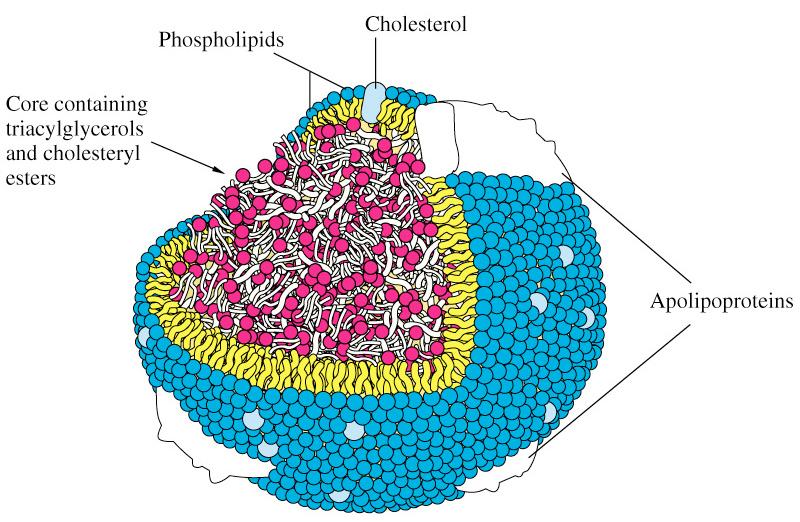 |
front 298 Diagram: large intestine | back 298 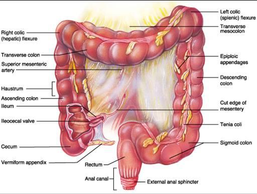 |
front 299 large intestine = colon | back 299 The portion of the gastrointestinal tract which extends from the ileum to the anus, forming an arch in the abdominal cavity around the convolutions of the small intestine and including the cecum, appendix, ascending colon, transverse colon, descending colon, sigmoid colon, rectum, and anal canal; it is a muscular membranous tube whose wall consists of an inner simple columnar epithelium, a lamina propria of loose fibrous connective tissue, a muscularis, and a serosa; it functions to complete the final absorption (less than 5%) of nutrients from food, to reabsorb the water added to the chyme by the secretions of the digestive system, and to compress and compact the remaining wastes into feces, which it stores and excretes. nickname - large bowel |
front 300 mesocolon | back 300 The portion of the abdominal mesentery which holds the lower colon against the dorsal abdominal wall. |
front 301 cecum | back 301 The large blind pouch forming the beginning of the large intestine in the lower right quadrant of the abdominal cavity, and from which the appendix extends; its wall consists of an inner simple columnar epithelium, a lamina propria of loose fibrous connective tissue, a muscularis, and a serosa; it receives chyme from the small intestine via the ileocecal valve. |
front 302 appendix | back 302 A vestigial process which extends from the lower end of the cecum and which resembles a small worm-like pouch; its wall consists of an inner simple columnar epithelium, a lamina propria of loose fibrous connective tissue, a muscularis, and a serosa; it has no significant function. |
front 303 ascending colon | back 303 The part of the large intestine which ascends from the cecum to the transverse colon on the right side of the abdominal cavity; its wall consists of an inner simple columnar epithelium, a lamina propria of loose fibrous connective tissue, a muscularis, and a serosa; it functions to complete the final absorption (less than 5%) of nutrients from food, to reabsorb the water added to the chyme by the secretions of the digestive system, and to compress and compact the remaining wastes into feces. |
front 304 transverse colon | back 304 The part of the large intestine which lies across the upper part of the abdominal cavity; it begins at the right colic flexure as a continuation of the ascending colon and ends at the left colic flexure where the descending colon begins; its wall consists of an inner simple columnar epithelium, a lamina propria of loose fibrous connective tissue, a muscularis, and a serosa; it functions to complete the final absorption (less than 5%) of nutrients from food, to reabsorb the water added to the chyme by the secretions of the digestive system, and to compress and compact the remaining wastes into feces. |
front 305 descending colon | back 305 The part of the large intestine which descends from the transverse colon to the sigmoid colon on the left side of the abdominal cavity; its wall consists of an inner simple columnar epithelium, a lamina propria of loose fibrous connective tissue, a muscularis, and a serosa; it functions to complete the final absorption (less than 5%) of nutrients from food, to reabsorb the water added to the chyme by the secretions of the digestive system, and to compress and compact the remaining wastes into feces. |
front 306 sigmoid colon | back 306 The S-shaped section of the large intestine between the descending colon and the rectum on the lower left side of the abdominal cavity; its wall consists of an inner simple columnar epithelium, a lamina propria of loose fibrous connective tissue, a muscularis, and a serosa; it functions to complete the final absorption (less than 5%) of nutrients from food, to reabsorb the water added to the chyme by the secretions of the digestive system, and to compress and compact the remaining wastes into feces, which it stores prior to excretion. |
front 307 taeniae coli | back 307 The set of three ribbon-like bands of smooth muscle which form the interrupted external layer of the muscularis of the large intestine; they play a role in haustral churning, peristalsis, and mass peristalsis movements in the large intestine. |
front 308 haustra | back 308 The series of shallow pouches which form the gross structure of the wall of the large intestine; contraction of the smooth muscle in the walls of these pouches assists in extracting the water added to the chyme by the secretions of the digestive system, and compresses and compacts the remaining wastes into feces, a process called haustral churning. |
front 309 epiploic appendages | back 309 Small irregular pouches filled with adipose tissue which are situated along the outer wall of the large intestine beneath the visceral peritoneum. |
front 310 right colic flexure | back 310 The bend in the large intestine which is found at the juncture of the ascending and transverse portions. |
front 311 left colic flexure | back 311 The bend in the large intestine which is found at the juncture of the transverse and descending portions. |
front 312 rectum | back 312 The terminal portion of the large intestine, extending from the sigmoid colon to the anal canal; its wall consists of an inner simple columnar epithelium, a lamina propria of loose fibrous connective tissue, a muscularis, and a serosa; it functions to reabsorb the water added to the chyme by the secretions of the digestive system, and to compress and compact the remaining wastes into feces, which it stores prior to excretion. |
front 313 rectal valves | back 313 Large transverse folds in the mucosal wall of the rectum which support the weight of the feces to prevent premature release of feces, especially during bouts of flatus (gas expulsion). |
front 314 anal canal | back 314 The terminal section of the rectum where feces are stored prior to excretion; its wall consists of an inner simple columnar epithelium which changes to a stratified sqaumous epithelium in the distal portion, a lamina propria of loose fibrous connective tissue, a muscularis, and a serosa; it discharges feces through the anus with powerful peristaltic waves of contraction. |
front 315 anus | back 315 The opening at the lower end of the large intestine through which solid waste is eliminated from the body by the process of defecation; its wall consists of an inner simple columnar epithelium which changes to a stratified sqaumous epithelium in the distal portion, a lamina propria of loose fibrous connective tissue, a muscularis, and a serosa; its mucous membrane merges with the skin of the perineum exteriorly. |
front 316 external anal sphincter | back 316 The outer rings or bands of skeletal muscle extending from the coccyx to the central tendinous part of the perineum and surrounding the anus immediately beneath the skin; their contraction and relaxation are under voluntary control and are stimulated to initiate or control defecation. |
front 317 internal anal sphincter | back 317 The inner rings of smooth muscle formed by the thickening of the circular smooth muscle of the rectum; their contraction and relaxation are under involuntary control; its action contributes to the control of defecation. |
front 318 List: structures which increase the surface area of the large intestine. | back 318 haustrae (Note: technically also the rectal valves, but that is not their purpose) |
front 319 gastroileal reflex | back 319 One of the visceral reflexes involved in the regulation of digestion in which sensory signals from gastric distension as a meal is ingested stimulate increased motility = peristalsis in the ileum and relaxation of the ileocecal valve; therefore, chyme moves through the ileocecal valve into the large intestine; it is mediated by gastrin release from G cells in the stomach and possibly also supported by parasympathetic autonomic motor impulses. |
front 320 haustral churning | back 320 Slow segmenting movements within the large intestine; as each haustral pouch is filled with chyme, the walls contract which assists in reabsorbing the water added to the chyme by the secretions of the digestive system, and compressing and compacting the remaining wastes into feces; after the compaction movement, the chyme/feces is slowly moved forward to the next haustral pouch; it is mediated by parasympathetic autonomic motor impulses. |
front 321 mass peristalsis | back 321 Forcible peristaltic movements of short duration, occurring only three or four times a day, which move the contents of the large intestine from one division to the next, as from the ascending to the transverse colon; it is mediated by parasympathetic autonomic motor impulses; it is often stimulated by gastric filling, the gastrocolic reflex. |
front 322 gastrocolic reflex | back 322 One of the visceral reflexes involved in the regulation of digestion in which sensory signals from distension of the stomach stimulate a mass peristaltic (coordinated, rhythmic, smooth muscle contraction which acts to force food through the digestive tract) movement of the colon that often occurs fifteen to thirty minutes after eating a meal; it is mediated by parasympathetic autonomic motor impulses. |
front 323 feces | back 323 The excrement discharged from the intestines, consisting of bacteria, exfoliated from the intestines, secretions, chiefly of the liver and a small amount of food residue. aka - stool |
front 324 dietary fiber | back 324 The bulky residue of food, primarily cellulose, which cannot be broken down by enzymes in the small intestine of the digestive system; almost all natural fiber comes from plants; although fiber has little nutritional value, it offers other health benefits; people with a high-fiber diet experience lower rates of cancer because food passes through the body more rapidly; by adding bulk to the diet, fiber also prevents constipation, minimizes intestinal disorders, and may serve as an aid in dieting. [Note: The benefits of consuming foods high in fiber include lower blood levels of cholesterol and triglycerides. Foods high in fiber include legumes, green, leafy vegetables, whole fruits, and unrefined foods such as bran and sprouted seeds.] aka - roughage |
front 325 defecation reflex | back 325 One of the visceral reflexes involved in the regulation of digestion in which sensory signals from distension of the rectum as feces accumulate there (after peristalsis moves them from the sigmoid colon) stimulates the involuntary relaxation of the internal anal sphincter; if voluntary relaxation of the external anal sphincter follows, feces will be discharged; if there is no voluntary relaxation of the external anal sphincter, the feces move back to the sigmoid colon until the next peristaltic wave; it is mediated by parasympathetic autonomic motor impulses. |
front 326 DIagram: defecation reflex | back 326 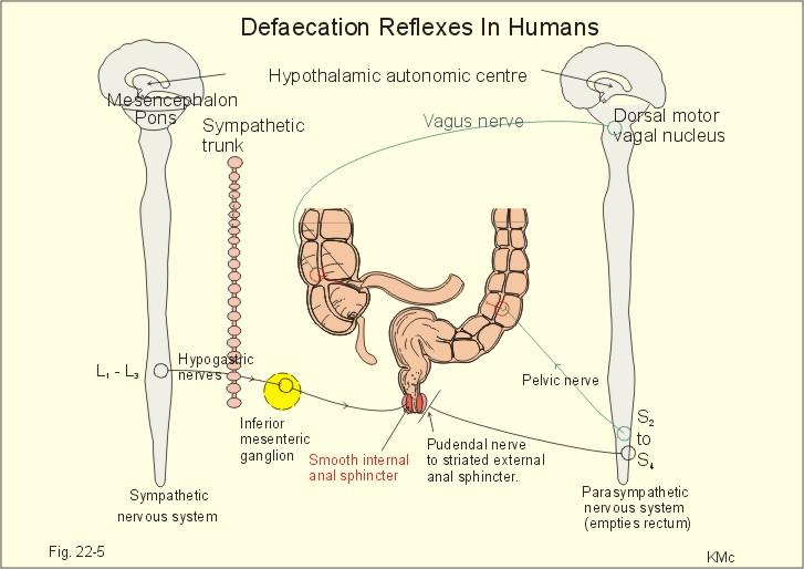 |
front 327 List:the functions of the large intestine. | back 327 (1) absorption of residual water and electrolytes from digestion
|