Instructions for Side by Side Printing
- Print the notecards
- Fold each page in half along the solid vertical line
- Cut out the notecards by cutting along each horizontal dotted line
- Optional: Glue, tape or staple the ends of each notecard together
Outside Hours Mosby's Essential Sciences for Therapeutic Massage
front 1 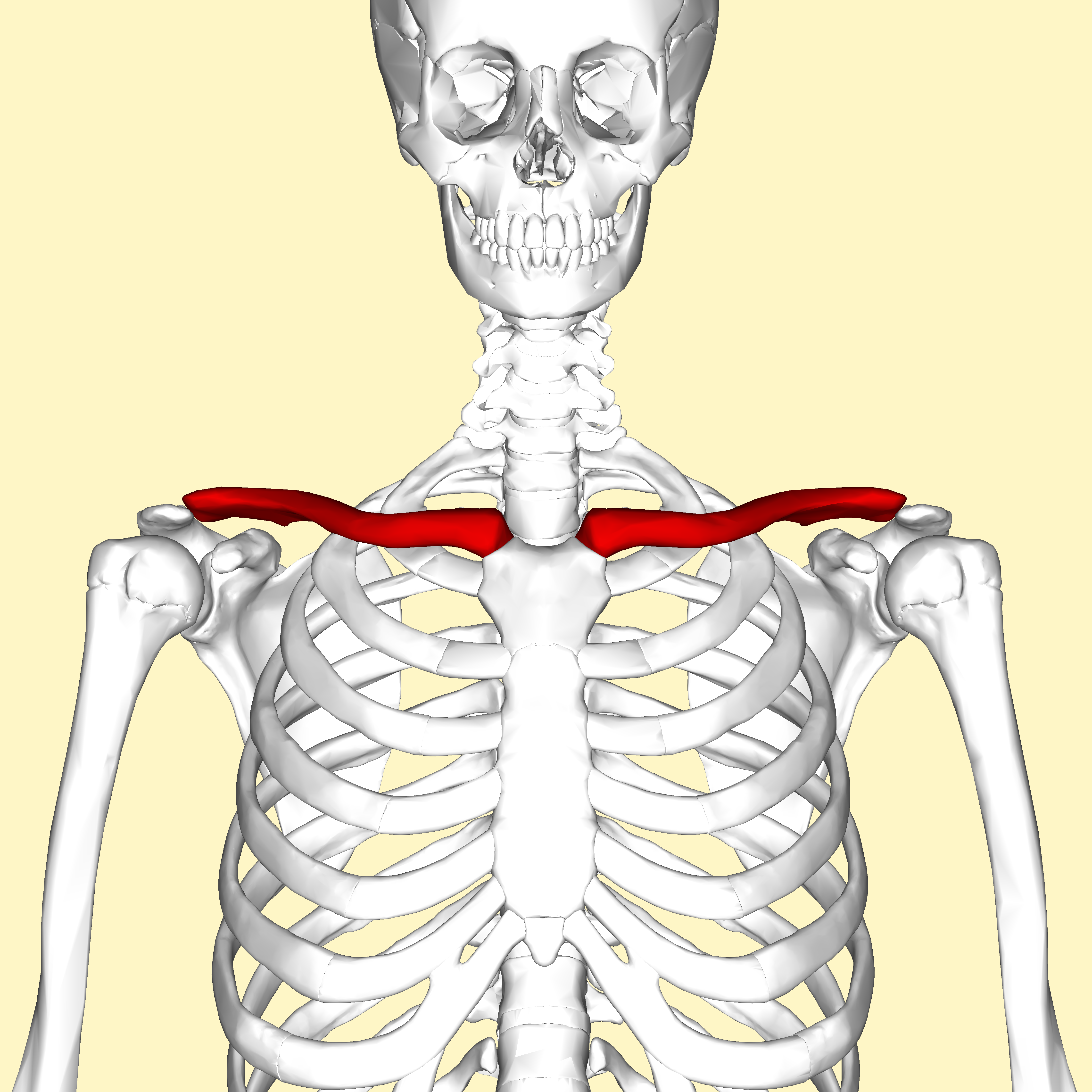 | back 1 1. sternal end 2. acromial end |
front 2  | back 2
|
front 3  | back 3
|
front 4  ulna | back 4
|
front 5 radius | back 5
|
front 6  | back 6 Carpals
|
front 7 | back 7 Metacarpals |
front 8 | back 8 Phalanges
|
front 9  | back 9
|
front 10  | back 10 cervical vertebrae
|
front 11  | back 11 thoracic vertebrae
|
front 12  | back 12 lumbar vertebrae |
front 13 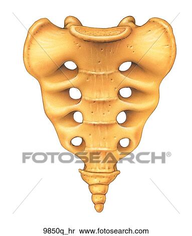 | back 13 sacrum
|
front 14  | back 14
|
front 15  | back 15
|
front 16  | back 16 Hyoid |
front 17  | back 17 Middle Ear
|
front 18  | back 18
|
front 19  | back 19
|
front 20  | back 20
|
front 21  | back 21
|
front 22  | back 22
|
front 23  | back 23 occipitofrontalis O: Two occipital bellies and two frontal bellies |
front 24 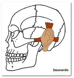 | back 24 auricularis anterior O: Temporal fascia |
front 25 | back 25 auricularis posterior O: Mastoid Process |
front 26 | back 26 auricularis superior O: Temporal fascia |
front 27  | back 27 orbicularis oculi O: frontal bone; medial palpebral ligament; lacrimal bone |
front 28  | back 28 orbicularis oris O: Maxilla and mandible |
front 29  | back 29 buccinator O: from the alveolar processes of the maxillary bone and mandible,
temporomandibular joint |
front 30 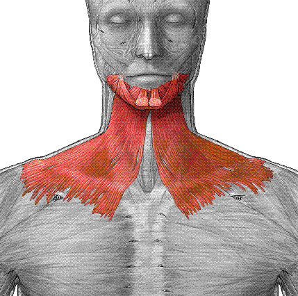 | back 30 platysma O: subcutaneous tissue of infraclavicular and supraclavicular
regions |
front 31  | back 31 masseter O: zygomatic arch and maxilla |
front 32  | back 32 temporalis O: Temporal lines on the parietal bone of the skull and the superior
temporal surface of the sphenoid bone. |
front 33 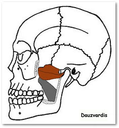 | back 33 lateral pterygoid O: Great wing of sphenoid and pterygoid plate |
front 34 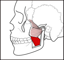 | back 34 medial pterygoid O: deep head: medial side of lateral pterygoid plate
behind the upper teeth, superficial head: pyramidal process
of palatine bone and maxillary tuberosity |
front 35  | back 35 sternocleidomastoid O: Manubrium sterni and medial portion of the clavicle |
front 36 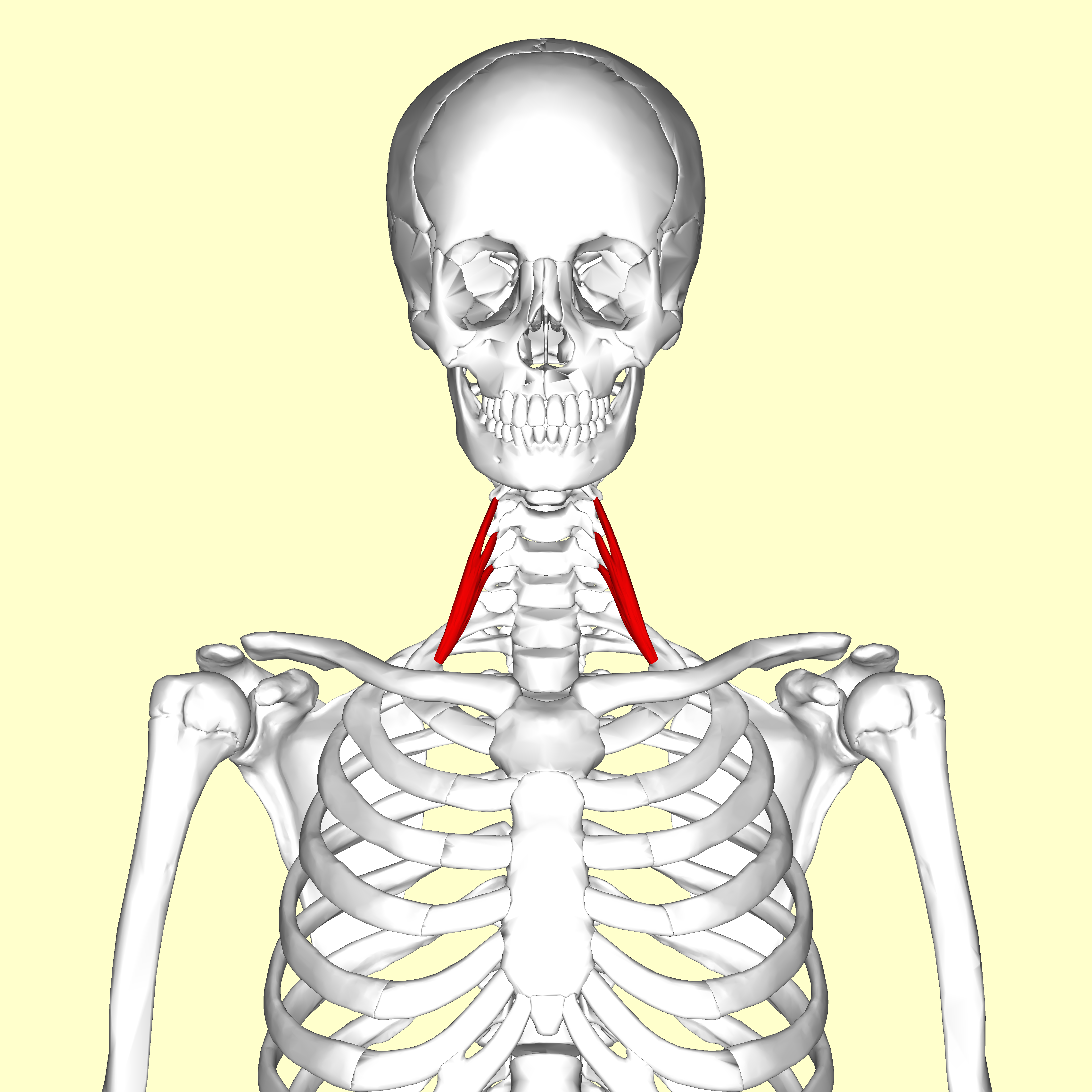 | back 36 anterior scalene O: Transverse processes of the third, fourth, fifth, and sixth
cervical vertebræ (C3, C4, C5 and C6) |
front 37  | back 37 middle scalene O: Posterior tubercles of the transverse processes of the lower six
cervical vertebræ (C2, C3, C4, C5, C6 and C7) |
front 38  | back 38 posterior scalene O: Transverse processes of C4, C5 and C6 |
front 39 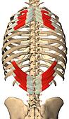 | back 39 serratus posterior superior O: Nuchal ligament (or ligamentum nuchae) and the spinous processes
of the vertebrae C7 through T3 |
front 40 | back 40 serratus posterior inferior O: Vertebrae T11 - L2 |
front 41  | back 41 diaphragm O: Attaches to the sternum and xiphoid process
anteriorly, the L1 through the L3 lumbar vertebrae and the arcuate
ligaments posteriorly, and the costal margin peripherally |
front 42 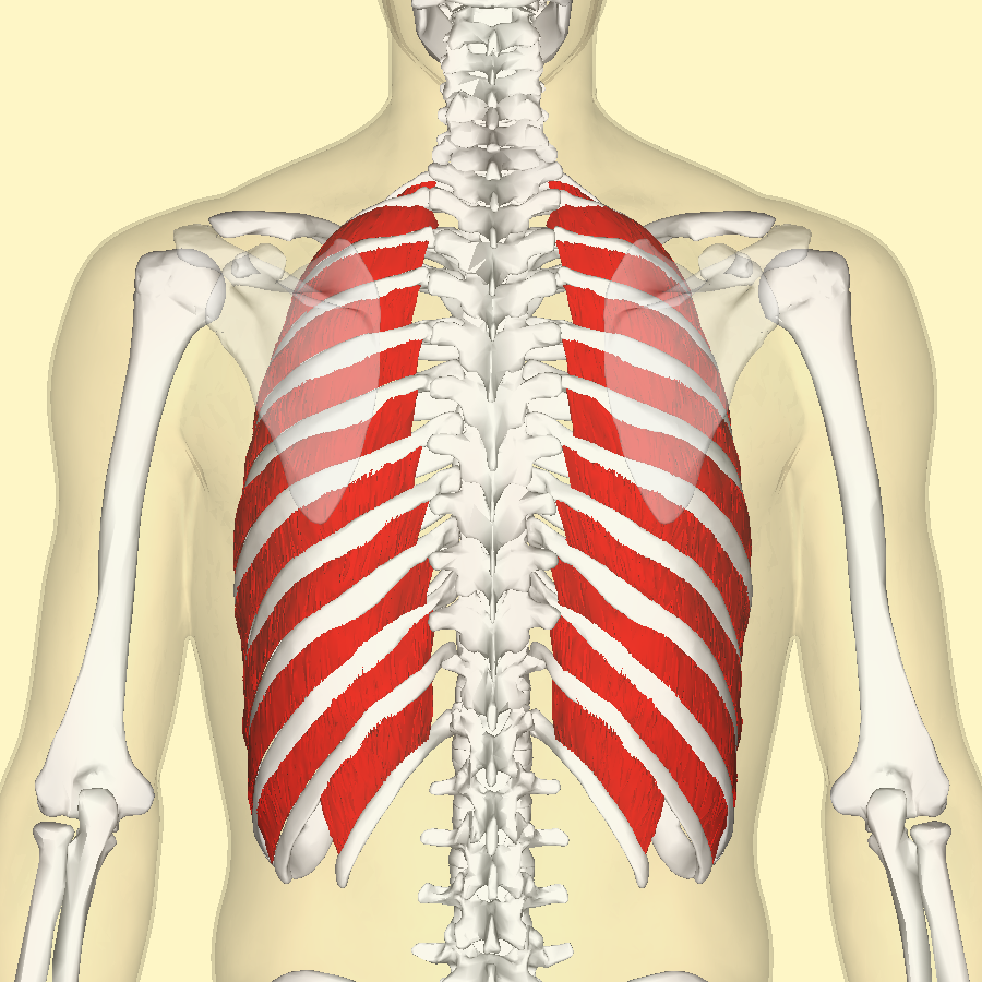 | back 42 external intercostals O: lower border of ribs |
front 43 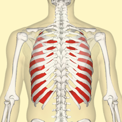 | back 43 internal intercostals O: Rib - inferior border |
front 44 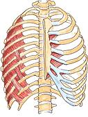 | back 44 transversus thoracis O: Costal cartilages of last 3-4 true ribs, body of sternum and
xiphoid process |
front 45 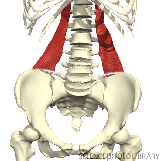 | back 45 quadratus lumborum O: iliac crest and iliolumbar ligament |
front 46 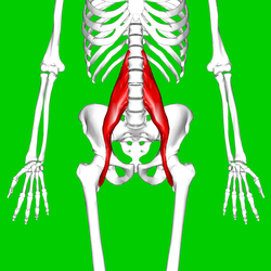 | back 46 Psoas Major (iliopsoas) O: Transverse processes of T12-L5 and the lateral aspects of the
discs between them |
front 47  | back 47 Iliacus (iliopsoas) O: upper two-third of the iliac fossa |
front 48  | back 48 1. external abdominal oblique O: Ribs 5-12 |
front 49 | back 49 2. internal abdominal oblique O: Inguinal ligament, Iliac crest and the Lumbodorsal
fascia. |
front 50 | back 50 3. transversus abdominis O: Iliac crest, inguinal ligament, thoracolumbar fascia, and costal
cartilages 7-12 |
front 51 | back 51 4. Rectus abdominis O: crest of pubis |
front 52  | back 52 splenius capitis O: Nuchal ligament and spinous process of C7-T3 |
front 53 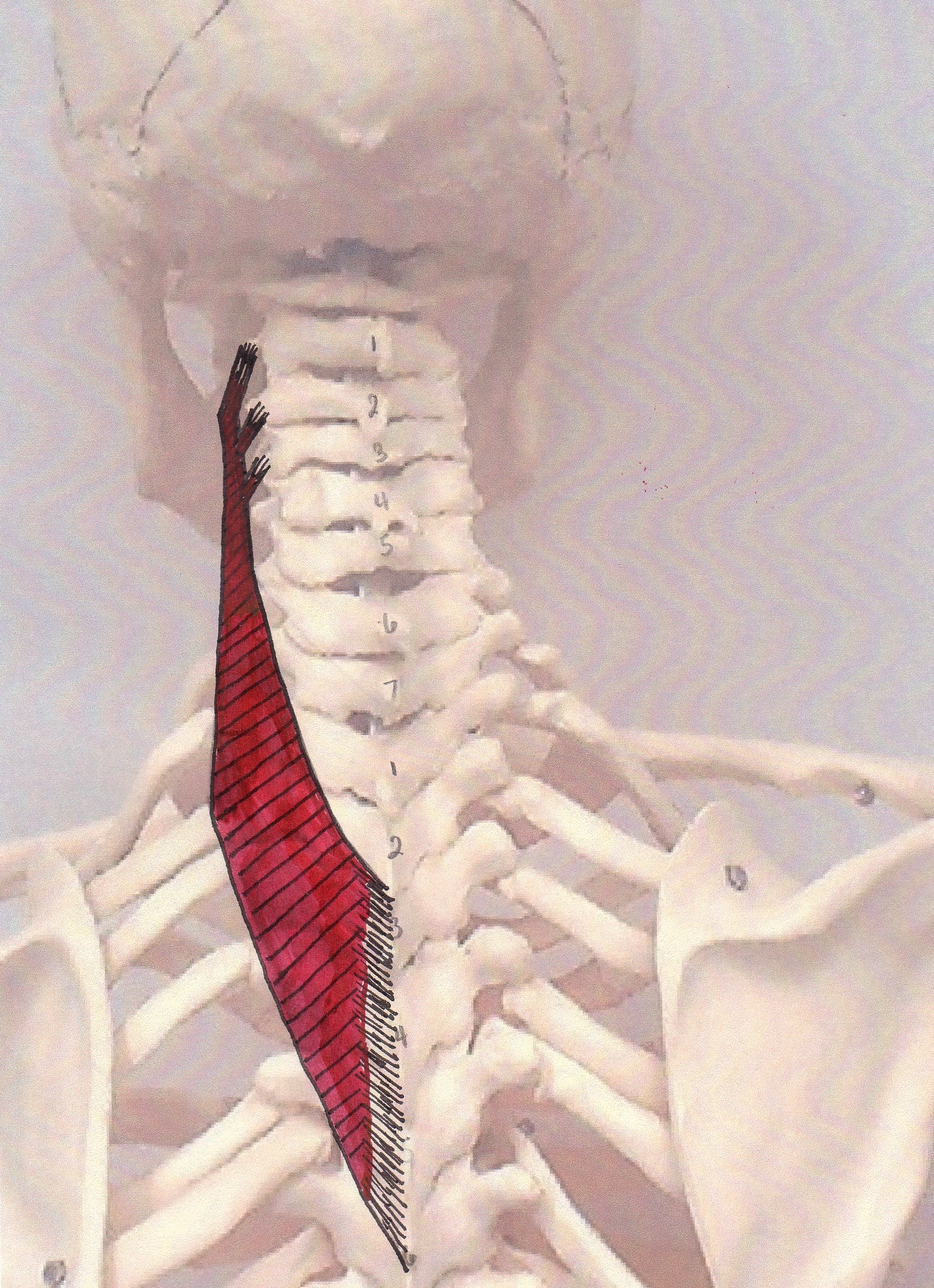 | back 53 splenius cervicis O: Spinous processes of T3-T6 |
front 54 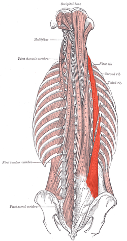 | back 54 iliocostalis (erector spinae group) O: Sacrum/Illiac Crest/Spinous Processes of lower lumbar/thoracic
vertebrae |
front 55  | back 55 longissimus (erector spinae group) O: transverse process |
front 56  | back 56 spinalis (erector spinae group) O: Thoracis: Spinous process of upper lumbar and lower thoracic
vertebrae. .Cervicis: nuchal ligament and spinous process of
C7 |
front 57  | back 57 semispinalis O: Transverse processes of lower cervical and higher thoracic
columna |
front 58 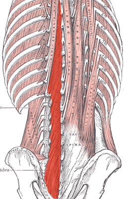 | back 58 multifidus O: Sacrum, Erector spinae Aponeurosis, PSIS, and Iliac
crest |
front 59  | back 59 suboccipital group |
front 60  | back 60 gluteus maximus O: Gluteal surface of ilium, lumbar fascia, sacrum, sacrotuberous
ligament |
front 61 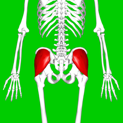 | back 61 gluteus medius O: Gluteal surface of ilium, under gluteus maximus |
front 62 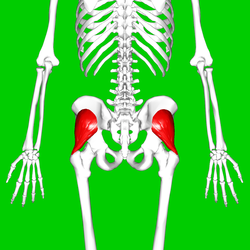 | back 62 gluteus minimus O: From area in between the anterior gluteal line and inferior
gluteal line of Gluteal surface ilium, under gluteus medius. |
front 63  | back 63 tensor fascia latae O: Iliac crest |
front 64  | back 64 piriformis (deep lateral rotator group) O: Sacrum |
front 65  | back 65 obturator internus (deep lateral rotator group) O: Ischiopubic ramus & obturator membrane |
front 66  | back 66 obturator externus (deep lateral rotator group) O: obturator foramen and obturatory membrane |
front 67 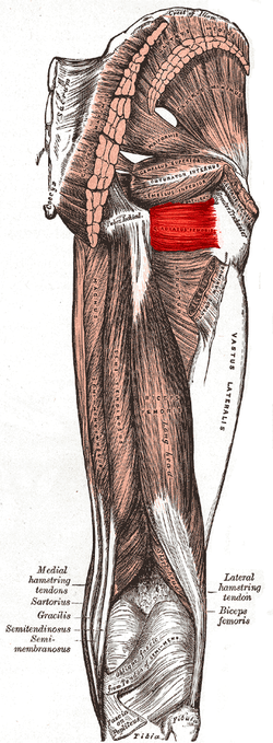 | back 67 quadratus femoris (deep lateral rotator group) O: Ischial tuberosity |
front 68  | back 68 gemellus superior (deep lateral rotator group) O: spine of the ischium |
front 69  | back 69 gemellus inferior (deep lateral rotator group) O: Ischial tuberosity |
front 70 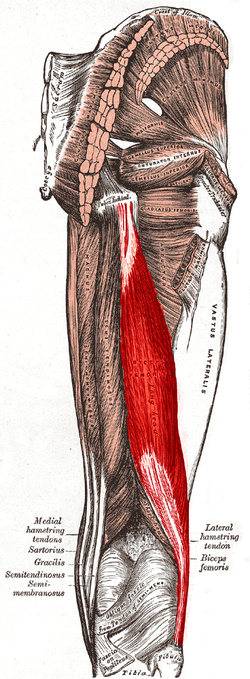 | back 70 biceps femoris (hamstring group) O: tuberosity of the ischium, linea aspera, femur |
front 71 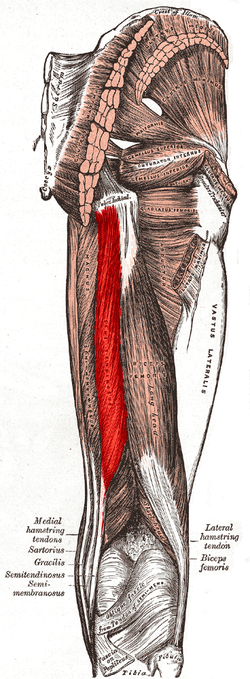 | back 71 semitendinosus (hamstring group) O: Tuberosity of the ischium |
front 72 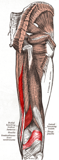 | back 72 semimembranosus O: Ischial tuberosity |
front 73 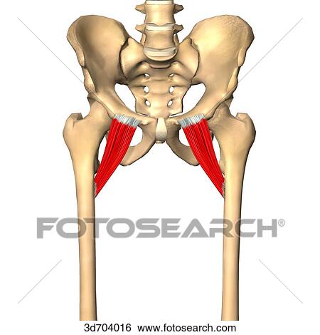 | back 73 pectineus O: Pectineal line of the pubic bone |
front 74  | back 74 adductor brevis (adductor group) O: anterior surface of the inferior ramus and body of the
pubis |
front 75  | back 75 adductor longus (adductor group) O: pubic body just below the pubic crest |
front 76  | back 76 adductor magnus (adductor group) O: Pubis, tuberosity of the ischium |
front 77  | back 77 gracilis O: ischiopubic ramus |
front 78 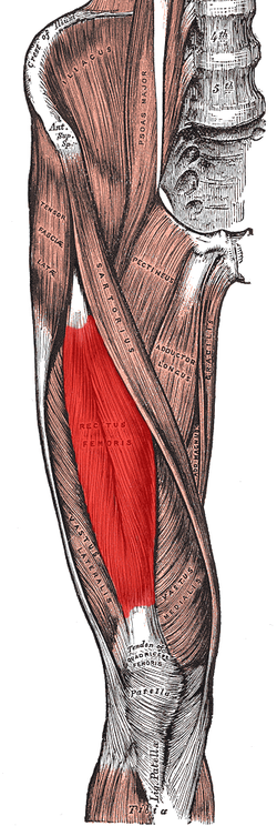 | back 78 rectus femoris (quadriceps group) O: anterior inferior iliac spine and the exterior surface of the
bony ridge which forms the groove on the iliac portion of the
acetabulum |
front 79  | back 79 vastus lateralis (quadriceps group) O: Greater trochanter, Intertrochanteric line, and Linea aspera
of the Femur |
front 80 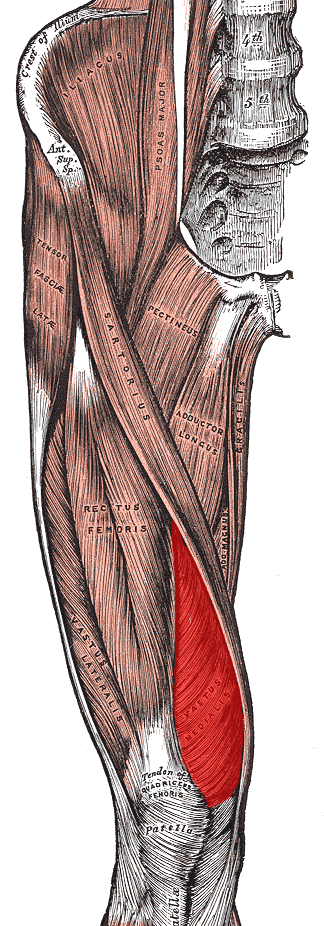 | back 80 vastus medialis (quadriceps group) O: Medial side of femur |
front 81  | back 81 vastus intermedius (quadriceps group) O: antero/ lateral femur |
front 82  | back 82 tibialis anterior O: Upper 1/2 & Lateral Condyle of Tibia |
front 83 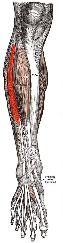 | back 83 extensor digitorum longus O: Anterior lateral condyle of tibia, anterior shaft of fibula
and superior 3⁄4 of interosseous
membrane |
front 84 | back 84 extensor hallicus longus O: Arises from the middle portion of the fibula on the anterior
surface and the interosseous membrane |
front 85  | back 85 fibularis (peroneus) tertius O: distal anterior surface of the fibula also the interosseous
membrane |
front 86  | back 86 fibularis (peroneus) longus O: Upper lateral shaft of fibula |
front 87  | back 87 fibularis (peroneus) brevis O: Lower two-thirds of lateral fibula |
front 88 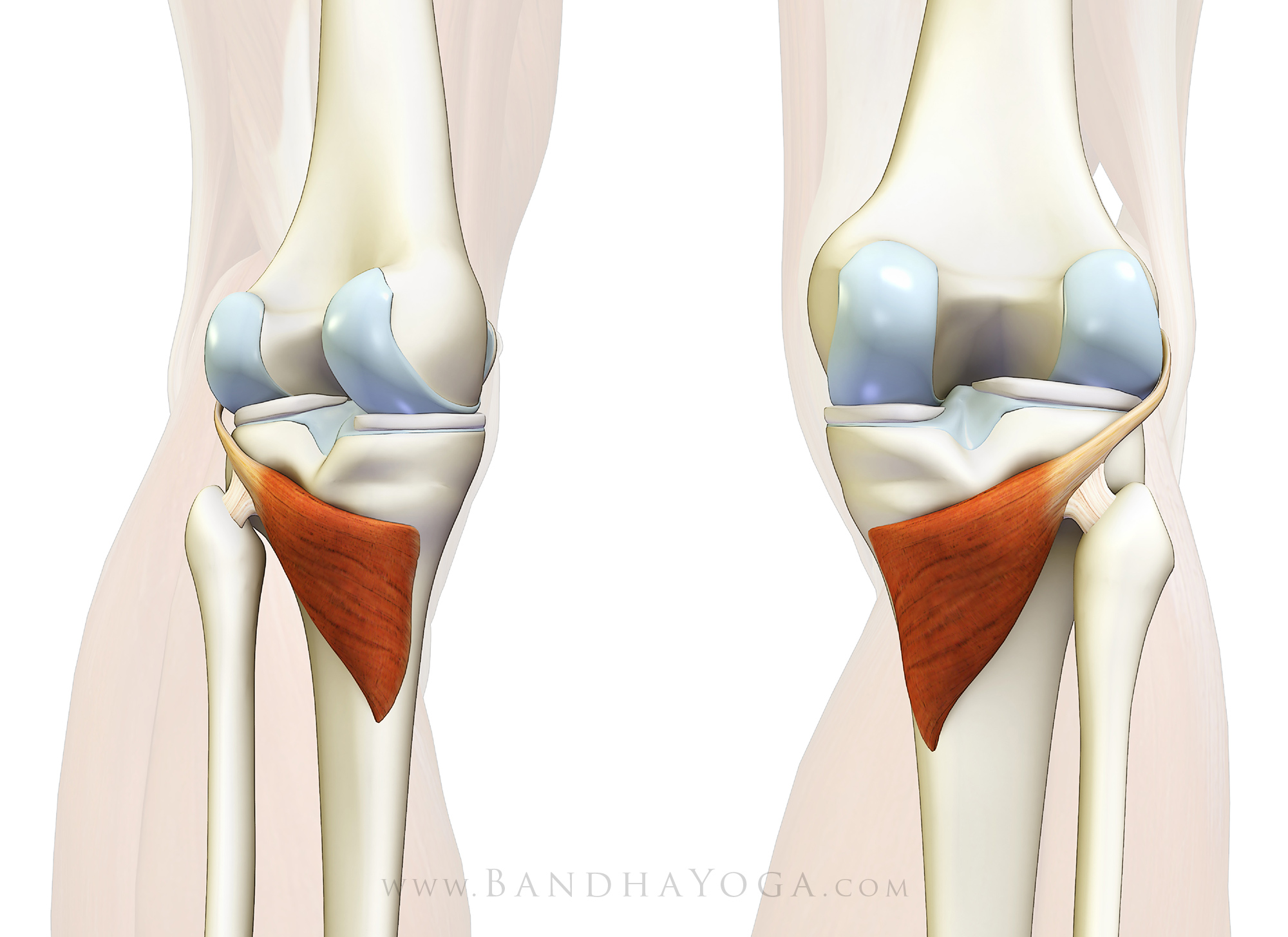 | back 88 popliteus O: Lateral condyle of the femur, and the lateral meniscus and
joint capsule |
front 89 | back 89 tibialis posterior O: Tibia and fibula |
front 90  | back 90 flexor digitorum longus O: Posterior surface of the body of the tibia |
front 91 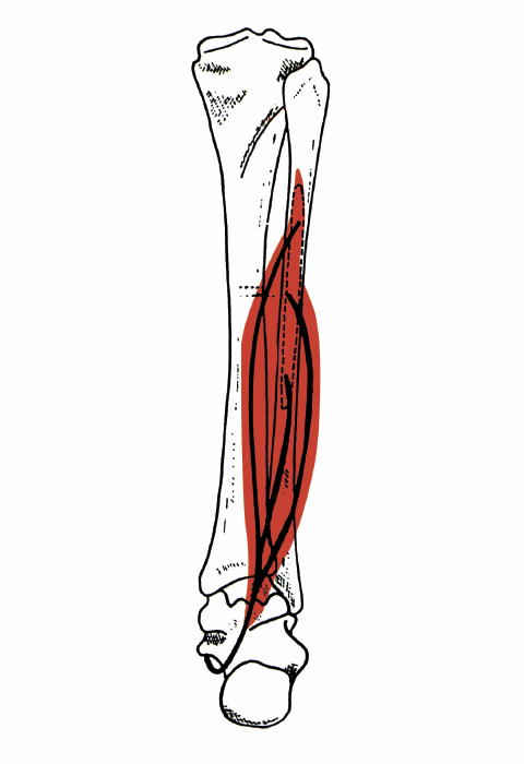 | back 91 flexor hallicus longus O: fibula, posterior aspect of middle 1/3 |
front 92 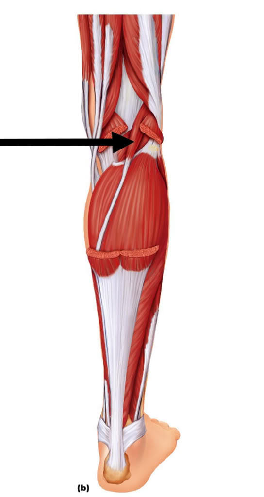 | back 92 plantaris O: Lateral supracondylar ridge of femur above lateral head of
gastrocnemius |
front 93 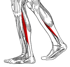 | back 93 soleus O: fibula, medial border of tibia (soleal line) |
front 94 | back 94 gastrocnemius O: superior to articular surfaces of lateral condyle of femur and
medial condyle of femur |
front 95 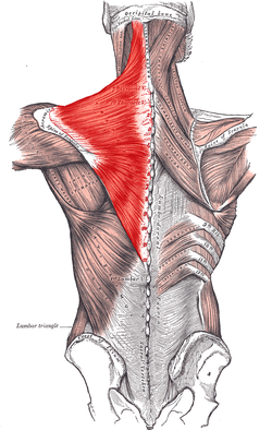 | back 95 trapezius O: external occipital protuberance, nuchal ligament, medial
superior nuchal line, spinous processes of vertebrae C7-T12 |
front 96  | back 96 rhomboid major O: spinous processes of the T2 to T5 vertebrae |
front 97 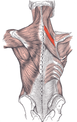 | back 97 rhomboid minor O: nuchal ligaments and spinous processes of C7–T1 |
front 98  | back 98 levator scapula O: Posterior tubercles of transverse processes of C1 - C4
vertebrae |
front 99 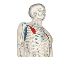 | back 99 pectoralis minor O: Third to fifth ribs, near their costal cartilages |
front 100 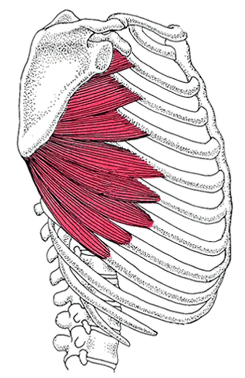 | back 100 serratus anterior O: fleshy slips from the outer surface of upper 8 or 9
ribs |
front 101 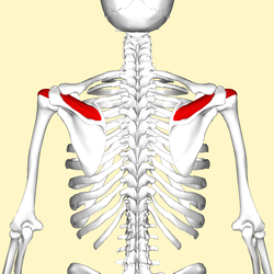 | back 101 supraspinatus O: supraspinous fossa of scapula |
front 102 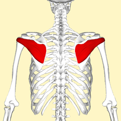 | back 102 infraspinatus O: infraspinous fossa of the scapula |
front 103  | back 103 teres minor O: lateral border of the scapula |
front 104 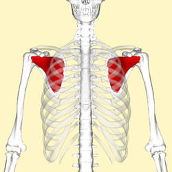 | back 104 subscapularis O: Subscapular fossa |
front 105  | back 105 deltoid O: the anterior border and upper surface of the lateral third of
the clavicle, acromion, spine of the scapula |
front 106  | back 106 pectoralis major O: Clavicular head: anterior surface of the
medial half of the clavicle. Sternocostal head:
anterior surface of the sternum, the superior six costal cartilages,
and the aponeurosis of the external oblique muscle |
front 107  | back 107 subclavius O: first rib and cartilage |
front 108  | back 108 latissimus dorsi O: Spinous processes of vertebrae T7-L5, thoracolumbar fascia,
iliac crest, inferior 3 or 4 ribs and inferior angle of
scapula |
front 109 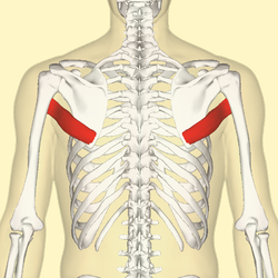 | back 109 teres major O: Posterior aspect of the inferior angle of the scapula |
front 110  | back 110 coracobrachialis O: Coracoid process of scapula |
front 111 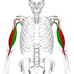 | back 111 biceps brachii O: Short head: coracoid process of the scapula
Long head: supraglenoid tubercle |
front 112 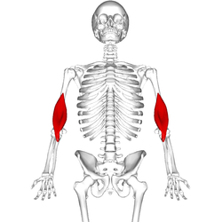 | back 112 brachialis O: anterior surface of the humerus, particularly the distal half
of this bone |
front 113  | back 113 brachioradialis O: Lateral supracondylar ridge of the humerus |
front 114  | back 114 pronator teres O: humeral head: medial epicondyle of humerus (common
flexor tendon) ulnar head: coronoid process of ulna |
front 115  | back 115 supinator O: Lateral epicondyle of humerus, supinator crest of ulna, radial
collateral ligament, annular ligament |
front 116  | back 116 pronator quadratus O: medial, anterior surface of the ulna |
front 117 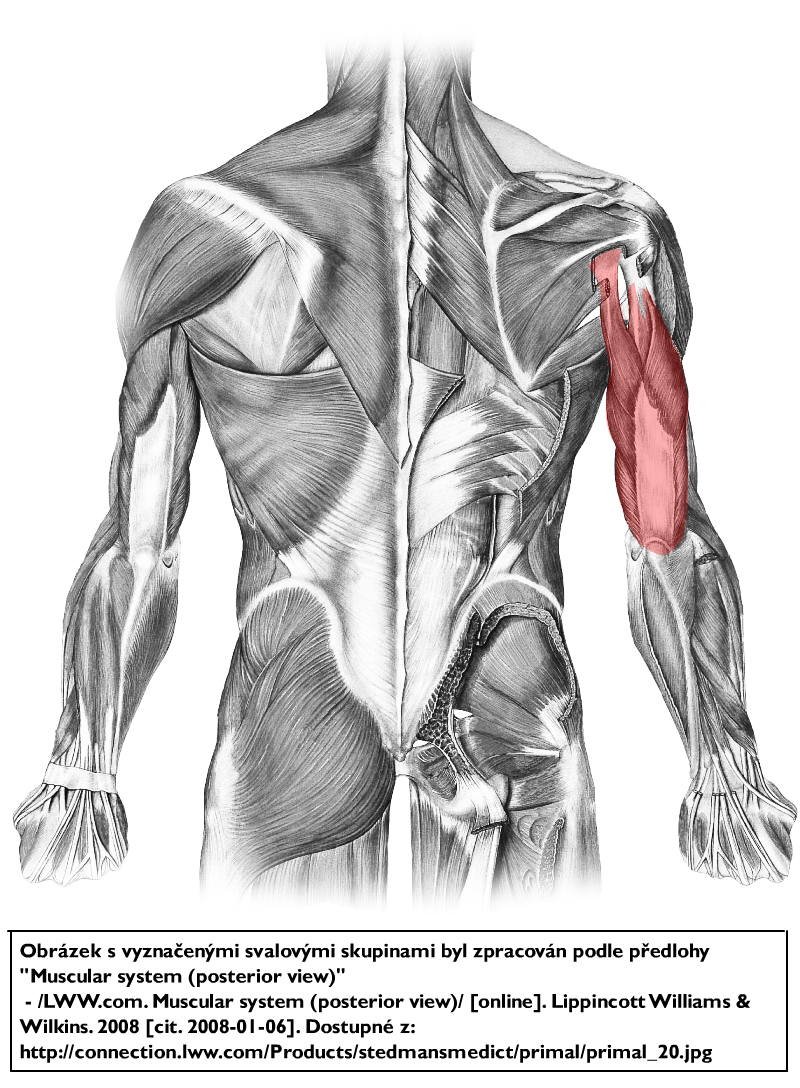 | back 117 triceps brachii O: Long head: infraglenoid tubercle of scapula
Lateral head: above the radial sulcus
Medial head: below the radial sulcus |
front 118  | back 118 anconeus O: lateral epicondyle of the humerus proximally |
front 119  | back 119 flexor carpi radialis O: medial epicondyle of humerus (common flexor tendon) |
front 120 | back 120 palmaris longus O: medial epicondyle of humerus (common flexor tendon) |
front 121  | back 121 flexor carpi ulnaris O: medial epicondyle (common flexor tendon) and medial margin on
olecranon of ulna |
front 122 | back 122 flexor digitorum superficialis O: medial epicondyle of the humerus (common flexor tendon) as well
as parts of the radius and ulna. |
front 123  | back 123 flexor digitorum profundus O: upper 3/4 of the volar and medial surfaces of the body of the
ulna, interosseous membrane and deep fascia of the forearm |
front 124  | back 124 flexor pollicis longus O: The middle 2/4 of the volar surface of the radius and the
adjacent interosseus membrane. |
front 125  | back 125 extensor carpi radialis longus O: lateral supracondylar ridge |
front 126  | back 126 extensor carpi radialis brevis O: humerus at the anterior of lateral epicondyle (common extensor
tendon) |
front 127  | back 127 extensor digitorum O: lateral epicondyle (common extensor tendon) |
front 128 | back 128 extensor digiti minimi O: the anterior portion of the lateral epicondyle of the humerus
(common extensor tendon) |
front 129  | back 129 extensor carpi ulnaris O: Common extensor tendon (lateral epicondyle), ulna |
front 130  | back 130 extensor pollicis brevis O: radius and the interosseous membrane |
front 131 | back 131 abductor pollicis longus O: ulna, radius, Interosseous membrane |
front 132  | back 132 extensor pollicis longus O: Middle third of posterior surface of ulna, interosseous
membrane |
front 133  | back 133 extensor indicis O: posterior distal third of ulna and interosseous membrane |