Instructions for Side by Side Printing
- Print the notecards
- Fold each page in half along the solid vertical line
- Cut out the notecards by cutting along each horizontal dotted line
- Optional: Glue, tape or staple the ends of each notecard together
Path: CNS
front 1 what is the CNS defined as | back 1 brain and spinal cord |
front 2 what are the cells of the CNS | back 2 neurons
|
front 3 what is the added dimension of disease in CNS | back 3 traumatic |
front 4 where is there nissl substance | back 4 dendrites but not axons |
front 5 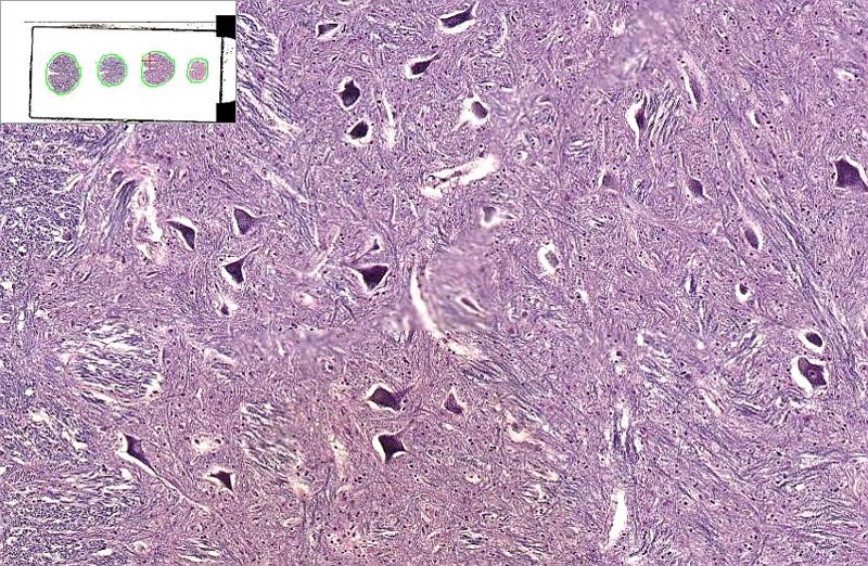 what is this section from | back 5 spinal cord |
front 6 are neurons or glial cells more common in the CNS | back 6 nerons |
front 7 what are the glial cells associated with the CNS | back 7  astrocyte
|
front 8 what do ependymal cells line | back 8 all ventricles and choroid plexus |
front 9 what are the most common glial cells | back 9 astrocyte |
front 10 what are the haloed cells in the CNS | back 10 oligodendrocytes |
front 11 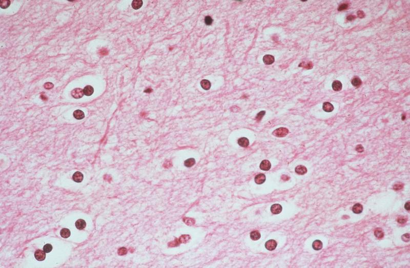 what are the cells that populate this screen | back 11 oligodendrocytes |
front 12 why would there be clear spaces around these oligodendrocytes | back 12 just like fat (myelin) washes out from Schwann glial cells |
front 13 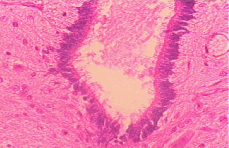 hat cells are lining this space in this frame | back 13 ependymal cells |
front 14 what cells do ependymal cells look like | back 14 ciliated columnar cells |
front 15 what is the only glial cell that looks epithelial | back 15 ependymal cells (simple columnar like in glands) |
front 16 what is the configureation of the choroid plexus | back 16 papillary in configuration |
front 17 what is the role of the choroid plexus | back 17 pump out CSF |
front 18 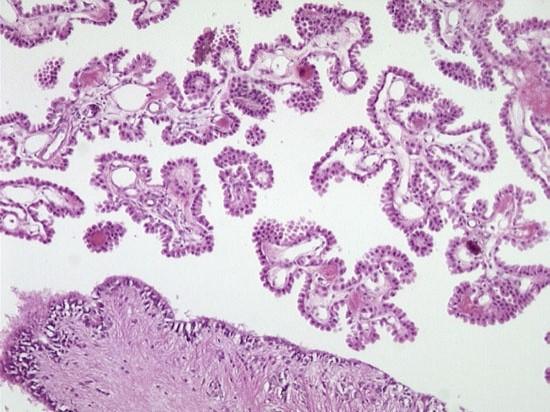 what is this | back 18 choroid plexus |
front 19 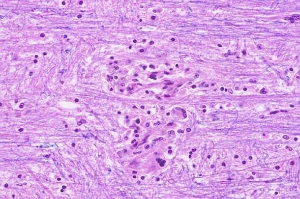 what type of cells is this a cluster of | back 19 microglia (MP of CNS) |
front 20 what are the cellular reactions of neurons | back 20 acute: red neuron=karyolysis
|
front 21 what are the cellular reactions of glia | back 21 gliosis
|
front 22 what all go hand in hand with gliosis in terms of nonspecific reaction to injury | back 22 GLIOSIS
|
front 23 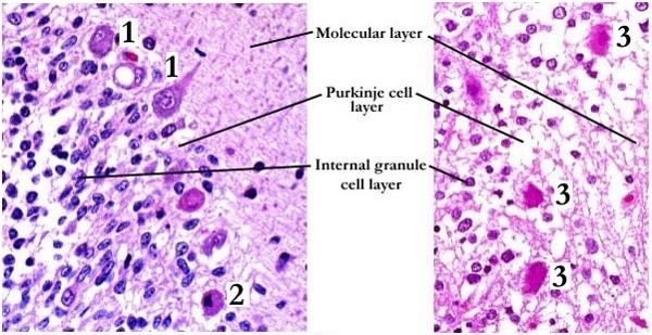 what kind of neuronal injury took place here | back 23 acute neuronal injury (red nucleus) |
front 24 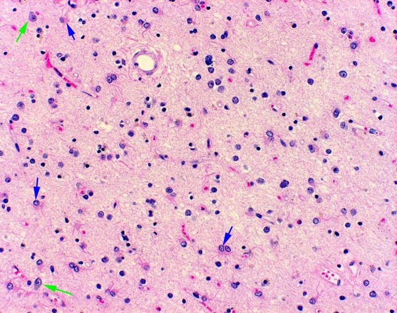 look for gliosis, neuronal loss in this picture | back 24 no data |
front 25 what types of cerebral edema can take place | back 25 vasogenic
|
front 26 what is the normal weight of the CNS | back 26 1200-1300 gms |
front 27 what happens in vasogenic cerebral edema | back 27 BBB is disturbed; intravascular to intercellular |
front 28 what type of cerebral edema is cytotoxic | back 28 intracellular |
front 29 where can cerebral edema go | back 29 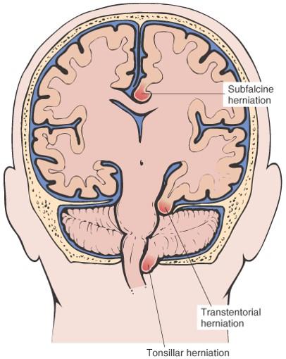 subfalcine (supratentorial)
|
front 30 what is the fourth obvious sign of cerebral edema | back 30 flattening of gyri and small sulci |
front 31 why do flattened gyri signify edema | back 31 compression against calvarium |
front 32 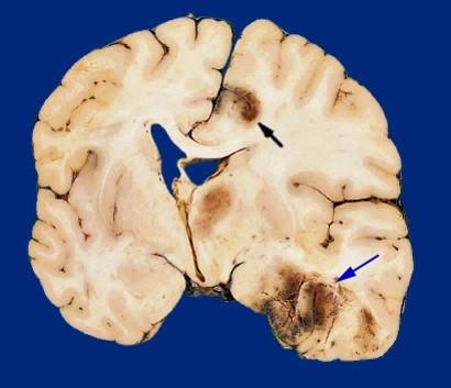 what was likely going on here | back 32 hemorrhage and edema |
front 33 what does notching of the cingulate gyrus indicate | back 33 cerebral edema |
front 34 what does cerebellar tonsil herniation indicate | back 34 cerebral edema |
front 35 what are the symptoms of cerebral edema | back 35 headache
|
front 36 what can cause hydrocephalus | back 36 impaired resorption
|
front 37 what is communicating vs noncommunicating hydrocephalus | back 37 entire head vs part of the head |
front 38 what types of pressures will someone have in hydrocephalus | back 38 high pressure and normal pressure |
front 39 what is the key factor in whether hydrocephalus will result in any cranial enlargment | back 39 fontanelle closure |
front 40 which fontanelle stays open for a while | back 40 anterior |
front 41 what can hydrocephalus on the CT look like | back 41 dilated ventricles |
front 42 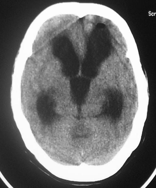 what is going on here | back 42 hydrocephalus |
front 43 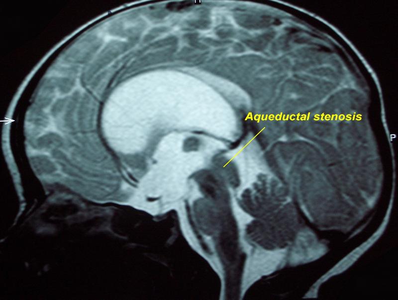 what is this aqueductal stenosis causing | back 43 hydrocephalus on MRI |
front 44 what space would be more dilated: space after aqueductal stenosis or before it | back 44 before |
front 45 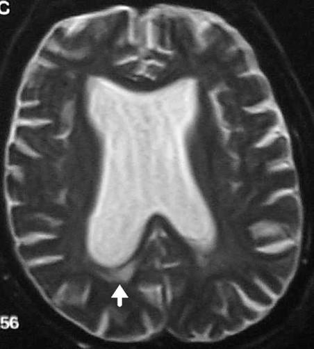 what type of imaging is this and what is being shown | back 45 MRI: water dense in MRI
|
front 46 would dilated ventricles at autopsy signal hydrocephalus | back 46 YEP |
front 47 where are some places where the CNS is malformed | back 47 neural tube
|
front 48 what are some neural tube malformations | back 48 anencephaly
|
front 49 what are some forebrain malformations | back 49 polymicrogyria
|
front 50 what are some posterior fossa malformations | back 50 arnold chiari (infratentorial herniation)
|
front 51 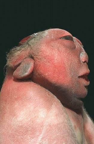 what is this | back 51 anencephaly |
front 52 what antigen is found when spina bifidas are present | back 52 AFP |
front 53 what type of cancer also has a penchant for AFP presence in the bloodstream | back 53 hepatoma |
front 54 where is spina bifida usually | back 54 bottom of the zipper |
front 55 what is polymicrogyria | back 55 small gyri |
front 56 what is holoprosencephaly | back 56 failure of the prosencephalon to develop and separate |
front 57 what is the prosencephalon | back 57 anterior/superior portion of the neural tube |
front 58 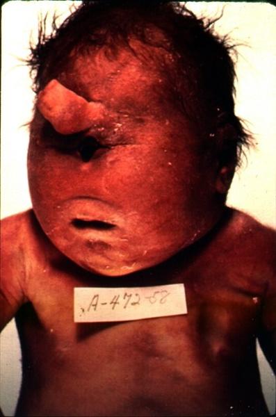 what is wrong with this baby | back 58 holoprosencephaly |
front 59 what is the limiting factor of the falx | back 59 corpus callosum |
front 60 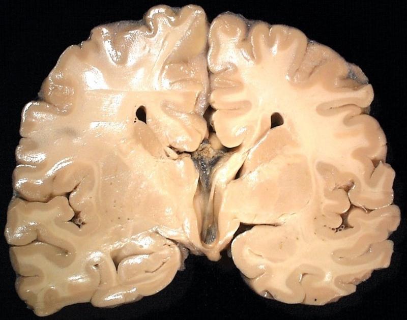 what is wrong with this brain | back 60 absent corpus callosum |
front 61 what is the range of symptoms with an absent corpus callosum | back 61 mild or partial cases are asymptomatic, severe cases result in severe retardation or fatality |
front 62 what is syrinx | back 62 dilation of the central canal of the spinal cord |
front 63 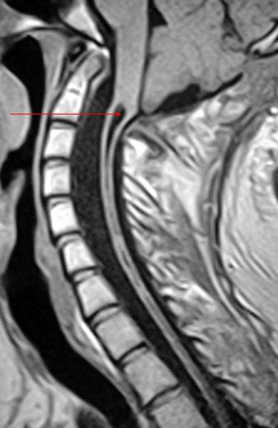 what is being pointed out | back 63 syrinx |
front 64 what are some perinatal brain injuries | back 64 intraparenchymal hemorrhage
|
front 65 can intracerebral hemorrhage extend intraparenchymally and vice versa | back 65 yep |
front 66 what types of things can CNS trauma cause | back 66 skull fractures
|
front 67 what are the brain traumas | back 67 contusion (bruise)
|
front 68 is a concussion clinical or pathological | back 68 clinical |
front 69 what is the hallmark of contusion | back 69 hemorrhage |
front 70 what are the types of skull fractures | back 70 hairline
|
front 71 what are skull fractures associated with | back 71 epidural hematoma |
front 72 if there is contact but no skull fracture what is next on the list of thoughts | back 72 subdural hematoma |
front 73 what do subarachnoid hematomas result from | back 73 some sort of arterial leak, no trauma |
front 74 how does an intraparenchymal hemorrhage go | back 74 any way |
front 75 how does an intraventricular hematoma transpire | back 75 no trauma, rare in adults, common in premies |
front 76 what does the most superficial layer of the dura blend with | back 76 periosteum |
front 77 what can subdural hematomas be related to | back 77 congenital aneurysms |
front 78 where does a subarachnoid hemorrhage occur | back 78 big intracranial arteries |
front 79 what 2 things commonly cause subarachnoid hemorrhage | back 79 hemorrhagic CVAs due to arterial wall rupture
|
front 80 could HTN be a risk factor for subarachnoid hemorhage | back 80 yep |
front 81 could intraparenchymal hemorrhage extend/dissect both ways if it is big enough | back 81 yes ventricular and subarachnoid |
front 82 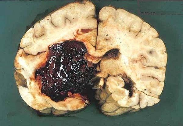 which way is this intraparenchymal hemorrhage dissecting | back 82 intraventricularly |
front 83 could an intraventricular hemorrhage diseaect intraparenchymally | back 83 yes |
front 84 what are the 3 main CNS trauma sequelae | back 84 hydrocephalus
|
front 85 what is the most common CNS trauma sequelae | back 85 hydrocephalus |
front 86 what is the more often sequelae of CNS trauma and what is it from | back 86 hydrocephalus from impaired reabsorption (hemorrhage that affected reabosrption parasagittally |
front 87 is dementia a specific syndrome | back 87 no |
front 88 what is difffuse axonal injury from | back 88 repeated trauma damaging white matter |
front 89 what does spinal cord trauma parallel | back 89 brain patterns of injury (cellularly) |
front 90 what is spinal cord trauma secondary to | back 90 spinal column displacement |
front 91 what does the level of injury of the spinal cord mirror | back 91 motor loss (death, quadriplegia, paraplegia |
front 92 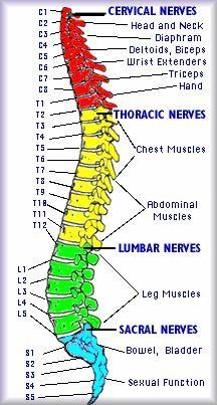 know this fucking chart | back 92 no data |
front 93 what are the cereebrovascular diseases usualyl called | back 93 stroke |
front 94 what are the types of stroke | back 94 ischemic and hemorrhagic |
front 95 what is another name for ischemic stroke | back 95 thrombotic |
front 96 what is decreased in ischemic stroke | back 96 blood and O2 |
front 97 what happens in acute ischemic stroke | back 97 edema to neuronal microvacuolization to pyknosis to karyorrhexis to neutrophils |
front 98 what happens in chronic ischemic stroke | back 98 MP to gliosis |
front 99 what is hemorrhagic stroke due to usually | back 99 rupture of artery/aneurysm |
front 100 what does the middle cerebral artery supply | back 100 lateral brain |
front 101 what does the posterior cerebral artery supply | back 101 medial, some posterior and anterior |
front 102 what does the anterior cerebral artery supply | back 102 medial, some posteiror and anterior |
front 103  what has happened here | back 103 thrombotic MCA |
front 104 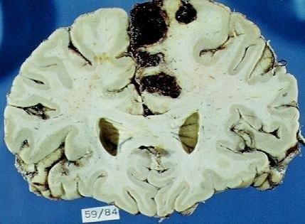 what has happened here | back 104 hemorrhagic ACA |
front 105 what is the only exception in the brain to normal progression of inflammatory response | back 105 usually no fibrosis in the brain, gliosis |
front 106 where would the CNS be sensitive to HTN | back 106  intracerebral area
|
front 107 what type of stroks would be more likely in a hypertensive issue | back 107 hemorrhagic |
front 108 can edema be felt better than seen on gross brain | back 108 yep |
front 109 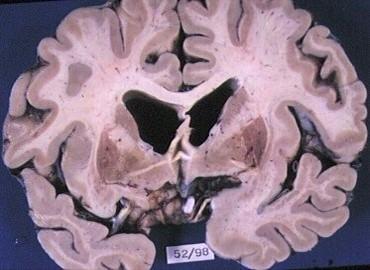 what type of infarct is this | back 109 lacunar infarct |
front 110 are lacunar infarcts asymptomatic? why? | back 110 yes because of very small pinpoint size |
front 111 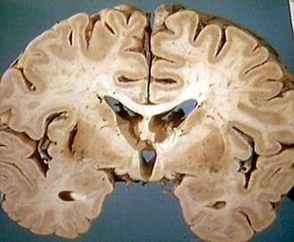 what type of hemorrhage is this | back 111 slit hemorrhage |
front 112 what usually ruptures in a subarachnoid hemorrhage | back 112 large intracerebral arteries which are the primary branches of the circle of willis |
front 113 what are some causes of subarachnoid hemorrhage | back 113 congenital (berry aneurysm)
|
front 114 what is young woman dropping dead instantly for not reason | back 114 berry aneurysm |
front 115 are berry aneurysms or atherosclerotic aneurysms more common | back 115 athero by a LOT |
front 116 where are the most common places for berry aneurysms | back 116 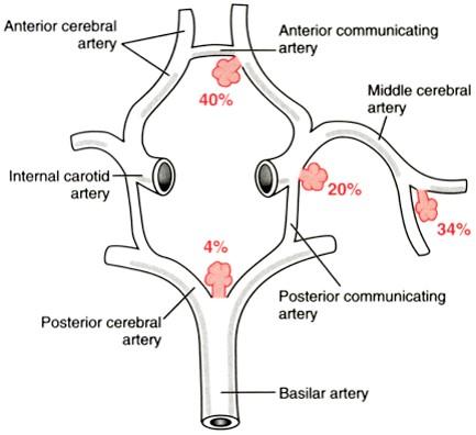 |
front 117 what are the types of hypertensive encephalopathy | back 117 acute
|
front 118 what are the symptoms of acute hypertensive encephalopathy | back 118 headaches
|
front 119 what are the symptoms of chronic hypertensive encephalopathy | back 119 dementia
|
front 120 what are some basal ganglia symptoms of hypertensive encephalopathy | back 120 tremors
|
front 121 what are some various CNS infections | back 121 acute meningitis
|
front 122 what are the main infections of the CNS | back 122 meningitis
|
front 123 what causes meningitis (generally) | back 123 bacterial |
front 124 what causes encephalitis (generally) | back 124 viral |
front 125 what are the specific causes of meningitis | back 125 e. coli, strep b
|
front 126 what is sign of meningitis infection | back 126 PMNs in CSF, increased protein, reduced glucose |
front 127 what particular viruses cause encephalitis | back 127 arboviruses
|
front 128 what will be present in the virchow robbins spaces in encephalitis | back 128 lymphs and MPs |
front 129 what is the big CNS invader when someone has HIV | back 129 toxoplasma |
front 130 how will leptomeninges look for in CNS infection | back 130 cloudiness |
front 131 what are some acute focal suppurative CNS infections | back 131 cerebral abscesses
|
front 132 where can cerebral abscesses take effect | back 132 local (mastoiditis, sinusitis)
|
front 133 what do subdural empyemas take placce in | back 133 sinusitis |
front 134 where do extradural abscesses take place i n | back 134 osteomyelitis |
front 135 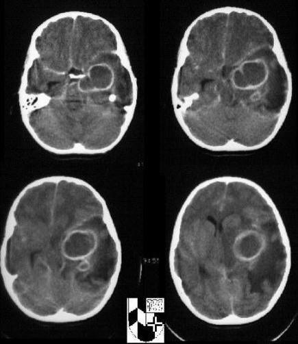 what is shown here | back 135 abscesses with satellits
|
front 136 what is the difference between capsules or pseudocapsules | back 136 true capsules are lined with epithelium |
front 137 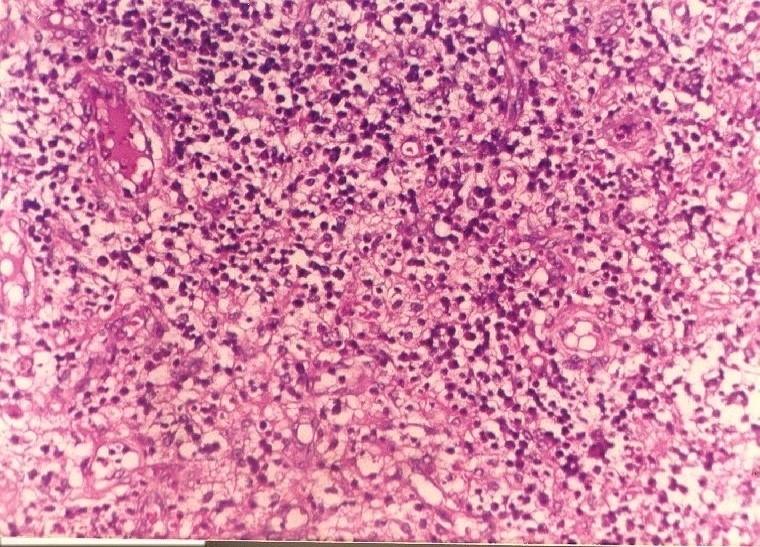 whats all over this abscess | back 137 neutrophils |
front 138 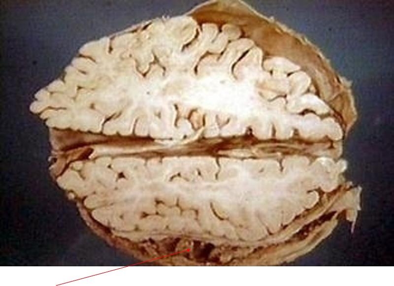 what is being pointed out here | back 138 subdural empyema |
front 139 why are many epidural abscesses in the spinal cord secondary to surgery | back 139 bone trauma |
front 140 what are the chronic bacterial meningoencephalitises | back 140 TB, brain and meninges
|
front 141 what is a gumma | back 141 large granuloma from syphillis |
front 142 can large granulomas look like tumors? | back 142 yep: tuberculoma |
front 143 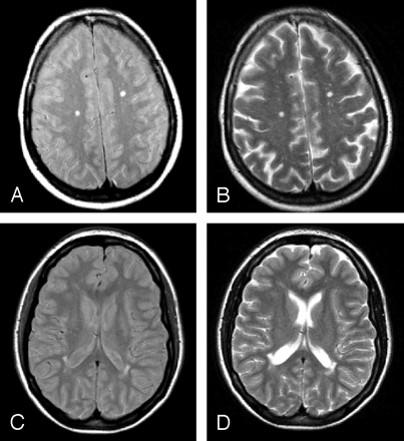 what is going on here | back 143 NeuroBorreliosis: encephalitis BACTERIAL THIS TIM |
front 144 why would encephlo-meningitis be a better term than meningo-encephalitis | back 144 viruses usually involve CNS parenchyma rather than meninges |
front 145 what is th hallmark of viral encephalitis | back 145 perivascular lymphocytic cuffing |
front 146 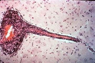 wahts going on here | back 146 viral encephaitis |
front 147 what is bitemporal encephalitis until proven otherwise | back 147 HSV |
front 148 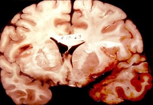 what is going on here | back 148 bitemporal encephalitis |
front 149 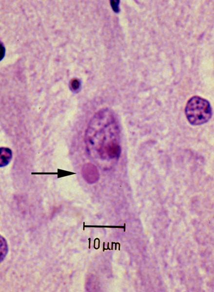 what is labeled by the arrow | back 149 eosinophilic negri body of rabies |
front 150 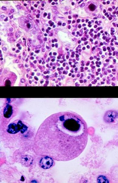 what is this shit | back 150 CMV basophilic inclusins |
front 151 what if you see perivascular giant cells in white matter | back 151 HIV ENCEPHALITIS |
front 152 what is the cause of PML (progressive multifocal leukoencephalopathy) | back 152 JC polyoma virus |
front 153 what does PML primarily affect | back 153 oligodendrocytes |
front 154 what is the main feature of PML | back 154 demyelination |
front 155 what is demyelination associated with | back 155 gliosis and edema |
front 156 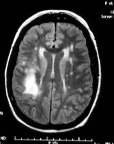 what is this on the MRI | back 156 demyelinization |
front 157 what is gliosis associated with | back 157 demyelinazation and edema |
front 158 what is edema associated with | back 158 demyelininzation and gliosis |
front 159 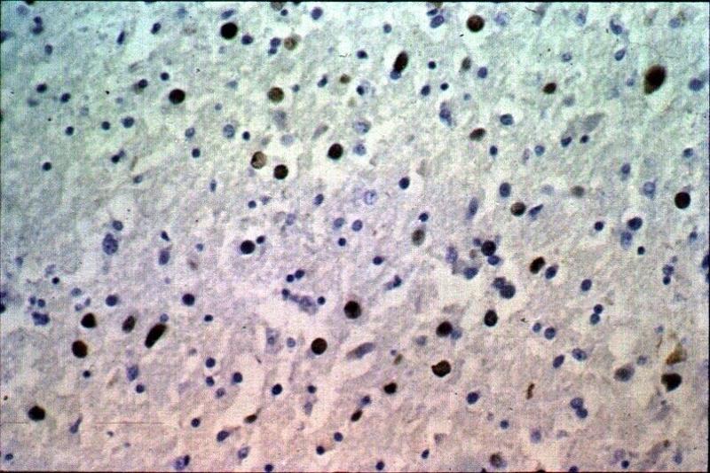 what is shown here | back 159 gliosis |
front 160 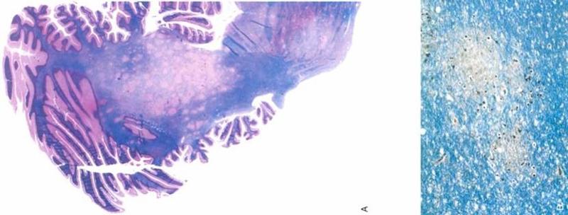 what is this | back 160 myelin/fat stain of PML: large area in middle with no myelin |
front 161 what is subacute sclerosing panencephalitis associated with | back 161 measles virus |
front 162 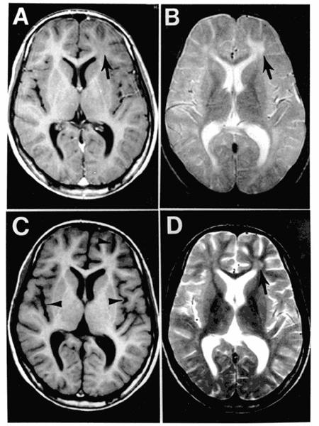 what happened here | back 162 subacute sclerosing panencephalitis |
front 163 what are the fungal meningo-encephalities | back 163 cryptococcus
|
front 164 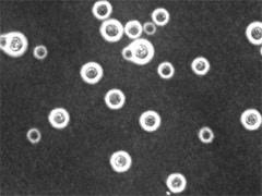 what type of stain is this | back 164 India ink |
front 165 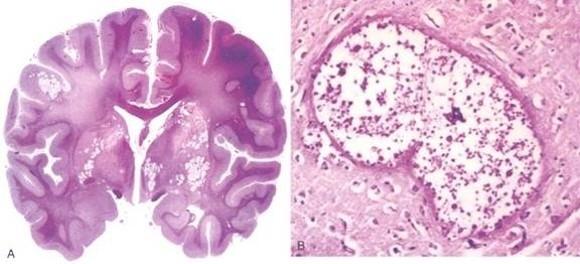 what are these microabscesses caused by | back 165 cryptococus |
front 166 what simple fungal stain could you use for abnormal areas in the brain | back 166 PAS |
front 167 what are the "other" things that can infect the CNS | back 167 malaria
|
front 168 what are the prion diseases | back 168 creutzfeldt-jakob diseasee
|
front 169 what are some common features of prion diseases | back 169 infectious agents with no DNA
|
front 170 why are prion diseases called spongiform | back 170 due to spaces between the cells caused by conformational changes |
front 171 what is prion replication due to | back 171 protein undergoes conformational change to induce neighboring proteins to become like it |
front 172 are prion proteins normally found in humans | back 172 yes |
front 173 what chromosome are prion proteins on | back 173 20 |
front 174 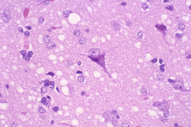 what is this a dead giveaway of | back 174 spongiform (prion disease) |
front 175 is CJD epidemic | back 175 no |
front 176 what happens in CJD | back 176 rapidly progressive dementia
|
front 177 what is affected in CJD | back 177 grey matter |
front 178 what are some of the demyelinating disaess | back 178 MS
|
front 179 if not for edema associated with demyelination, would the plaques be seen on MRI | back 179 no |
front 180 what is the cause of MS | back 180 nobody knows |
front 181 does MS affect females or males more | back 181 females |
front 182 when does MS usually take course | back 182 30-40 |
front 183 what is MS a disease against | back 183 white matter: plaquing of the nerves |
front 184 what is increased in the CSF with MS | back 184 CSF gamma globulin/oligoclonal bands |
front 185 what does MS often present with | back 185 visual probelms |
front 186 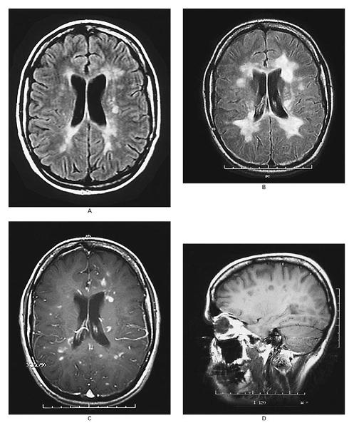 what is happeneing here | back 186 demyelination: MS |
front 187 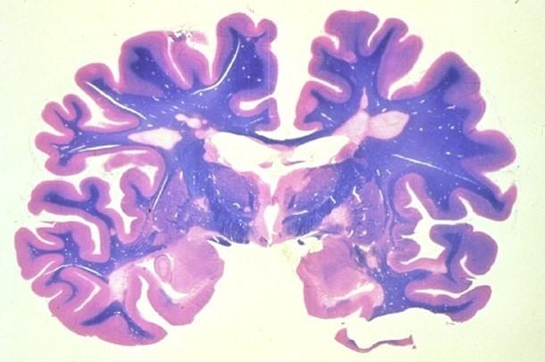 what is stained blue here | back 187 myelinated white matter |
front 188 grossly, what would plaques look like in contrast to better myelinated areas | back 188 less white |
front 189 what would plaques look like microscopicallly | back 189 demyelination, edema, gliosis, preseravtion of nerve fibers, inflammtory cells |
front 190 what are some degenerative disaeses of the CNS | back 190 cortex
|
front 191 what are CNS degenerative disaess of the cortex | back 191 dementias |
front 192 what are CNS degenerative diseass of the BG and brainstem | back 192 parkinsonian diseass |
front 193 what are CNS degenerative diseass of the spinocerebellar tract | back 193 ataxias |
front 194 what are CNS degenerative diseass of the motor neurons | back 194 muscle atrophy |
front 195 what are some cortical degenerative diseas | back 195 alzheimer's diseas
|
front 196 what is the most common CNS cortical degenerative diseass | back 196 alzheimer's |
front 197 what are most of the dementias | back 197 tauopathies |
front 198 what happens to the cortex (grey matter) in alzhemiers | back 198 atropphy |
front 199 what is present in alzheimers | back 199 neuritic plaques
|
front 200 are neuritic plaques just like MS's plaques | back 200 NO |
front 201 are the sulci or gyri prominent in cortical atrophy | back 201 cortical loss |
front 202 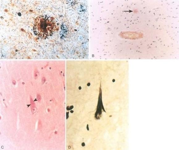 what is being shown here | back 202 plaques and tangles and beta amyloid of alzhemier's sieas |
front 203 what is aneuritic plaque | back 203 cluster of nerve fibers surrounding a substance that ends up being beta amyloid |
front 204 what is a tangle | back 204 phosphorylated MTs around indivdiual neurons |
front 205 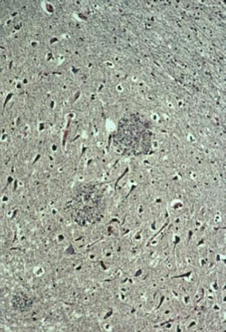 what is this | back 205 neuritic plaque |
front 206 what type of stain can be used to find beta amyloid | back 206 immunohistochemical stain |
front 207 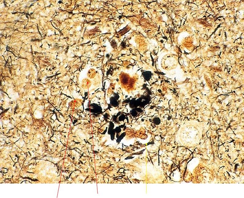 what is ponted out in red and yelow here | back 207 red: plaques
|
front 208 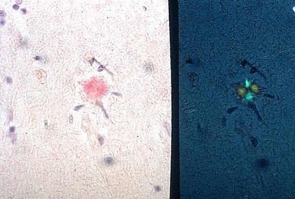 what stains are used here | back 208 congo red on left
|
front 209 is there alpha amyloid | back 209 yes with Igproliferative disaess like myelomas |
front 210  what is being ponited out here | back 210 neurons with tangles displacing nucleus |
front 211 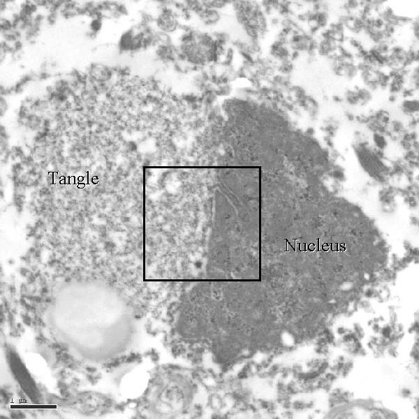 | back 211 no data |
front 212 what is a tangle | back 212 hyperphosphorylation of a neuron microtubule, causing it to precipitate |
front 213 what are the "other" cortical dementias | back 213 tau gene proteins/tauopathies
|
front 214 what are most cortical dementias known as | back 214 tauopathies |
front 215 what is vascular dementia associated with | back 215 multiple infarcts: lacunar, cortical, embolic |
front 216 what is the second commonest form of dimentia after alzheimer | back 216 vascular dimentia |
front 217 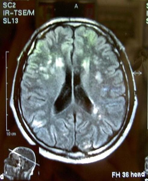 what is this and how do we know | back 217 MID because MS is purely white matter loss |
front 218 what are the CNS degenerative diseases in the basal ganglia and brainstem | back 218 parkinsonism
|
front 219 is parkinsonism a clinical syndrome or a diseas | back 219 clinical syndrome |
front 220 what are the featurs of parkinson syndrome | back 220 diminished facial expression
|
front 221 what is the clinical finding of parkinsonism | back 221 substantia nigra pathologic state |
front 222 what are some other substantia nigra diseass | back 222 parkinson diseas
|
front 223 what is the key idea in parkinsonsim | back 223 not enough dopamine |
front 224 where is there pallor in parkinson's disease | back 224 substantia nigra and locus ceruleus
|
front 225 what are lewy bodies | back 225 alpha-synuclien protein |
front 226 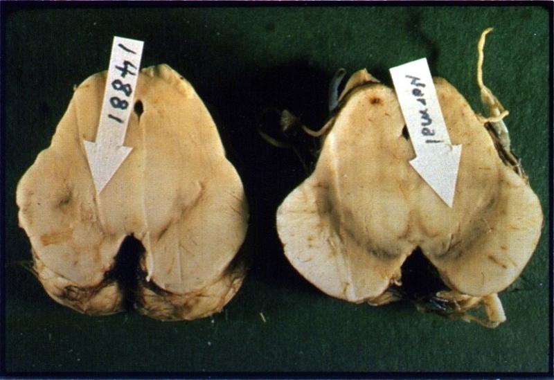 what happened here | back 226 parkinson's diseas as seen in SN |
front 227 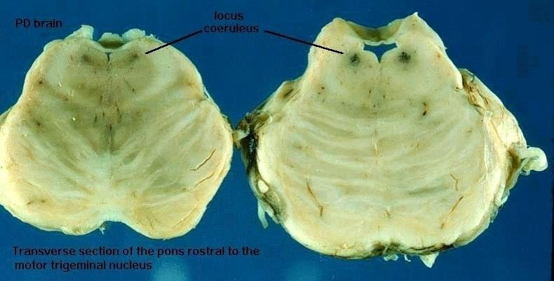 what happened here | back 227 parkinsons diseas as seen in LC |
front 228  which patient has parkinson's | back 228 right becuase of decreased dopamine |
front 229 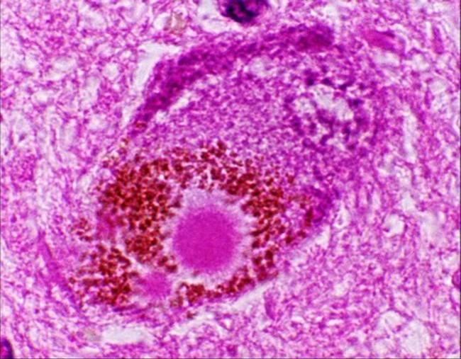 what is this and what is it from (disease) | back 229 lewy body
|
front 230 along iwth the parkinson symptoms, what does parkinsons disaese involve | back 230 progressive symptoms
|
front 231 what happens in multiple system atrophy | back 231 wide spectrum of diseasses
|
front 232 what are the clinical symptoms of multiple system atrophy | back 232 parkinsonism
|
front 233  what is this | back 233 alpha synuclein stains |
front 234 what type of disease is Huntingon's | back 234 genetic |
front 235 what happens in Huntingon's | back 235 progressive motor loss and dementia
|
front 236 what types of disesases are spinocerebellar degenerations | back 236 ataxias |
front 237 what are some spinocerebellar degenerations | back 237 spinocerebellar ataxias
|
front 238 what are some motor neuron diseass | back 238 ALS
|
front 239 what is the etiology of ALS | back 239 unknown |
front 240 what is ALS | back 240 progressive muscle atrophy due to motor neuron loss |
front 241 what does ALS progress from | back 241 hand weaknes to diaphragm |
front 242 where does ALS take place | back 242 anteiror horn cells reduece and gliotic
|
front 243 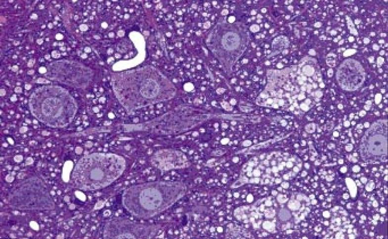 what is this a pic of | back 243 ALS |
front 244 what are the categories of genetic metabolic diseass | back 244 neuronal storage diseases
|
front 245 what trait do the neuronal storage diseases have | back 245 classical autosomal recessive enzyme deficiencies |
front 246 whta do the leukodystrophies present | back 246 abnormal myelin synthesis |
front 247 what happens with mitochondrial encephalopathies | back 247 mitochondrial gene mutations |
front 248 what are the major leukodystrophies | back 248 Krabbe
|
front 249 what are leukodystorphies | back 249 a group of disorders characterized by progressive degeneration of the myelinated white matter of the brain |
front 250 what are leukodystrophies caused by | back 250 imperfect growth/development of myelin due to genetic defects in the enzymes required for proper myelin production and maintenance |
front 251 what are the acquired toxic/metabolic CNS diseases | back 251 vitamin B1 def (Wernicke-Korsakoff)
|
front 252 what is the showing of wernicke korsakoff syndrome | back 252 hemorrhagic mamillary bodies |
front 253 what column will demyelinate in B12 deficiency | back 253 posterior column (subacute combined degeneration) |
front 254 what is earliest clinical symptom of Wernicke Korsakoff | back 254 loss of vibratory sense |
front 255 what are some CNS tumors | back 255 gliomas of astrocytes, oligodendrogliomas, ependymomas
|
front 256 what do ependymal cells look like | back 256 glandular |
front 257 what do oligodendrocytes have around them | back 257 halos |
front 258 what do meningiomas have the constistency of | back 258 super ball |
front 259 are primary lymphomas of the brain rare? when are they common | back 259 yes
|
front 260 if there is just one lesion in the brain, what is the probablity it is primary | back 260 50/50 |
front 261 if there is a nonprimary tumor in the brain, where did it pprobably come from | back 261 lung |
front 262 what are the symptoms of CNS tumors | back 262 headache
|
front 263 do CNS tumors present abruptly | back 263 no very subtle, only appear after tumor is sizeable |
front 264 what is the routine workup of CNS tumors | back 264 history
|
front 265 what are the questions to be asked of a CNS tuomor | back 265 benign or malignant
|
front 266 how old are people that normally get CNS tumors | back 266 younger |
front 267 what do certain CNS tumors have an abundant amount of | back 267 Ca |
front 268 what do you do an angiogram for in CNS tumors | back 268 vascularity determinatino (how much this thing has grown) |
front 269 gliosis vs. glioma, how to tell | back 269 age
|
front 270 what is a good indication of malignancy | back 270 vascularity and necrosis in high amounts usually indicate malignancy |
front 271 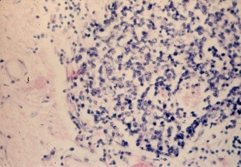 gliiosis or glioma | back 271 could be either one |
front 272 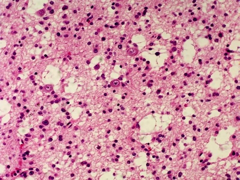 what is this | back 272 glioma, intermediate grade |
front 273 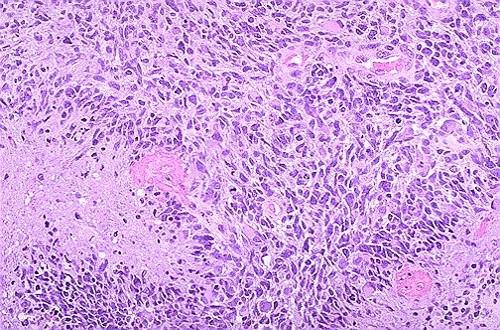 what is this | back 273 glioma high grade |
front 274 what is needed for the diagnosis of a high grade glioma | back 274 necrosis |
front 275 what are the higher grade gliomas called | back 275 gliobastome multiforme |
front 276 what is often seen in GBM | back 276 perivascular growth pattern |
front 277 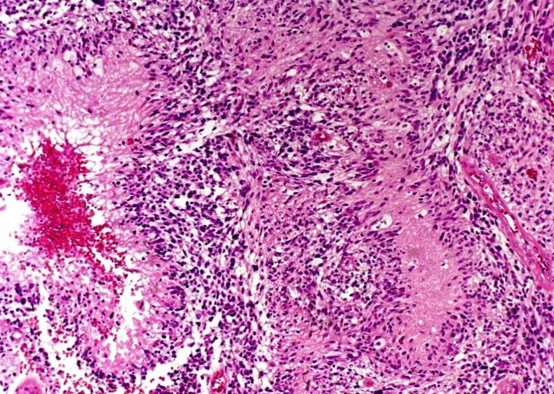 what is this | back 277 GBM
|
front 278 what is the sign of rapid growth in a tumor | back 278 central necrosis |
front 279 why would there be central necrosis in a tumor | back 279 outgrows blood supply and liquiefies centrally, like abscess |
front 280 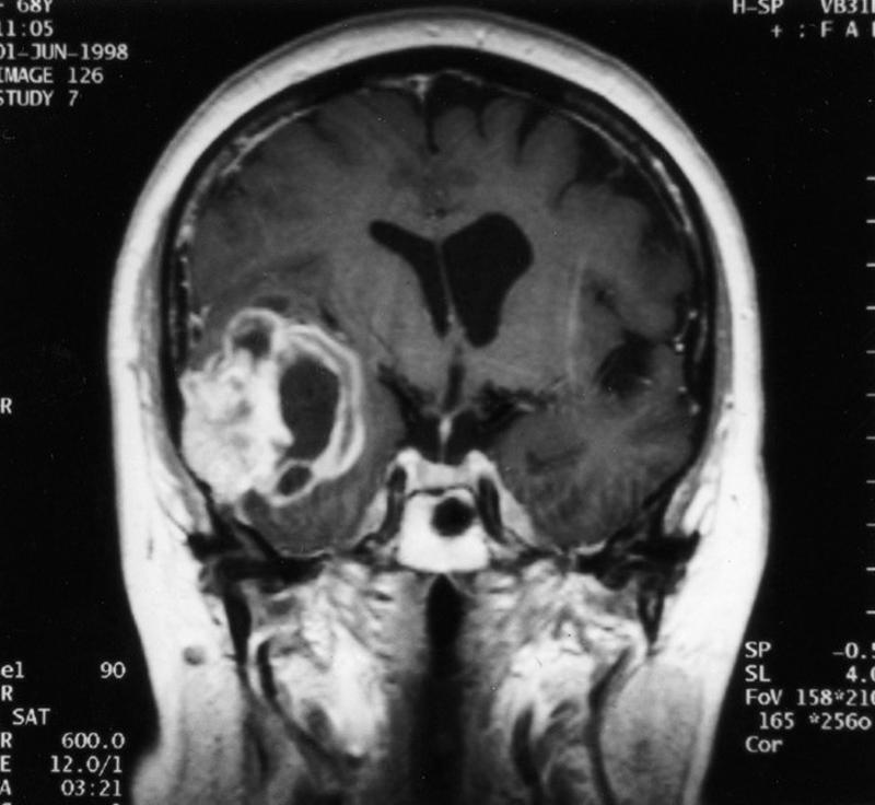 what is this | back 280 central necrosis |
front 281 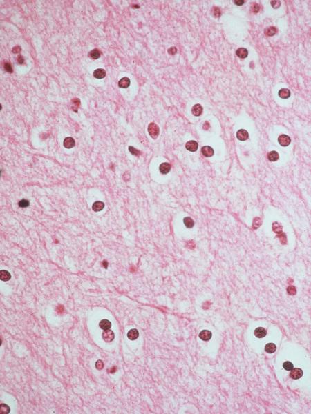 what kinds of glial cells are these | back 281 oligodendrocytes |
front 282 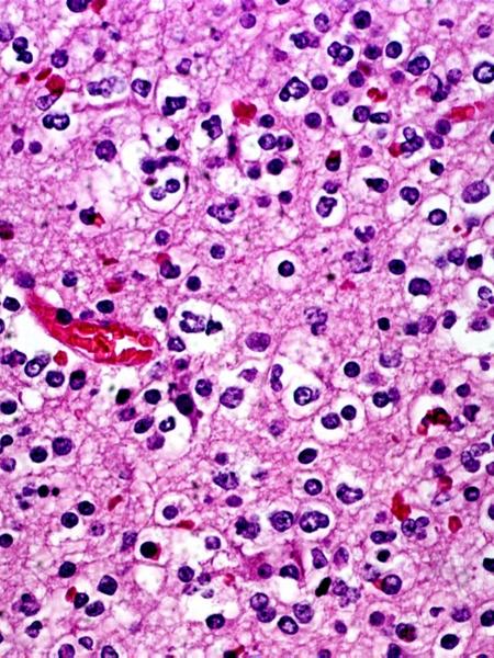 what is this | back 282 oligodendroglioma |
front 283 where do oligodendroglomas frequently occur | back 283 frontal or temporal lobes |
front 284 what are oligodendroglioma classifications | back 284 low or high grade |
front 285 who are oligodendrogliomas commmon in | back 285 men and women 20-40, but also children |
front 286 are oligodendrogliomas more common in men or wome | back 286 men |
front 287 how many of brain tumors are oligodendrogliomas | back 287 2% |
front 288 what chromosome losses are associated with oligodendrogliomas | back 288 1p or 19q |
front 289 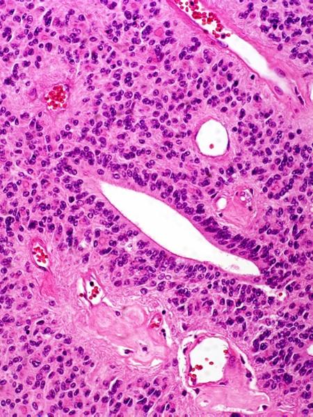 what is this | back 289 ependymomas |
front 290 would a choroid plexus tumor be a type of ependymoma | back 290 yep |
front 291 would an ependymoma result in an overproduction of CSF | back 291 yep |
front 292 are ependymomas diffuse in the brain | back 292 no, localized |
front 293 where do ependymomas develop from | back 293 cells that line the hollow cavities at bottom of brian and canal containing the spinal cord |
front 294 do ependymomas grow slow or fast | back 294 either |
front 295 where are ependymomas located | back 295 ventricles |
front 296 where do ependymomas extend | back 296 spinal cord |
front 297 what can ependymomas do | back 297 block ventricles causing hydrocephalus |
front 298 what does occurrence peak in ependymomas | back 298 5 and 34 |
front 299 how many brain cancer are ependymomas | back 299 2% |
front 300 what do ependymomas look most like | back 300 adenocarcinoma |
front 301  what type of cancer is this | back 301 neuroblastoma (rosettes) |
front 302 what is any midline cerebellum tumor in a child until proven otherwise | back 302 medulloblastoma |
front 303 are medulloblastomas PNET tumors | back 303 yes |
front 304 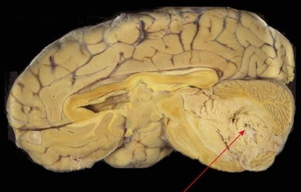 what is this | back 304 medulloblastoma |
front 305 where do meningiomas occur | back 305 where dura is |
front 306 are meningiomas vascular | back 306 yes |
front 307 are meningtiomas benign | back 307 yes but can be invasive |
front 308 what do meningiomas invade | back 308 areas adjacent to dura, parasagittal, falx, tentorium, venous sinuses |
front 309 what are meningiomas like | back 309 well defined like a superball |
front 310 what are often meningiomas have | back 310 psammoma bodies |
front 311 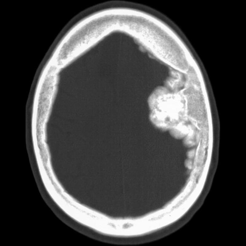 what is part of this meningioma denser than | back 311 bone |
front 312 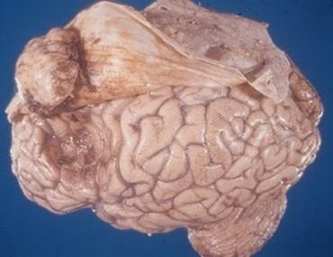 what is this | back 312 meningioma |
front 313 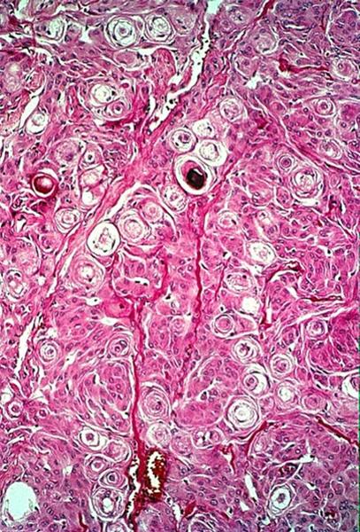 other than meningiomas what are psammoma bodies diagnostic of | back 313 papillary carcinomas |
front 314 what CNS diseases are common in AIDS | back 314 toxoplasmosis
|
front 315 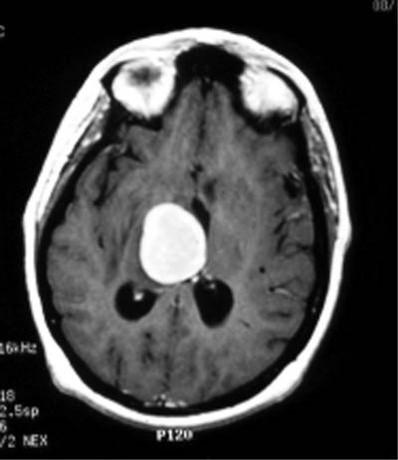 what is this | back 315 toxoplasmoma |
front 316 what are the metastatic CNS tumors that go to the brain | back 316 lung
|
front 317 is a solitary brain mass more likely to be metastatic or primary | back 317 same odds |
front 318 what are the paraneoplastic syndromes | back 318 small cell, lung
|
front 319 what are the familial CNS tumor syndromes | back 319 NF1 (neurofibromas and gliomas)
|