Instructions for Side by Side Printing
- Print the notecards
- Fold each page in half along the solid vertical line
- Cut out the notecards by cutting along each horizontal dotted line
- Optional: Glue, tape or staple the ends of each notecard together
SWM Module 18 Wound Care Other Wounds
front 1 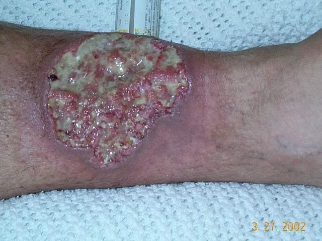 _____________________, or PG is a rare chronic inflammatory disease characterized by painful skin ulcers (Mayo Clinic, 2022b). PG is frequently noted on the lower extremities but can be present elsewhere, including surgical sites, minor traumas, and peristomal areas. | back 1 Pyoderma Gangrenosum |
front 2 Pyoderma Gangrenosum : Etiology - The exact cause of PG is unknown, but... | back 2 it is multifactorial with contributions from neutrophil dysfunction, genetic predispositions, and immune system dysregulation |
front 3 Those at risk for PG are patients who have (Mayo Clinic, 2022b): | back 3
Timely recognition of PG is crucial for appropriate management (NORD, 2023). |
front 4 PG Assessment * Providers rule out other conditions before diagnosing the patient with PG (Mayo Clinic, 2022b). Other conditions that can mimic PG include: | back 4
|
front 5 Most common, starts as an erythematous lesion that progresses rapidly to a deep ulcer commonly found on the leg | back 5 Classic or Ulcerative |
front 6 Superficial blisters on the hands, often associated with hematological cancers | back 6 Atypical or Bullous |
front 7 Painful bumps on the legs and arms that develop into ulcers, often associated with inflammatory bowel disease | back 7 Pustular |
front 8 Mildly painful ulcerations in various locations | back 8 Vegetative |
front 9 Individuals with a history of inflammatory bowel disease may experience ______ _____ around their stoma (NORD, 2023). These lesions can be very painful for patients. | back 9 pyoderma gangrenosum |
front 10 Treatment | back 10 Corticosteroids are a common first-line systemic treatment for PG (Mayo Clinic, 2022b; NORD, 2023). For limited disease, where the ulcers are smaller, or less severe, topical treatments such as corticosteroids and immunosuppressive drugs, like tacrolimus, are effective treatment options (Wu & Shinohara, 2023). |
front 11 What is a sign of the CLASSIC variant classification of pyoderma gangrenosum? | back 11 Deep ulcers on the leg * In the classic variant classification of pyoderma gangrenosum, the wound appears as deep ulcers on the leg. Atypical or bullous appear as superficial blisters on the hands, pustular presentation appears as painful bumps on extremities, and vegetative presentation appears with non-painful ulcerations. |
front 12 _____ _________ Key Points
| back 12 Pyoderma Gangrenosum |
front 13 Infection with the human herpesvirus 8, also known as Kaposi sarcoma-associated herpes, or KSHV, cause KS. KS present with red, pink, purple, or brown nodules or patches | back 13 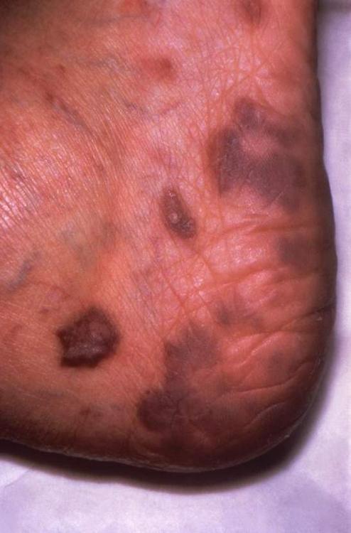
|
front 14 KS Treatment | back 14 Management includes immunotherapy and improving the overall immune system function (Johns Hopkins Medicine, 2023; Mayo Clinic, 2023). Local therapies may also be beneficial, including (Johns Hopkins Medicine, 2023):
|
front 15 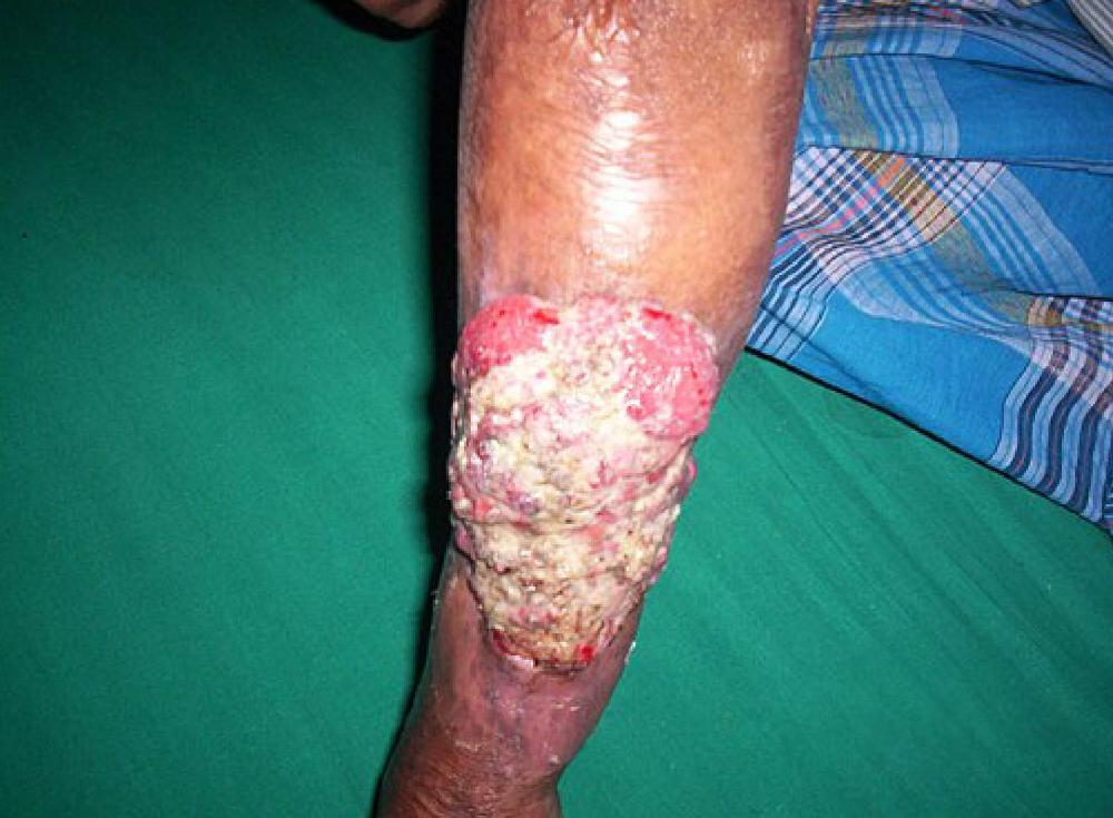 Marjolin Ulcer: | back 15 A Marjolin ulcer is a type of squamous cell carcinoma that occurs at a site of chronic inflammation or where a patient has had a scarred or poorly healed wound |
front 16 Marjolin Ulcer: Etiology Marjolin ulcers develop on traumatized skin tissue. On average, marjolin ulcers tend to appear about 30 years after the initial scarring injury (Junaid, 2017). Why these ulcers develop is not known, but one theory is that the presence of scar tissue, chronic inflammation, and trauma to the site are contributing factors. Underlying causes might include: | back 16
|
front 17 Marjolin Ulcer: Appearance Marjolin ulcers typically appear in the head, neck, legs, or feet (Junaid, 2017). Clinical features of Marjolin ulcer may include: | back 17
|
front 18 Marjolin Ulcer: Diagnosing | back 18 Typically involves an incisional biopsy to confirm the presence of cancerous cells. Tissue biopsy is an important differential diagnostic tool, as these ulcers are commonly mistaken for |
front 19 Marjolin Ulcer: Treatment 1 of 2 Treatment of Marjolin ulcers usually involves wide excision of the affected area, which is necessary to mitigate the spread and prevent recurrence (Junaid, 2017). Depending on the severity and extent of the cancer, additional treatments may include: | back 19
|
front 20 Marjolin Ulcer: Treatment 2 of 2 After surgical excision, local wound care to promote healing will be ongoing. Wound care clinicians should educate patients and their families on the following (Vera, 2018): | back 20
|
front 21 A _____ ______ wound is a cancerous tumor that grows under the skin and eventually breaks through the surface, forming an ulcerative lesion (Cancer Research UK, 2023). These wounds are often associated with advanced cancers and can cause significant physical and emotional distress. | back 21 fungating malignant |
front 22 Wounds are classified as ______ when they do not arise from common causes such as pressure injuries, venous insufficiency, arterial disease, diabetes, or surgical complications. Wound care clinicians must be able to identify wounds with uncommon etiologies as effective management begins with understanding and addressing the underlying conditions. | back 22 Atypical |
front 23
| back 23 Assessment Fungating malignant tumors often present with a cauliflower or fungus-like appearance (Cancer Research UK, 2023). Other symptoms include: |
front 24 True or False: The Kaposi sarcoma-associated herpes virus will always develop into Kaposi sarcoma, so early intervention is essential. | back 24 False Getting the Kaposi sarcoma-associated herpes virus does not necessarily mean the patient will develop Kaposi sarcoma. It occurs most frequently in men and immuno-compromised patients. |
front 25 Kaposi Sarcoma Key Points: 1 | back 25
|
front 26 Kaposi Sarcoma Key Points: 2 | back 26
|
front 27 Kaposi Sarcoma Key Points: 3 | back 27
|
front 28 Kaposi Sarcoma Key Points: 4 | back 28
|
front 29 Pemphigus | back 29 Pemphigus is a rare autoimmune skin disorder that attacks the epidermis and mucous membranes (Johns Hopkins Medicine, 2019; NIAMS, 2024). |
front 30 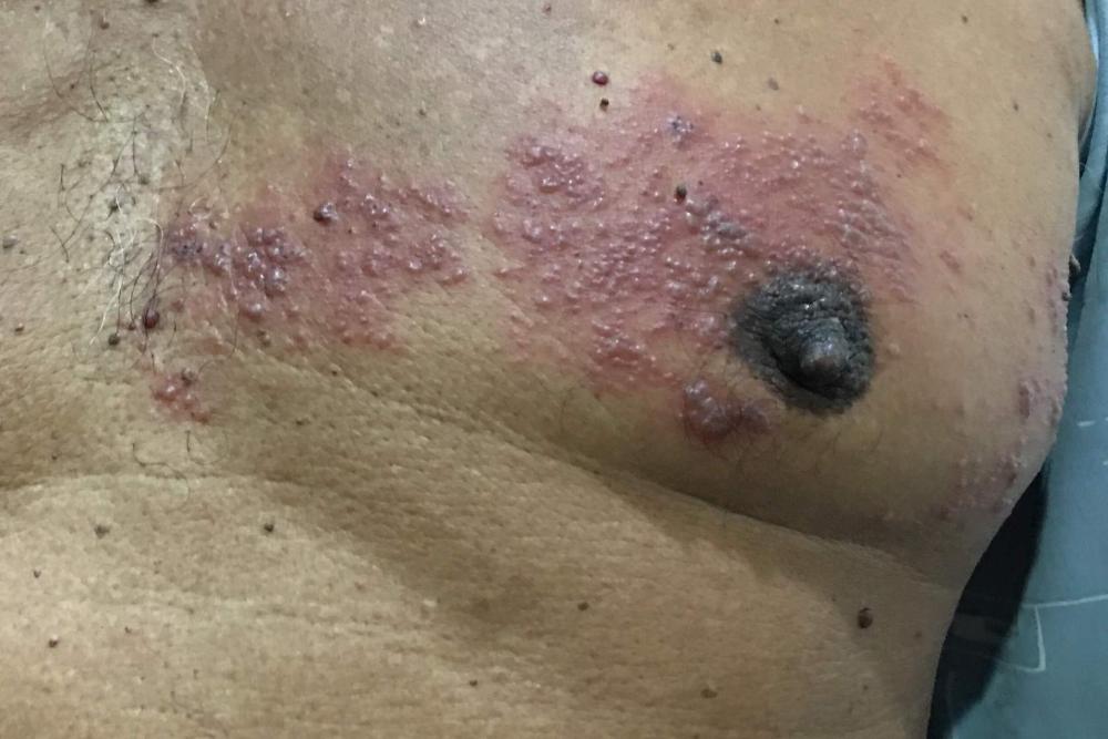 Herpes Zoster (Shingles) | back 30 Herpes zoster, more commonly referred to as shingles, is a viral infection that creates a painful rash or blisters on the body |
front 31 Pemphigus Etiology: In pemphigus, the body produces antibodies that fight against desmogleins, creating fragile skin and blistering under the skin. The disease is more likely to occur if you have certain risk factors, such as | back 31 Ethnicities- People of Indian, Middle Eastern, Southeast European, and Jewish descent are more susceptible. Location- Rural regions of Tunisia and Brazil have a higher incidence of pemphigus. Age- People between 40 and 60 tend to get the disease more frequently, with women having onset between the ages of 50 and 60. Genetics- People with certain variations in the HLA immune system genes tend to get pemphigus at a higher rate. Medications- Taking medications from certain chemical groups has been associated with drug-induced pemphigus. |
front 32 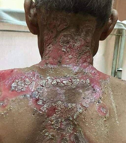 Blister and skin erosion on the neck and upper back of a patient w/pemphigus vulgaris. Pemphigus are categorized into common and rare types: | back 32
|
front 33
| back 33 Most common type in the U.S.; blistering in the mouth and other mucous membranes and deep in the epidermis |
front 34 Pemphigus Foliaceus: | back 34 Less common, affecting only the skin; blisters in the upper epidermis |
front 35 Bullous Pemphigoid (BP): | back 35 More common, has prodromal phase of weeks to months of pruritic, eczema-like skin rash, followed by widespread tense bullae. |
front 36 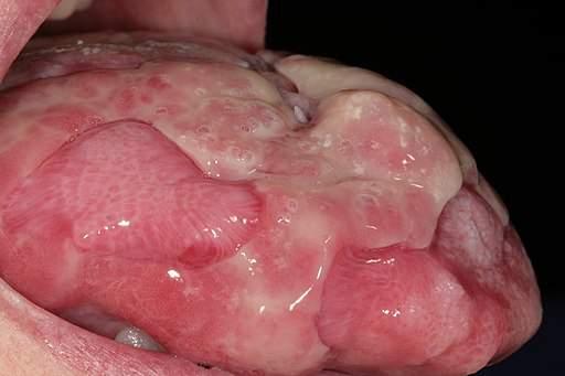 Chronic oral lesions on tongue of patient with Paraneoplastic Pemphigus | back 36 Sores form in the mouth and lips, blistering on the skin and mucous membranes; usually related to having a tumor |
front 37
| back 37 Caused by the IgA antibody; blisters or pimples appear on the skin in rings or groups |
front 38 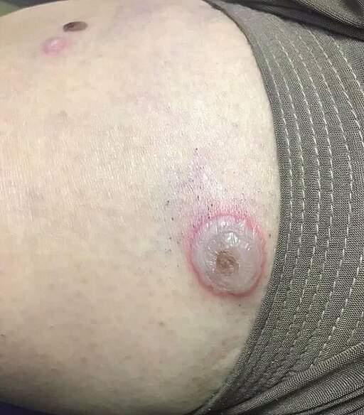
| back 38 Medications such as blood pressure medicines, antibiotics, and medications containing thiol cause sores or blisters; resolve once the medication discontinued |
front 39 Drug-induced Pemphigus: Treatment | back 39
|
front 40 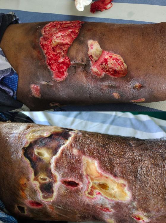 Necrotizing Fasciitis | back 40 Necrotizing fasciitis, also known as “flesh-eating bacteria” infection, is a rapidly spreading infection of soft tissues. It is also a type of necrotizing soft tissue infections (NSTIs) that are aggressive, life-threatening infections characterized by rapid tissue destruction and systemic illness. Most commonly caused by the bacteria Streptococcus pyogenes but often involves multiple types of bacteria working together, including methicillin-resistant Staphylococcus aureus, or MRSA, Clostridium perfringens, and bacteria found in marine environments |