Instructions for Side by Side Printing
- Print the notecards
- Fold each page in half along the solid vertical line
- Cut out the notecards by cutting along each horizontal dotted line
- Optional: Glue, tape or staple the ends of each notecard together
BMD 315 Module 6 Study Guide/Learning Objectives
front 1 Describe the characteristics of sensory receptors. | back 1 1. Modality-Specific Each receptor is sensitive to a specific type of stimulus (called a modality), such as light, sound, pressure, or temperature. Example: Photoreceptors respond to light; mechanoreceptors respond to pressure or touch. 2. Location Receptors can be:
3. Threshold Receptors require a minimum intensity of stimulus (called the threshold) to activate and produce a response. 4. Transduction Receptors convert the stimulus (e.g., heat, light) into an electrical signal (action potential or graded potential). 5. Adaptation Some receptors adapt over time:
6. Intensity Coding The strength of a stimulus is encoded by:
7. Receptive Field
|
front 2 How are sensory receptors categorized? | back 2 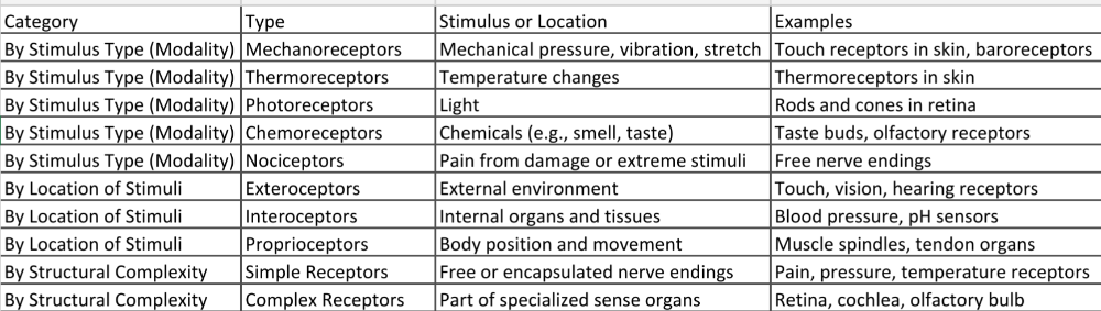 |
front 3 Describe the nature of the receptor (generator) potential. | back 3 A receptor (or generator) potential is a graded electrical response produced in a sensory receptor when it detects a stimulus.
|
front 4 Describe the significance of receptor (generator) potential. | back 4 1. Initiates Sensory Signaling:
2. Determines Action Potential Firing:
3. Encodes Stimulus Intensity: Stronger receptor potentials → more frequent action potentials → brain interprets as a stronger sensation (e.g., more intense pain or pressure). 4. Occurs in Both Specialized Receptors and Free Nerve Endings:
|
front 5 Describe the types of cutaneous receptors | back 5 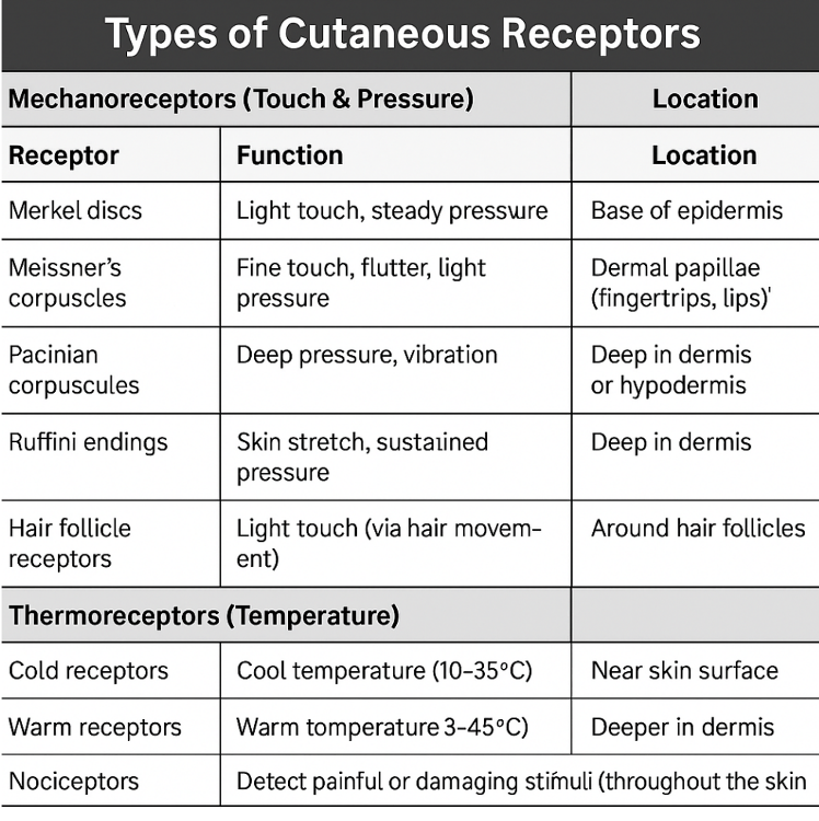 Extreme temperatures activate nociceptors instead of thermoreceptors. 3. Nociceptors (Pain)
4. Proprioceptors (Less common in skin) Mostly found in muscles and joints, but some may sense skin stretch to help detect body position. |
front 6 Define sensory acuity and explain how it is affected by receptor density and lateral inhibition. | back 6 Sensory Acuity: It means how well you can feel small details — like how close two touches can be before they feel like one. 1. Receptor Density
2. Lateral Inhibition
|
front 7 Identify the modalities of taste and explain how they are produced. | back 7 1. Sweet Stimulus: Sugars (e.g., glucose, sucrose), artificial sweeteners. How it's produced: Sweet molecules bind to G-protein-coupled receptors (GPCRs) on taste cells. This triggers a signaling cascade, leading to neurotransmitter release. 2. Salty Stimulus: Sodium ions (Na⁺), mainly from table salt (NaCl). How it's produced: Na⁺ ions enter directly through ion channels in the taste cell membrane. This causes depolarization, which triggers a signal to the brain. 3. Sour Stimulus: Hydrogen ions (H⁺) from acidic substances (like lemon juice). How it's produced: H⁺ ions enter the cell or block potassium channels, causing depolarization and a signal to the brain. 4. Bitter Stimulus: Toxic or bitter compounds (e.g., caffeine, quinine). How it's produced: Bitter compounds bind to GPCRs on taste cells. This starts a chemical cascade that alerts the brain — often as a warning signal. 5. Umami (Savory) Stimulus: Glutamate (found in meats, cheeses, soy sauce). How it's produced: Glutamate binds to specific GPCRs on taste cells. Triggers a signal that the food is protein-rich. How the Brain Gets the Signal: The taste receptor cells activate sensory neurons, which carry signals to the brain via: Facial nerve (VII), Glossopharyngeal nerve (IX), and Vagus nerve (X) |
front 8 Explain how odorant molecules stimulate their receptors and describe how the information is conveyed to the brain. | back 8 1. Detection: Odorant Binds to Receptor
2. Receptor Activation (Signal Transduction) Each olfactory receptor is a G-protein-coupled receptor (GPCR). When an odorant binds:
3. Signal Travels to the Brain
4. Processing in the Brain In the olfactory bulb, the signal goes to structures called glomeruli, where it is sorted. From there, the signal travels to:
|
front 9 Describe the structures of the vestibular apparatus and explain how they function to produce a sense of equilibrium. | back 9 The vestibular apparatus is a part of the inner ear that helps maintain balance and spatial orientation (equilibrium). It detects head position and movement using fluid, hair cells, and gravity. 1. Semicircular Canals
How they work:
2. Utricle and Saccule (part of the otolith organs)
How they work:
|
front 10 Explain how sound waves result in movements of the oval window and then the basilar membrane. | back 10 1. Sound Waves Enter the Ear Sound waves enter through the external auditory canal and strike the tympanic membrane (eardrum), causing it to vibrate. 2. Vibration Passes Through the Ossicles
3. Movement of the Oval Window
4. Fluid Waves Move the Basilar Membrane Fluid waves travel through the scala vestibuli and scala tympani (chambers in the cochlea). This motion causes the basilar membrane (in the middle chamber) to vibrate at specific locations, depending on sound frequency:
5. Activation of Hair Cells
|
front 11 Explain how movements of the basilar membrane at different sound frequencies (pitches) affect hair cells. | back 11 The Basilar Membrane Responds to Pitch (Frequency)
Why This Happens:
How This Affects Hair Cells: Hair cells sit on the basilar membrane, topped with tiny projections called stereocilia. When the membrane vibrates:
|
front 12 Describe how action potentials are produced, and their neural pathways. | back 12 How Action Potentials Are Made An action potential is an electrical signal that a nerve cell (neuron) uses to send messages. Steps: 1. Resting: Neuron is at rest: inside is more negative than outside. 2. Stimulus Happens: A signal (like touch or smell) causes sodium (Na⁺) channels to open. Na⁺ rushes in → inside gets less negative. 3. Threshold is Reached: If enough Na⁺ enters, the neuron hits –55 mV and fires. 4. Depolarization: More Na⁺ enters → inside becomes positive (~+30 mV). 5. Repolarization: Na⁺ channels close, potassium (K⁺) channels open. K⁺ flows out → inside goes back to negative. 6. Reset: The neuron returns to resting state, ready to fire again. (Neural Pathway): How the signal travels through the body: 1. Sensory Neuron: Detects something (like heat, pressure) and Sends signal to the spinal cord or brain 2. Processing: The brain or spinal cord understands the signal. 3. Motor Neuron: Sends a signal out to muscles or glands to respond. |
front 13 Describe the structures of the eye, and how these focus light onto the retina. | back 13 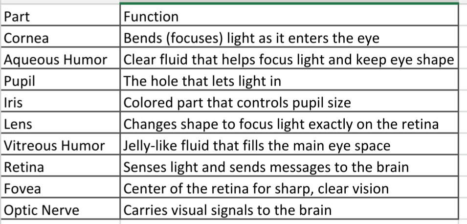 How Light is Focused: 1. Light enters the eye through the cornea, which bends the light. 2. The pupil lets in more or less light (controlled by the iris). 3. The lens fine-tunes the focus: Flattens to see far away, and Bulges to see up close 4. Light goes through the vitreous humor. 5. The light lands on the retina, where photoreceptors detect the image. 6. The optic nerve sends the picture to your brain. |
front 14 Explain how accommodation at different distances is accomplished. | back 14 Accommodation is the process by which the eye changes the shape of the lens to focus on objects at different distances. This is done by the ciliary muscles, suspensory ligaments (zonules), and the lens. How It Works: For Near Objects: Ciliary muscles contract, Suspensory ligaments loosen, and Lens becomes rounder (thicker) This increases the lens's curvature to bend light more, focusing the image on the retina. For Distant Objects: Ciliary muscles relax, Suspensory ligaments tighten, and Lens flattens (thins) This reduces the curvature of the lens, bending light less to focus distant images on the retina. |
front 15 Explain common disorders of refraction | back 15 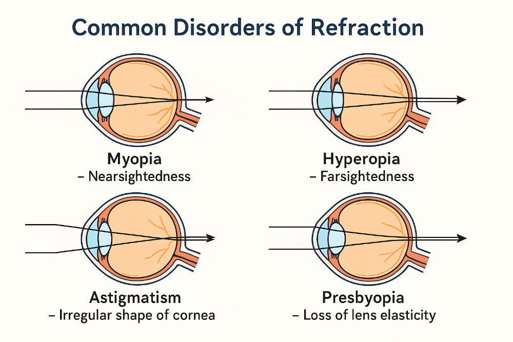 1. Myopia (Nearsightedness)
2. Hyperopia (Farsightedness)
3. Astigmatism
4. Presbyopia
|
front 16 Describe the structure of the retina and how light affects rhodopsin | back 16 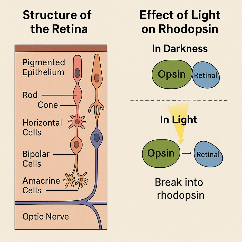 Rhodopsin and Light: Rhodopsin is found in rod cells. It consists of: Opsin (protein) and Retinal (light-sensitive molecule from vitamin A) In Darkness: Rhodopsin is whole and active. Rods depolarize and release glutamate, which inhibits bipolar cells. No signal is sent to the brain (no vision signal). In Light: Light causes rhodopsin to split → opsin + retinal. Rods hyperpolarize and stop releasing glutamate. This activates bipolar cells, which excite ganglion cells. Signal is sent via optic nerve to the brain → visual perception. |
front 17 Explain how light affects synaptic activity in the retina and describe the neural pathways of vision. | back 17 Light & Synaptic Activity in Retina: In Darkness: Photoreceptors (rods) are active. They release glutamate. Glutamate blocks the signal → no vision signal In Light: Light stops glutamate release. Bipolar cells are activated. Ganglion cells send signals to the brain Neural Pathway of Vision (Simple Steps): 1. Light hits photoreceptors in the retina 2. Signal goes to bipolar cells 3. Then to ganglion cells 4. Ganglion cells form the optic nerve 5. Optic nerve goes to the optic chiasm (fibers cross) 6. Continues through the optic tract 7. Reaches the thalamus (lateral geniculate nucleus) 8. Ends in the primary visual cortex (in the brain’s occipital lobe) |
front 18 Compare the function of rods and cones and describe the significance of the fovea centralis. | back 18 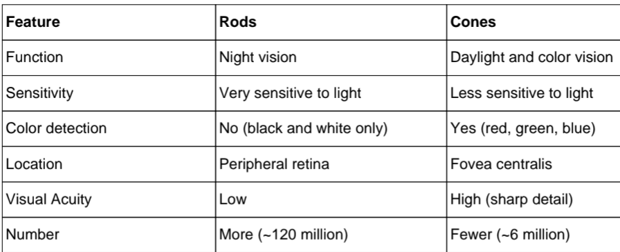 Fovea Centralis (Importance):
|