Instructions for Side by Side Printing
- Print the notecards
- Fold each page in half along the solid vertical line
- Cut out the notecards by cutting along each horizontal dotted line
- Optional: Glue, tape or staple the ends of each notecard together
BMD 315 Module 5 Study Guide/Learning Objectives
front 1 Describe the different types of neurons and their functions | back 1 Neurons send messages through your body. 1. Sensory Neurons: Carry messages from the body to the brain. Example: You touch something hot → message goes to your brain. 2. Motor Neurons: Carry messages from the brain to the body. Example: Brain tells your hand to move away. 3. Interneurons: Found in the brain and spinal cord. They connect sensory and motor neurons. Help with quick reactions, like reflexes. Parts of a Neuron
|
front 2 What are the supporting cells of neuron and their functions? | back 2 Supporting Cells (Help Neurons) These cells don’t send messages. They support and protect neurons. In the Brain & Spinal Cord (CNS):
In the Rest of the Body (PNS):
|
front 3 • Identify the myelin sheath and describe how it is formed in the CNS and PNS | back 3 The myelin sheath is a fatty coating around a neuron's axon. It helps messages travel faster and protects the nerve. In the Brain and Spinal Cord (CNS)
In the Rest of the Body (PNS)
⚡ Why It’s Important
Think of it like rubber coating on a wire—it keeps the signal strong and fast! |
front 4 • Describe the nature and significance of the blood-brain barrier. | back 4 The blood-brain barrier is a wall of protection around the brain. What Does It Do?
What Is It Made Of?
Why It’s Important
|
front 5 • Explain the electrical activity associated with action potentials | back 5 an electrical signal sent along a neuron. It’s how neurons communicate with each other and with muscles 1. Resting: Neuron is quiet. Inside = negative, Outside = positive 2. Depolarization: Message starts Sodium (Na⁺) rushes in → inside becomes positive 3. Repolarization: Potassium (K⁺) moves out → inside goes back to negative 4. Reset
Rest → Sodium in → Potassium out → Reset It’s like flipping a light switch ON and then OFF again! |
front 6 o Explain, step-by-step, how an action potential is produced. | back 6 Step 1: Resting Potential
Step 2: Stimulus Reaches Threshold
Step 3: Depolarization
Step 4: Peak of Action Potential
Step 5: Repolarization
Step 6: Hyperpolarization
Step 7: Return to Resting Potential
|
front 7 What features of neurons establish and maintain the resting membrane potential? | back 7 The resting membrane potential is when the neuron is
off (not sending a signal). 3 Main Things That Keep It That Way: 1. Sodium-Potassium Pump: Pushes 3 sodium (Na⁺) out and pulls 2 potassium (K⁺) in → More positive charges go out than in 2. Leaky Potassium Channel: Potassium
slowly leaks out 3. Negative Stuff Inside: Big negatively
charged proteins stay inside |
front 8 What causes changes in membrane potential? | back 8 The charge changes when tiny particles called ions move in or out of the cell. Caused by: Ion channels open (like little doors) Sodium (Na⁺) goes in → inside gets more positive Potassium (K⁺) goes out → inside gets more negative |
front 9 What does it mean that an action potential is “all or none?” | back 9 It means the nerve either fires completely or not at all — there’s no halfway.
"All-or-none" = Full signal or no signal — never a weak or partial one. |
front 10 Define the terms depolarization and repolarization and be able to illustrate graphically. | back 10 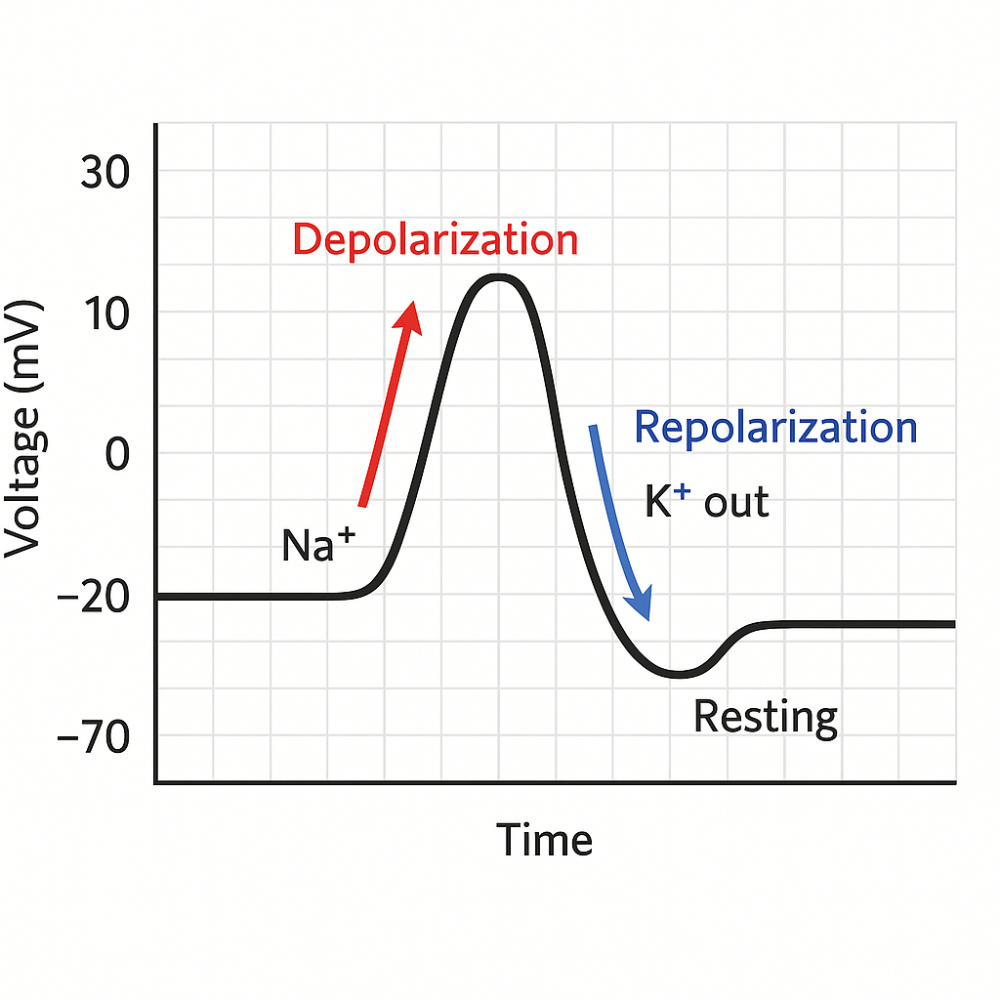 Depolarization
Repolarization
|
front 11 Describe the structure and function of electrical and chemical synapses. | back 11 Electrical Synapse: Two neurons are connected by gap junctions (tiny tunnels).
Chemical Synapse: There's a small gap between the two neurons.
|
front 12 Identify the nature of excitatory and inhibitory postsynaptic potentials. | back 12 Excitatory Postsynaptic Potential (EPSP): A small positive change in the postsynaptic neuron's charge.
Inhibitory Postsynaptic Potential (IPSP): A small negative change in the postsynaptic neuron's charge.
|
front 13 Describe the characteristics of action potentials | back 13 1. All-or-None Response: The neuron either fires completely or not at all. 2. Threshold: A certain level of stimulation (usually –55 mV) must be reached to trigger an action potential. 3. Depolarization: Sodium (Na⁺) channels open → Na⁺ rushes in. Inside of the neuron becomes positive. 4. Repolarization: Potassium (K⁺) channels open → K⁺ flows out. Inside becomes negative again. 5. Hyperpolarization: The neuron becomes more negative than resting (goes below –70 mV). Caused by K⁺ leaving too much. 6. Resting Membrane Potential: The neuron returns to its normal state (about –70 mV) using sodium-potassium pumps. 7. Unidirectional: Action potentials move in one direction down the axon (from cell body to axon terminal). 8. Refractory Period: Brief time when the neuron can’t fire again, or needs a stronger stimulus to do so. Ensures signals don’t go backward. 9. Self-propagating: Once started, the action potential continues along the axon without fading. |
front 14 Explain how these characteristics are conducted by unmyelinated and myelinated axons. | back 14 Unmyelinated Axons
Myelinated Axons
|
front 15 Explain how ligand-gated channels produce synaptic potentials, using nicotinic ACh receptors as an example. | back 15 They’re like locked doors on a neuron. A special key (neurotransmitter) opens them. Example: Nicotinic ACh Receptor 1. ACh is released: A nerve sends acetylcholine (ACh) into the gap between two neurons. 2. ACh binds to the receptor: ACh is the key that opens the nicotinic receptor (a special door). 3. The channel opens: When the door opens, sodium ions (Na⁺) rush into the next neuron. 4. The inside gets more positive: This small positive change is called an EPSP (excitatory postsynaptic potential). 5. Signal might continue: If enough sodium comes in, it might start a new action potential in the next neuron. 6. ACh is broken down: An enzyme removes ACh, and the door closes again. In Short:
|
front 16 Explain how G-protein-coupled channels produce synaptic potentials, using the muscarinic ACh receptor as an example. | back 16 It’s a receptor that works indirectly. It doesn’t open a channel by itself. Instead, it activates a helper inside the cell called a G-protein. Muscarinic ACh Receptor (Example) 1. ACh is released: Acetylcholine (ACh) is sent into the gap between two neurons. 2. ACh binds to the muscarinic receptor: The receptor is on the next neuron — this turns on a G-protein inside the cell. 3. G-protein takes action: It tells ion channels (like potassium channels) to open or close. 4. Ions move → charge changes
5. Signal is slow but lasts longer: It takes more time to start, but the effect stays longer. In Short:
|
front 17 Describe the action and significance of acetylcholinesterase. | back 17 ⚙️ What does it do? 1. ACh sends the message from one neuron to the next. 2. Acetylcholinesterase breaks ACh into small pieces. 3. This stops the message so the neuron or muscle can rest. 4. The pieces are reused to make more ACh later. ❗ Why is it important?
> Acetylcholinesterase stops the signal by breaking down ACh, so the nerve or muscle can relax. |
front 18 Compare EPSPs and action potentials, identify where each is produced in a neuron | back 18 EPSP (Excitatory Postsynaptic Potential): A small, temporary increase in the neuron's charge (makes inside more positive). It helps the neuron get closer to firing. Where it happens: At the dendrites or cell body (soma) of the neuron — where the neuron receives signals. Action Potential: A large, fast electrical signal that travels down the axon. It's all-or-none — once it starts, it goes all the way. Where it starts:
|
front 19 Explain how action potentials can be stimulated by EPSPs. | back 19 1. EPSPs are small signals
2. EPSPs add up
Temporal summation – EPSPs from the same place, quickly one after another. Spatial summation – EPSPs from multiple places at the same time. 3. Threshold is reached If the combined EPSPs make the membrane potential reach threshold (about –55 mV), an action potential is triggered. 4. Action potential starts It begins at the axon hillock and travels down the axon to send the signal. In short: > EPSPs make the neuron more positive. If enough of them happen at once, they can reach the threshold and trigger an action potential. |
front 20 Identify the monoamine neurotransmitters | back 20 These are chemical messengers made from amino acids. The main types include: 1. Dopamine (DA) 2. Norepinephrine (NE) – also called noradrenaline 3. Epinephrine (E) – also called adrenaline 4. Serotonin (5-HT) 5. Histamine |
front 21 Explain how monoamine neurotransmitters are inactivated at the synapse. | back 21 Once they’ve sent their message, they must be cleared to stop the signal. This happens in two main ways: 1. Reuptake (Recycling)
Example: Dopamine transporter (DAT), Serotonin transporter (SERT). Think: "Vacuuming up the neurotransmitter." 2. Enzymatic Breakdown Enzymes break the neurotransmitter into inactive parts. Main enzymes:
Think: "Chemical scissors that cut up the message." |
front 22 Describe the steps involved with release from the presynaptic neuron and reaction of the postsynaptic neuron. | back 22 In the Presynaptic Neuron (Sending Side): 1. Action potential arrives A nerve signal (action potential) reaches the axon terminal. 2. Calcium channels open The signal causes calcium ions (Ca²⁺) to enter the axon terminal. 3. Neurotransmitters are released Calcium causes vesicles (tiny bubbles) filled with neurotransmitters to fuse with the membrane and release them into the synaptic cleft (gap between neurons). In the Postsynaptic Neuron (Receiver): 4. Neurotransmitters bind to receptors: The chemicals float across the gap and attach to receptors on the next neuron. 5. Ion channels open: Receptors cause ion channels to open, letting ions in or out. 6. Neuron’s charge changes: More positive = Excitatory (EPSP) → helps start a new signal, More negative = Inhibitory (IPSP) → blocks a new signal 7. New signal may start: If enough EPSPs happen, the postsynaptic neuron will fire its own action potential. |
front 23 Explain the action and significance of GABA and glycine as inhibitory neurotransmitters. | back 23 GABA (gamma-aminobutyric acid) and glycine are inhibitory neurotransmitters. Their job is to calm down or stop nerve signals in the brain and spinal cord. How They Work (Action)
This is called an inhibitory postsynaptic potential (IPSP). Why Are They Important? (Significance) They prevent overexcitation of the nervous system. Help with: Muscle control, calming anxiety, and balance between excitation and inhibition Without them, the brain could become overactive, leading to seizures, spasms, or anxiety. |
front 24 Explain the nature of spatial and temporal summation at the synapse. | back 24 Spatial and temporal summation are mechanisms by which a neuron integrates multiple synaptic inputs to determine whether it will generate an action potential. Spatial Summation : The combined effect of simultaneous inputs from multiple synapses at different locations on the postsynaptic neuron. How it works:
Ex: Neuron A, B, and C all form synapses on one neuron and fire together—this collective input may push the postsynaptic neuron past the threshold. Temporal Summation : The summation of multiple inputs from a single synapse over time. How it works:
Ex: Neuron A fires several times rapidly—each EPSP adds to the next, increasing the chance of action potential generation. |
front 25 Describe the organization of autonomic motor neurons. | back 25 What is the Autonomic Nervous System (ANS)?
⚙️ How It's Organized: Two-Neuron Pathway Autonomic motor pathways always use 2 neurons in a row: 1. Preganglionic Neuron
2. Postganglionic Neuron
Releases either: ACh (in parasympathetic system), or Norepinephrine (NE) (in sympathetic system). |
front 26 Describe how neural regulation of smooth and cardiac muscles differs from neural regulation of skeletal muscles. | back 26 Skeletal Muscle
Smooth and Cardiac Muscles
Sympathetic: Norepinephrine (NE) Parasympathetic: Acetylcholine (ACh) → These act on muscarinic receptors or adrenergic receptors (not nicotinic).
|
front 27 Describe the structure of the sympathetic nervous system, locating the ganglia and the preganglionic and postganglionic neurons. | back 27 The sympathetic nervous system (SNS) is part of the autonomic nervous system and is responsible for the “fight or flight” response (e.g., increased heart rate, dilated pupils, inhibited digestion). Structure Overview 1. Preganglionic Neurons Cell bodies are located in the lateral horns of the thoracic and upper lumbar spinal cord (T1–L2). These neurons send out short axons that exit the spinal cord via the ventral root and then enter the sympathetic chain (also called paravertebral ganglia) through a structure called the white ramus communicans. 2. Sympathetic Ganglia There are two main types of ganglia where pre- and postganglionic neurons synapse: Paravertebral Ganglia → Also called the sympathetic chain ganglia → Form a chain on either side of the spinal cord → Connected like beads on a string → These ganglia run from the neck to the pelvis Prevertebral (Collateral) Ganglia → Located in front of the spinal column, near major abdominal arteries (e.g., celiac, superior mesenteric, inferior mesenteric ganglia) → Preganglionic axons must pass through the sympathetic chain without synapsing to reach these 3. Postganglionic Neurons
Path Summary (Typical Route): 1. Lateral horn of spinal cord (T1–L2) 2. Preganglionic axon exits via ventral root → white ramus 3. Enters sympathetic chain May synapse immediately, may travel up/down chain and then synapse, or may pass through to a prevertebral ganglion 4. Postganglionic neuron sends axon to target tissue |
front 28 Explain the relationship between the sympathetic nervous system and the adrenal medulla. | back 28 What is the Adrenal Medulla?
How They Work Together: 1. The sympathetic nervous system sends a message to the adrenal medulla. 2. The adrenal medulla acts like a special nerve center. 3. It releases adrenaline and norepinephrine into the blood. 4. These hormones travel all over the body to help with "fight or flight" (like faster heart rate, more energy). Sympathetic nerves → adrenal medulla → adrenaline in blood Prepares your whole body to deal with stress quickly. |
front 29 Describe the structure and innervation pathways of the parasympathetic division of the autonomic system. | back 29 The parasympathetic system controls “rest and digest” functions — like slowing the heart rate, increasing digestion, and calming the body after stress. Where It Starts Comes from the brainstem and the sacral part of the spinal cord (that’s why it's called craniosacral outflow) Key Parts 1. Preganglionic Neurons
2. Ganglia Small clusters of nerve cells close to or inside the organs they control (e.g., heart, stomach, bladder). 3. Postganglionic Neurons
Pathways (Cranial Nerves Involved) 1. Oculomotor (III) → eyes (pupil constriction) 2. Facial (VII) → salivary and tear glands 3. Glossopharyngeal (IX) → parotid salivary gland 4. Vagus (X) → most organs in the chest and abdomen (heart, lungs, stomach, intestines) From the sacral spinal cord (S2–S4): Goes to the lower organs like the bladder, rectum, and reproductive organs. |
front 30 Identify the neurotransmitters of the sympathetic and parasympathetic divisions, and the hormone released by the adrenal medulla. | back 30 Parasympathetic Division Neurotransmitter (both neurons): → Acetylcholine (ACh) Used by: Preganglionic neurons Postganglionic neurons Acts on: Nicotinic receptors (at ganglia) and Muscarinic receptors (on target organs) Sympathetic Division Preganglionic neurotransmitter: → Acetylcholine (ACh) (acts on nicotinic receptors in ganglia) Postganglionic neurotransmitter:
Adrenal Medulla (Sympathetic-related) Releases hormones (not neurotransmitters) directly into the blood:
|
front 31 Define adrenergic and cholinergic and use these terms to describe the neurotransmitters of different autonomic nerve fibers. | back 31 Cholinergic = uses acetylcholine (ACh) Adrenergic = uses norepinephrine (NE) or epinephrine (adrenaline) Parasympathetic Nerves Use ACh for both steps → So they are cholinergic Sympathetic Nerves Preganglionic neurons use ACh → cholinergic Postganglionic neurons usually use NE → adrenergic Exception: Sweat glands use ACh → cholinergic Adrenal Medulla Gets ACh from a sympathetic nerve → cholinergic Then releases epinephrine and norepinephrine into the blood → acts like adrenergic |
front 32 Describe the effects of adrenergic stimulation on different organs and identify the types of adrenergic receptors involved. | back 32 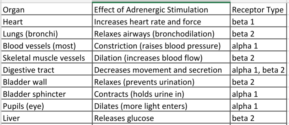 It happens when norepinephrine (NE) or epinephrine (adrenaline) bind to adrenergic receptors on organs. This is part of the sympathetic “fight or flight” response. There are two main types of adrenergic receptors:
|
front 33 List the effects of sympathoadrenal stimulation on different effector organs. In each case, indicate whether the effect is due to alpha- or beta-receptor stimulation. | back 33 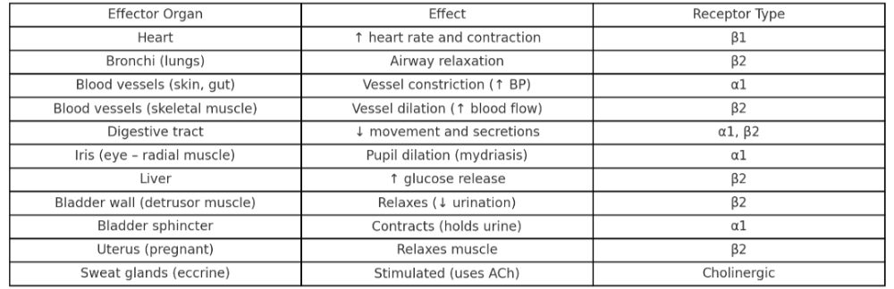 |
front 34 Describe the effects of parasympathetic nerve regulation and explain how atropine and related drugs affect this regulation. | back 34 What the Parasympathetic System Does
What Atropine Does
Parasympathetic = chill Atropine = blocks the chill → speeds things up |
front 35 Describe and give examples of antagonistic, cooperative, and complementary actions of the sympathetic and parasympathetic divisions of the autonomic system. | back 35 1. Antagonistic = Opposite effects The sympathetic and parasympathetic systems do the opposite things to the same organ. Examples: Heart: Sympathetic = speeds it up Parasympathetic = slows it down Pupils: Sympathetic = makes them bigger Parasympathetic = makes them smaller Digestion: Sympathetic = slows it down Parasympathetic = speeds it up 2. Complementary = Help the same thing in different ways Both systems do different jobs that work together. Example: Saliva: Parasympathetic = makes watery saliva Sympathetic = makes thick, sticky saliva Together, they help with chewing and swallowing. 3. Cooperative = Work together for one result Both systems are needed for a full response. Example: Male reproduction: Parasympathetic = erection Sympathetic = ejaculation |