Instructions for Side by Side Printing
- Print the notecards
- Fold each page in half along the solid vertical line
- Cut out the notecards by cutting along each horizontal dotted line
- Optional: Glue, tape or staple the ends of each notecard together
Anatomy JV Exam 3: Eye Micro
front 1 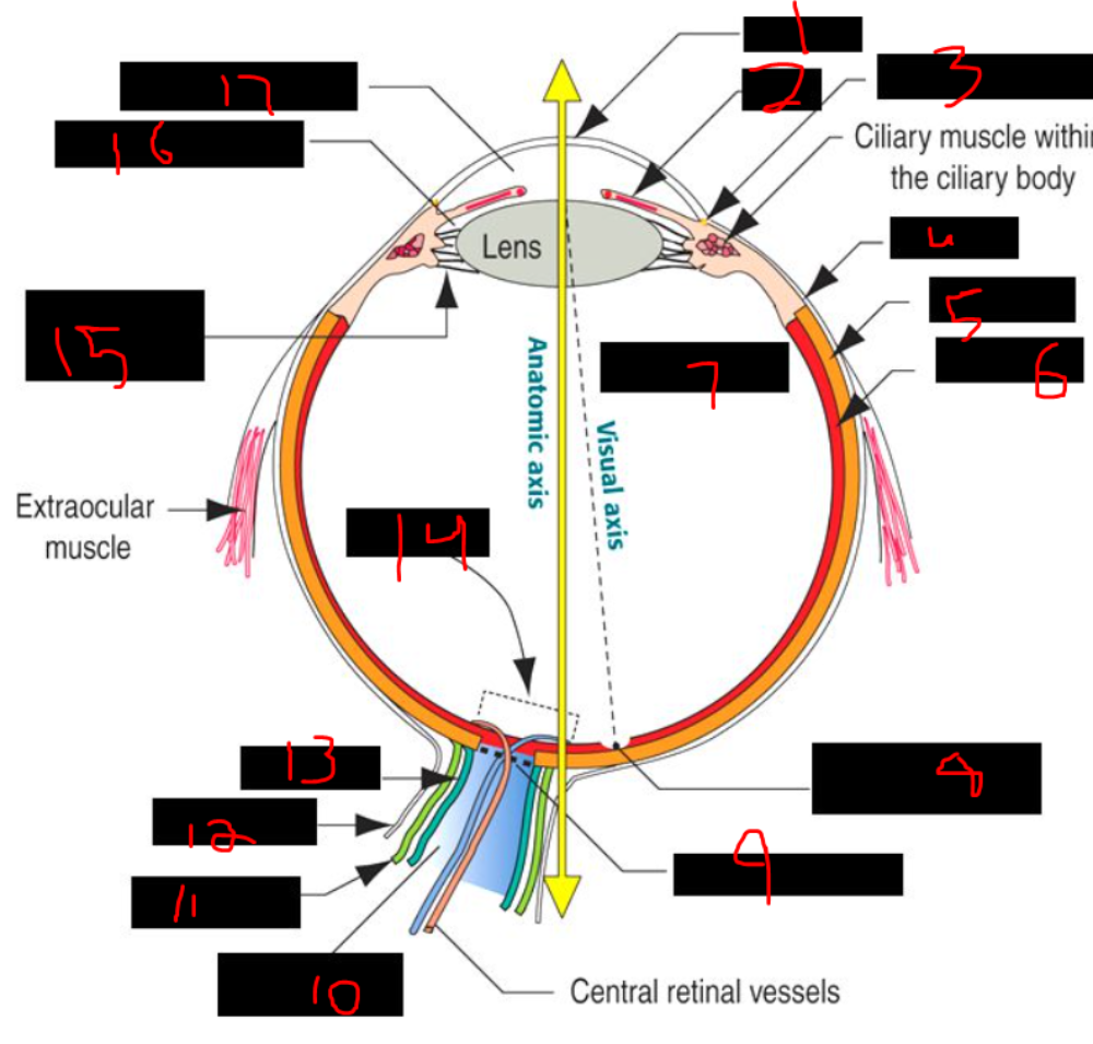 What is 1? | back 1 cornea |
front 2 What is 2? | back 2 iris |
front 3 What is 3? | back 3 canal of schlemm |
front 4 What is 4? | back 4 sclera |
front 5 What is 5? | back 5 choroid |
front 6 What is 6? | back 6 retina |
front 7 What is 7? | back 7 vitreous cavity |
front 8 What is 8? | back 8 fovea centralis |
front 9 What is 9? | back 9 lamina cribrosa |
front 10 What is 10? | back 10 optic nerve |
front 11 What is 11? | back 11 arachnoid |
front 12 What is 12? | back 12 dura mater |
front 13 What is 13? | back 13 pia mater |
front 14 What is 14? | back 14 optic disk |
front 15 What is 15? | back 15 suspensory ligaments |
front 16 What is 16? | back 16 posterior chamber |
front 17 What is 17? | back 17 anterior chamber |
front 18 at one point in embryo do you first notice signs of eye development? ______ days of gestation | back 18 22 days of gestation |
front 19 The optic vesicles form through the ______ of the developing ______, which gives rise to the optic ______. | back 19 evagination, forebrain, cup |
front 20 As the optic vesicle contacts the ______ ______, it induces the formation of ______ ______, a specialization that gives rise to the lens. | back 20 surface ectoderm, lens placodes |
front 21 The lens placodes originate from the ______ ______ and ultimately develop into the ______ of the ______. | back 21 surface ectoderm, lens, eye |
front 22 The lens placode is a ______ of the ______ ______ that ultimately gives rise to the ______. | back 22 specialization, surface ectoderm, lens |
front 23 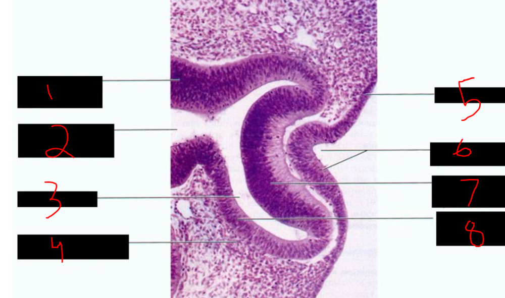 What is 1? | back 23 wall of optic stalk |
front 24 What is 2? | back 24 cavity of optic stalk |
front 25 What is 3? | back 25 intraretinal space |
front 26 What is 4? | back 26 mesenchyme |
front 27 What is 5? | back 27 surface ectoderm |
front 28 What is 6? | back 28 lens pit |
front 29 What is 7? | back 29 inner layer of optic cup |
front 30 What is 8? | back 30 outer layer of optic cup |
front 31 The intraretinal space is a ______ space, present even in ______, and is the site of ______ ______. | back 31 potential, adults, retinal detachment |
front 32 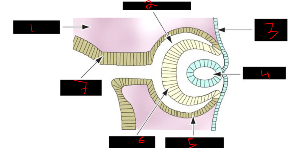 What is 1? | back 32 mesenchyme |
front 33 What is 2? | back 33 intraretinal space |
front 34 What is 3? | back 34 surface ectoderm |
front 35 What is 4? | back 35 lens vesicle |
front 36 What is 5? | back 36 pigmented layer |
front 37 What is 6? | back 37 neural layer |
front 38 What is 7? | back 38 optic stalk |
front 39 The formation of the optic fissure is significant because it allows the ______ ______ to reach the ______ ______ of the developing ______. | back 39 hyaloid artery, inner chamber, eye |
front 40 The hyaloid artery eventually becomes the ______ ______ of the ______. | back 40 central artery, retina |
front 41 The cornea is derived from ______ and mostly from ______, with the epithelium forming due to induction from the developing ______. | back 41 ectoderm, mesoderm, lens |
front 42 The developing lens induces the overlying ______ to form the ______ ______, while the rest of the cornea originates from ______. | back 42 ectoderm, corneal epithelium, mesoderm |
front 43 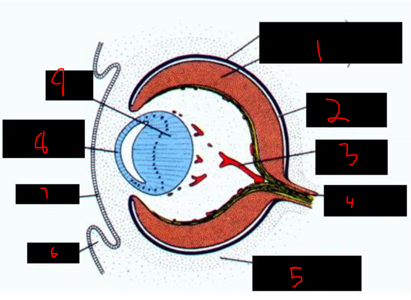 This is about how far along? | back 43 ~7 weeks |
front 44 What is 1? | back 44 pigment layer neural layer of the retina |
front 45 What is 2? | back 45 intrarenal space |
front 46 What is 3? | back 46 hyaloid vessel |
front 47 What is 4? | back 47 optic nerve fibers |
front 48 What is 5? | back 48 undifferentiated mesenchyme |
front 49 What is 6? | back 49 eyelid |
front 50 What is 7? | back 50 ectoderm |
front 51 What is 8? | back 51 anterior lens epithelium |
front 52 What is 9? | back 52 lens fibers |
front 53 The undifferentiated mesenchyme around the eye primordium gives rise to the ______ layer, which is continuous with the ______ and ______ layers of the optic nerve. | back 53 choroid, pia, arachnoid |
front 54 The scleral layer forms from loose mesenchyme and is continuous with the ______ ______ of the ______ ______. | back 54 dura mater, optic nerve |
front 55 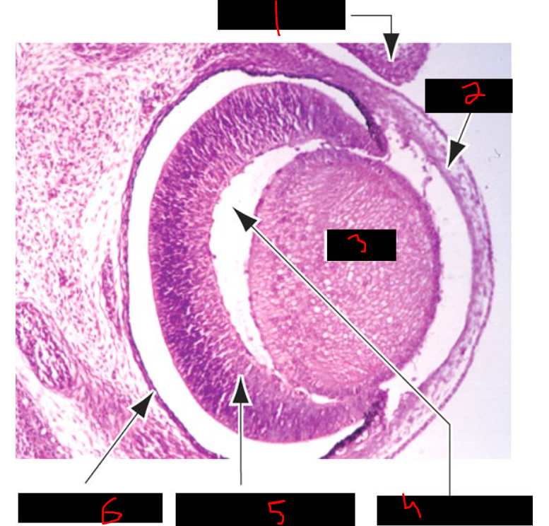 What is 1? | back 55 future eyelid |
front 56 What is 2? | back 56 cornea |
front 57 What is 3? | back 57 lens |
front 58 What is 4? | back 58 vitreous chamber |
front 59 What is 5? | back 59 neural layer (retina) |
front 60 What is 6? | back 60 pigmented layer |
front 61 The outermost layer of the eye is the ______ coat, composed of the ______, a dense connective tissue forming the white of the eye, and the ______, which is anterior, transparent, and avascular. | back 61 fibrous, sclera, cornea |
front 62 The middle layer of the eye is the ______ coat or ______ tract, consisting of the ______, ______ ______, and ______ from posterior to anterior. | back 62 vascular, uveal, choroid, ciliary body, iris |
front 63 The innermost layer of the eye is the ______, which lies internal to the vascular coat and functions as the light-sensitive ______ layer. | back 63 retina, neural |
front 64 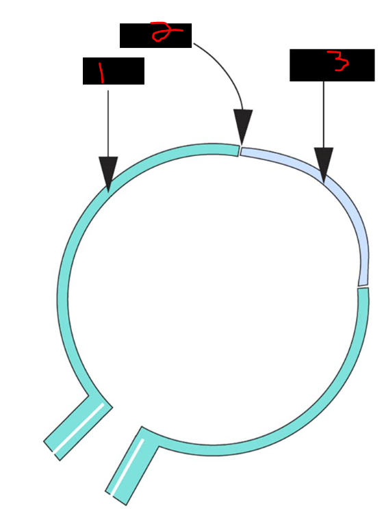 which layer of the eye? | back 64 outermost - fibrous coat |
front 65 What is 1? | back 65 sclera |
front 66 What is 2? | back 66 limbus |
front 67 What is 3? | back 67 cornea |
front 68 The middle layer of the eye, also called the ______ ______ or ______, includes the ______ posteriorly, the ______ ______ anteriorly, and the ______ at the most anterior position. | back 68 vascular coat, uvea, choroid, ciliary body, iris |
front 69  which layer of the eye? | back 69 middle - vascular coat, uvea, uveal tract |
front 70 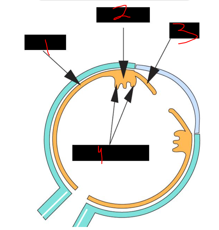 What is 1? | back 70 choroid |
front 71 What is 2? | back 71 ciliary body |
front 72 What is 3? | back 72 iris |
front 73 What is 4? | back 73 ciliary process |
front 74 The innermost layer of the eye, called the ______, includes the ______, which is part of the CNS with ______ layers, and the ______ ______, the transition zone from 10 to ______ layers, marking the anterior limit of the neuroretina. | back 74 retina, neuroretina, 10, ora serrata, 2 |
front 75 The ora serrata is the area of transition from a ______-layered sensory retina to a ______-layered non-sensory ______. | back 75 10, 2, retina |
front 76 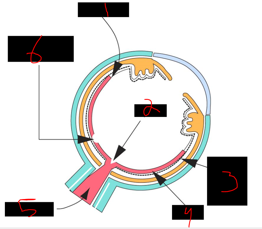 which layer of the eye? | back 76 innermost - retina |
front 77 What is 1? | back 77 ora serrata |
front 78 What is 2? | back 78 papilla |
front 79 What is 3? | back 79 outer pigmented layer |
front 80 What is 4? | back 80 retina |
front 81 What is 5? | back 81 optic nerve |
front 82 What is 6? | back 82 macula lutea and fovea |
front 83  What is 1? | back 83 vitreous chamber |
front 84 What is 2? | back 84 bv |
front 85 What is 3? | back 85 pigmented layer |
front 86 What is 4? | back 86 sclera |
front 87 What is 5? | back 87 choroid |
front 88 What is 6? | back 88 retina |
front 89 The five layers of the cornea, from outermost to innermost, are the ______ _______, ______'s layer (basement membrane), the ______ (connective tissue), ______'s membrane, and the corneal ______. | back 89 corneal epithelium, Bowman, stroma, Descemet, endothelium |
front 90 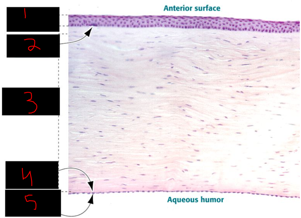 What is 1? | back 90 corneal epithelium |
front 91 What is 2? | back 91 Bowman's layer |
front 92 What is 3? | back 92 Stroma |
front 93 What is 4? | back 93 descemet's membrane |
front 94 What is 5? | back 94 corneal endothelium |
front 95 The cornea is innervated by ______ nerves that are ______ in the stroma and become ______ after crossing ______'s layer, supplied by the ______ nerve (CN ______). | back 95 myelinated, unmyelinated, Bowman, trigeminal, V |
front 96 The layers of the choroid from inside to outside begin with the ______ ______, also called ______'s membrane, which contains ______ layers. | back 96 lamina elastica, Bruch, 5 |
front 97 Following Bruch's membrane, the choroid includes the ______, which contains capillaries essential to the outer retina, the ______ layer with larger vessels, and the ______, which is closest to the ______. | back 97 choriocapillaris, vessel, epichoroid, sclera |
front 98 which layer of the choroid is nearest the sclera? ______ | back 98 epichoroid |
front 99 Bruch's membrane is a ______-layered basement membrane of the inner layer of the ______ and serves as the ______ ______ ______. | back 99 5, choroid, blood retinal barrier |
front 100 Bruch's membrane is composed of the basal lamina of the ______ ______ of the retina, ______ fibers, ______ fibers, more ______ fibers, and the basal lamina of the ______ ______. | back 100 pigmented epithelium, collagen, elastic, collagen, choroidal capillaries |
front 101 The ciliary body is a specialized structure of the ______ layer of the eye, also called the ______ ______ or ______, containing the ______ muscle with smooth muscle in two orientations, ______ fibers that suspend the lens posteriorly, and ______ processes that produce aqueous humor. | back 101 middle, vascular coat, uvea, ciliary, zonule, ciliary |
front 102 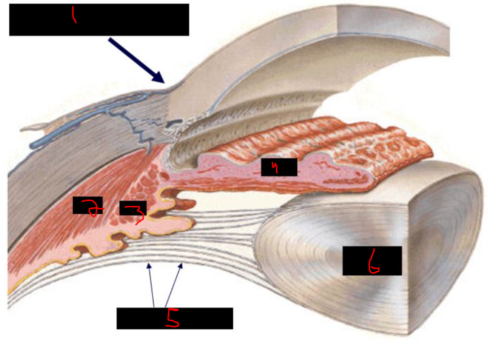 What is 1? | back 102 limbus corneae |
front 103 What is 2? | back 103 SM |
front 104 What is 3? | back 104 SM |
front 105 What is 4? | back 105 Iris |
front 106 What is 5? | back 106 zonule fibers |
front 107 What is 6? | back 107 lens |
front 108 what structures produce aqueous humor? _____ _____ - projections from the _____ _____ | back 108 ciliary processes - projections from the ciliary body |
front 109 Ciliary muscles are innervated by ______ postganglionic parasympathetics from the ______ ganglion, whose preganglionic neurons originate in the ______-______ nucleus of the oculomotor complex in the ______. | back 109 postganglionic, ciliary, Edinger-Westphal, midbrain |
front 110 The ciliary body contains ______ processes that produce ______ ______, and the ______ muscle, which contains ______ muscle that contracts under ______ influence during the ______ reflex. | back 110 ciliary, aqueous humor, ciliary, smooth, parasympathetic, accommodation |
front 111 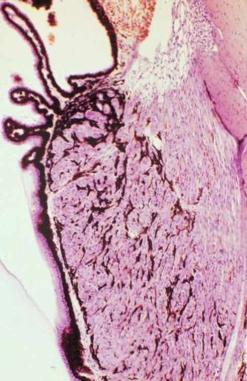 what is shown here? | back 111 ciliary body w/ ciliary processes |
front 112 The most anterior part of the middle layer of the eye, also called the ______ ______ or ______ ______, is the ______. | back 112 vascular coat, uveal tract, iris |
front 113 Eye color is determined by the number of ______: few melanocytes result in ______ eyes, many melanocytes cause ______ eyes, and an intermediate number leads to ______ or ______ eyes. | back 113 melanocytes, blue, brown, green, gray |
front 114 what is the central aperture of the iris? ______ | back 114 pupil |
front 115 In dim light, ______ sympathetic nerve fibers innervate the ______ ______, causing the pupil to ______; in bright light, ______ parasympathetic nerve fibers from the ______ ganglion innervate the ______ ______, causing the pupil to ______. | back 115 postsynaptic, dilator pupillae, dilate, postsynaptic, ciliary, sphincter pupillae, constrict |
front 116 The constrictor pupillae muscle, which is ______ muscle, is innervated by ______ parasympathetic fibers from the ______ ganglion, with preganglionic neurons in the ______ -______ nucleus of the ______ complex in the ______. | back 116 smooth, postganglionic, ciliary, Edinger-Westphal, oculomotor, midbrain |
front 117 Fixed and dilated pupils may indicate damage to the ______ involving the ______ -______ nucleus in the ______, leading to unopposed action of the ______ ______ muscle, which is innervated by the ______ nervous system. | back 117 brainstem, Edinger-Westphal, midbrain, dilator pupillae, sympathetic |
front 118 Preganglionic neurons that innervate the dilator pupillae muscle are located in the ______ ______ ______ of the ______ spinal cord. | back 118 intermediolateral cell column, T1 |
front 119 The dilator pupillae muscle is composed of ______ cells, which form an indeterminant layer just anterior to the ______ ______ ______ of the iris. | back 119 myoepithelial, posterior pigmented epithelium |
front 120 Horner's syndrome includes ______, or drooping of the upper eyelid due to paralysis of ______ muscle; ______, or loss of sweating on the affected side; ______, a small constricted pupil from unopposed ______ pupillae action; and flushing of the ______ and ______ due to vasodilation. | back 120 ptosis, Muller's, anhidrosis, miosis, sphincter, face, neck |
front 121 The lens is ______ and receives oxygen and nutrients from the ______ humor and ______ humor. | back 121 avascular, aqueous, vitreous |
front 122 The normal state of the lens is ______, as it would be for ______ vision; however, the ______ or ______ fibers can hold it in a ______ state. | back 122 thickened, near, suspensory, zonule, flattened |
front 123 For distant vision, the ciliary muscles ______, the zonule fibers become ______, and the lens is held in a ______ shape. | back 123 relax, tense, flat |
front 124 For near vision, ______ stimulation causes the ciliary muscles to ______, reducing tension on ______ fibers, allowing the lens to become ______, and the pupils to ______. | back 124 parasympathetic, contract, zonule, thickened, constrict |
front 125 Presbyopia is an age-related condition, typically after age ______, in which the lens loses ______ and can no longer ______ when ______ on the zonule fibers is released by contraction of the ______ muscles, resulting in difficulty focusing on ______ objects. | back 125 40, flexibility, thicken, tension, ciliary, near |
front 126 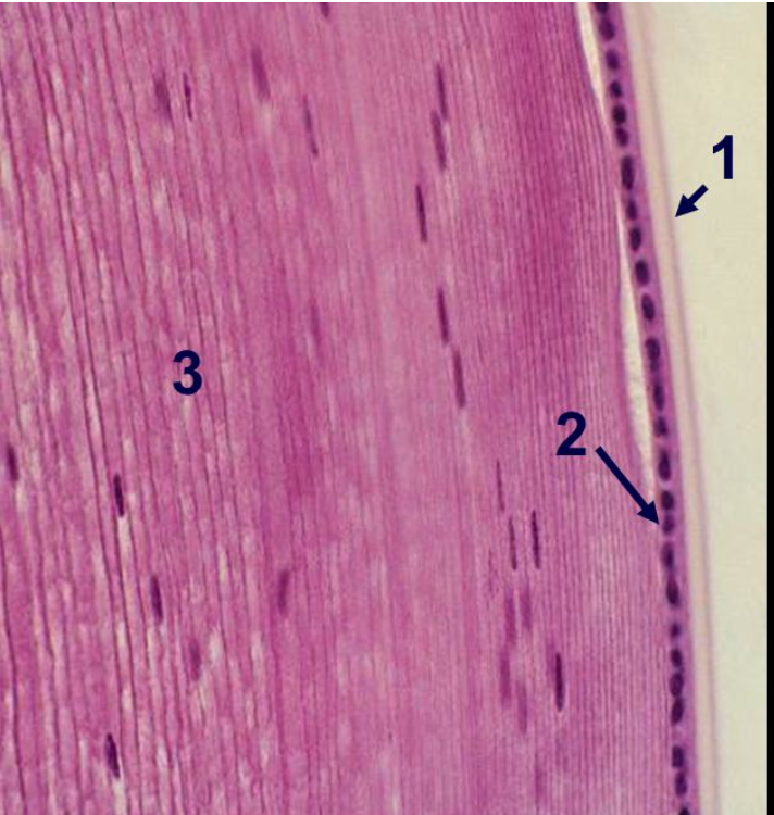 What is 1? | back 126 1) lens capsule |
front 127 What is 2? | back 127 2) subcapsular epithelium |
front 128 What is 3? | back 128 3) lens substance |
front 129 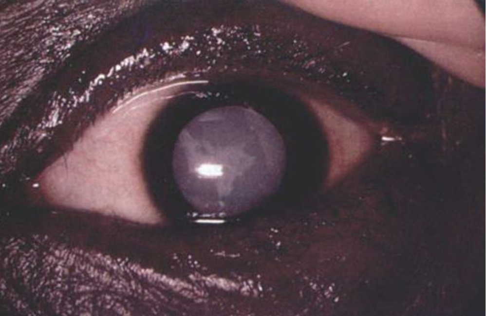 What is this? | back 129 cataracts |
front 130 Cataracts are an ______ of the ______, commonly associated with advancing ______ and leading to impaired vision. | back 130 opacity, lens, age |
front 131 In diabetes mellitus, high levels of ______ or ______ in the lens stroma draw ______ into the region, causing ______ and potentially thickening the lens, leading to diabetic ______. | back 131 glucose, sorbitol, water, cataracts, myopia |
front 132 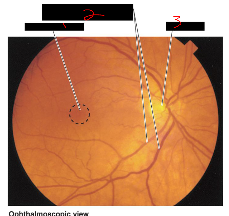 What is 1? | back 132 macula of retina |
front 133 What is 2? | back 133 branches of retinal vessels (arterioles and venules) |
front 134 What is 3? | back 134 optic disk |
front 135 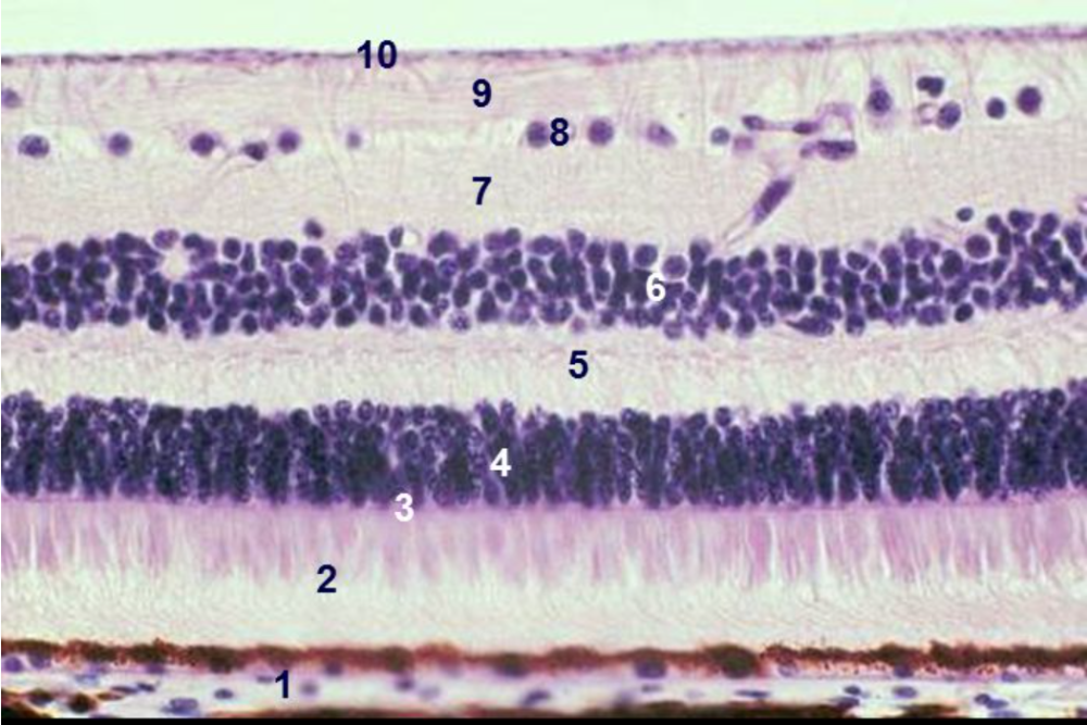 What are these? | back 135 layers of the neuroretina |
front 136 What is 1? | back 136 1) pigmented epithelium |
front 137 What is 2? | back 137 2) layer of rods & cones or outer segments of rods & cones |
front 138 What is 3? | back 138 3) external or outer limiting membrane |
front 139 What is 4? | back 139 4) external or outer nuclear layer |
front 140 What is 5? | back 140 5) external or outer plexiform layer |
front 141 What is 6? | back 141 6) internal or inner nuclear layer |
front 142 What is 7? | back 142 7) internal or inner plexiform layer |
front 143 What is 8? | back 143 8) ganglion cell layer |
front 144 What is 9? | back 144 9) optic nerve fiber layer |
front 145 What is 10? | back 145 10) inner limiting membrane |
front 146 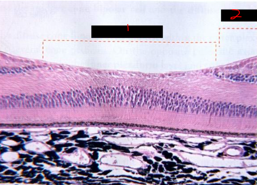 What is 1? | back 146 fovea centralis |
front 147 What is 2? | back 147 macula |
front 148 Layer 1 of the neuroretina is the ______ ______, a single layer of ______ cells that absorbs ______ to prevent reflection, stores and releases ______ ______ as a ______ precursor, and ______ membrane from photoreceptor lamellae. | back 148 pigmented epithelium, polygonal, light, vitamin A, rhodopsin, phagocytoses |
front 149 There are approximately ______ million rods and ______ million cones in the retina, making ______ the more numerous photoreceptor type. | back 149 130, 6.5, rods |
front 150 Rods are distributed throughout the ______, while cones are most concentrated in the region of the ______ ______. | back 150 neuroretina, fovea centralis |
front 151 Rhodopsin is found in the membrane-bound ______ or ______ of the cylindrical ______ segment of ______ photoreceptors, with about ______ discs present. | back 151 discs, lamellae, outer, rod, 1000 |
front 152 what type of vision are rods & cones important/responsible for? rods - ______ ______ (______ ______ ) cones - ______ ______ & ______ | back 152 twlight vision (low light) visual acuity & color |
front 153 what is this area called? | back 153 optic papilla |
front 154 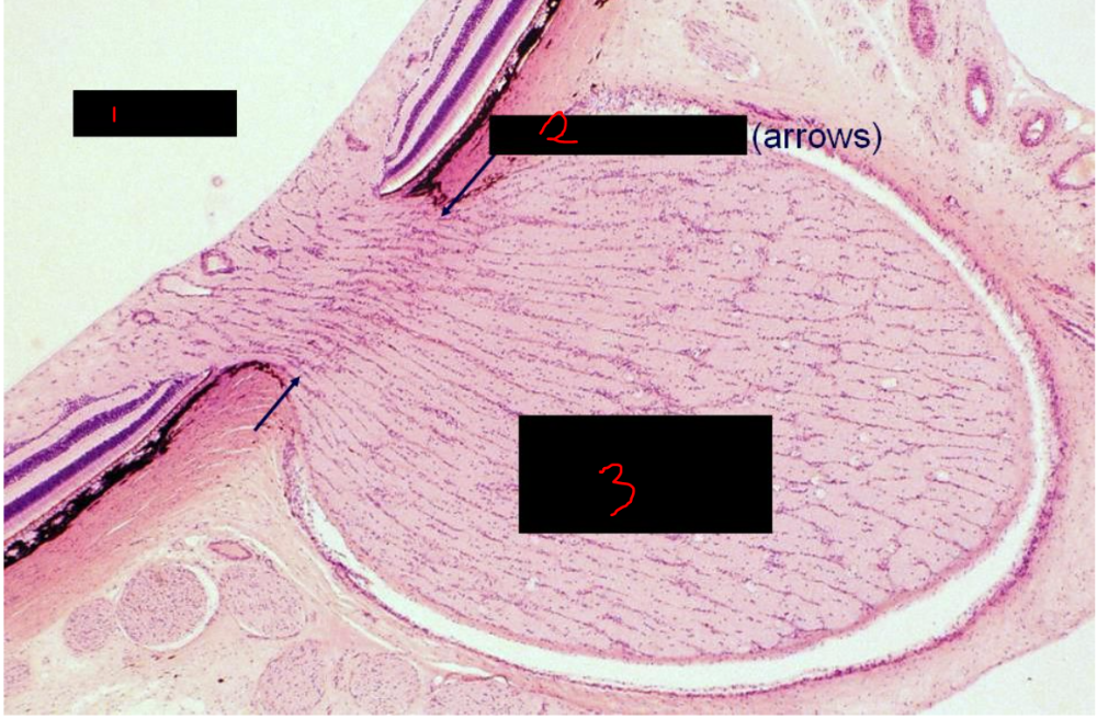 What is 1? | back 154 vitreous |
front 155 What is 2? | back 155 lamina cribosa (arrows) |
front 156 What is 3? | back 156 optic nerve (CNS myelinated by oligodendrocytes) |
front 157 The lamina cribosa is a layer of ______ found at the optic ______ that prevents ______ cells from passing, resulting in myelin and ______ being present only on the ______ nerve side, not the ______ side. | back 157 connective tissue, papilla, oligodendroglia, oligodendroglia, optic, retinal |
front 158 The retina receives blood from the ______ in the choroid layer, supplying the outer retina including the pigmented epithelium and receptors, and from branches of the ______ ______ artery, which supply the inner layers. | back 158 choriocapillaris, central retinal |
front 159 Occlusion of the ______ ______ artery causes ischemic death of ______ ______ cells and results in instant ______; this can be caused by emboli from ______ plaques or clots traveling to the artery. | back 159 central retinal, retinal ganglion, blindness, atherosclerotic |
front 160 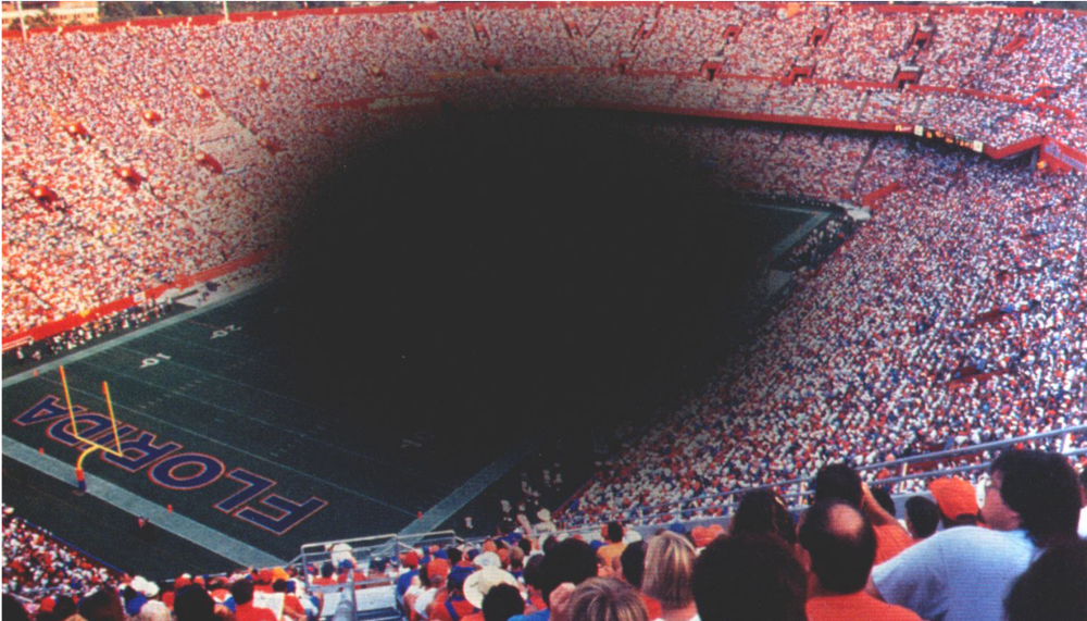 what condition does this patient have? | back 160 age related macular degeneration (ARMD) |
front 161 Age-related macular degeneration (ARMD) has two types: ______ ARMD, which accounts for about ______% of cases, and ______ ARMD, which makes up about ______% and causes the most severe vision loss. | back 161 dry, 90, wet, 10 |
front 162 age-related macular degeneration: both types ______ ______ from their ______ ______ | back 162 separate photoreceptors, blood supply |
front 163 Dry ARMD involves accumulation of ______ between ______'s membrane and the ______ ______, leading to gradual retinal damage. | back 163 drusen, Bruch, pigmented epithelium |
front 164 Wet ARMD is characterized by growth of new ______ ______ that push against the ______ ______, causing further separation and more severe vision loss. | back 164 blood vessels, pigmented epithelium |
front 165 how do you test for ARMD? ______ ______ | back 165 amsler grid |
front 166 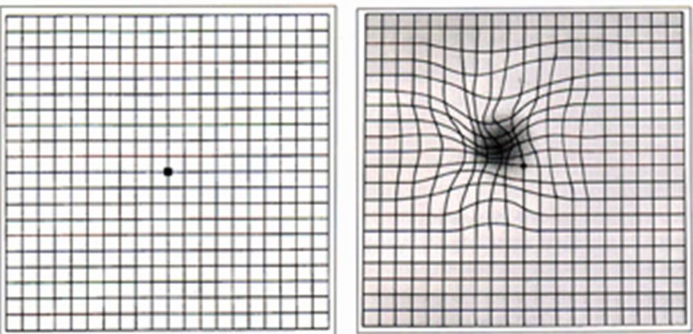 What is this? | back 166 amsler grid test |
front 167 Retinal detachment occurs between the ______ ______ and the photoreceptors of the ______ ______; this separation deprives photoreceptors of ______ and ______, leading to their death. | back 167 pigmented epithelium, neural retina, oxygen, nutrients |
front 168 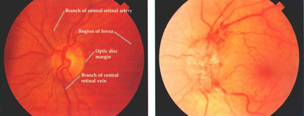 What condition is this? | back 168 papilladema |
front 169 Papilledema is the bulging of the ______ disc or papilla into the eye due to increased ______ ______. | back 169 optic, intracranial pressure |
front 170 The anterior chamber of the eye is bounded anteriorly by the ______, posteriorly by the ______, and laterally by the angle of the ______, occupied by the ______ ______ through which aqueous humor drains to the ______ of Schlemm. | back 170 cornea, iris, limbus, trabecular meshwork, canal |
front 171 The posterior chamber of the eye is bounded anteriorly by the ______, posteriorly by the ______ and ______ fibers, and peripherally by the ______ ______. | back 171 iris, lens, zonule, ciliary processes |
front 172 Aqueous humor drains by collecting in the ______ ______, then passing into the ______ of Schlemm, which encircles the eye, and ultimately entering the ______ circulation via the ______ veins. | back 172 trabecular meshwork, canal, venous, aqueous |
front 173 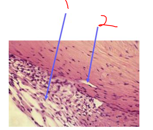 What is 1? | back 173 trabecular meshwork |
front 174 What is 2? | back 174 canal of schlemm |
front 175 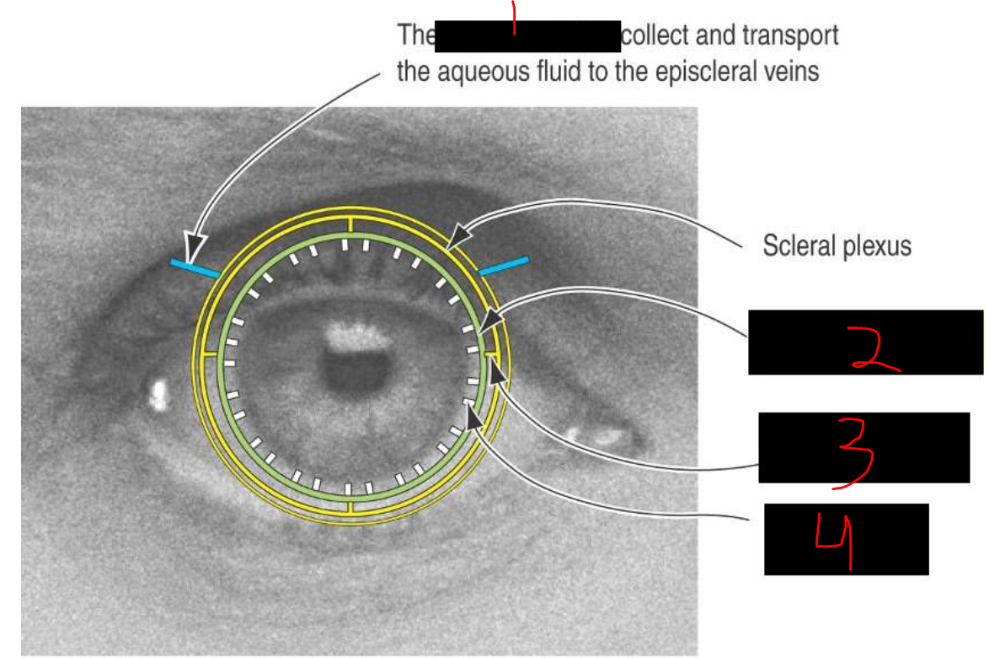 What is 1? | back 175 aqueous veins |
front 176 What is 2? | back 176 canal of schlemm |
front 177 What is 3? | back 177 external collecting channel |
front 178 What is 4? | back 178 trabecular meshwork |
front 179 The two main types of glaucoma are ______ ______ glaucoma, which is the most common and a major cause of blindness, and ______ ______ (also called ______-angle) glaucoma, which is rarer. | back 179 primary open-angle, primary closed-angle, narrow |
front 180 Primary open-angle glaucoma features a normal ______ of the anterior chamber, caused by slow blockage of the ______ of Schlemm, leading to a gradual, often unnoticed increase in intraocular pressure and eventual visual field ______. | back 180 angle, canal, defects |
front 181 Primary closed-angle glaucoma occurs when the angle of the anterior chamber is blocked by the ______; it opens when the pupil is ______ and closes when the pupil ______, causing rapid onset symptoms like ocular ______, blurred vision, and halos around ______. | back 181 iris, constricted, dilates, pain, lights |
front 182 Optic cupping is caused by increased ______ pressure and is a sign or symptom of ______. | back 182 intraocular, glaucoma |
front 183 In optic cupping, the optic disc appears ______ and ______, which can lead to ______ atrophy and neuronal ______. | back 183 pale, enlarged, retinal, death |
front 184 The vitreous body is a homogenous, transparent ______ that fills the large ______ chamber in the ______ segment of the eye, composed of about ______% water, collagen, and ______. | back 184 gel, vitreous, posterior, 99, GAGs |
front 185 The vitreous body contains ______, which synthesize collagen and GAGs, and functions to help maintain the ______ in its proper position. | back 185 halocytes, retina |
front 186 Floaters are ______ in the vitreous, usually aggregates of ______ proteins, seen as fine ______ particles moving about; they are typically benign but a sudden increase can indicate serious ______ disease. | back 186 deposits, vitreal, dust-like, eye |
front 187 During a vitrectomy, the ______ or ______ vitreous humor is removed and replaced with ______ to maintain the eye’s shape and keep the ______ in position. | back 187 cloudy, bloody, saline, retina |
front 188 Age-related macular degeneration (ARMD) specifically affects the ______ and the ______ ______ of the eye. | back 188 macula, fovea centralis |