Instructions for Side by Side Printing
- Print the notecards
- Fold each page in half along the solid vertical line
- Cut out the notecards by cutting along each horizontal dotted line
- Optional: Glue, tape or staple the ends of each notecard together
Anatomy JV Exam 3: Blood Supply to Brain, Dual Sinuses, CSF Flow
front 1 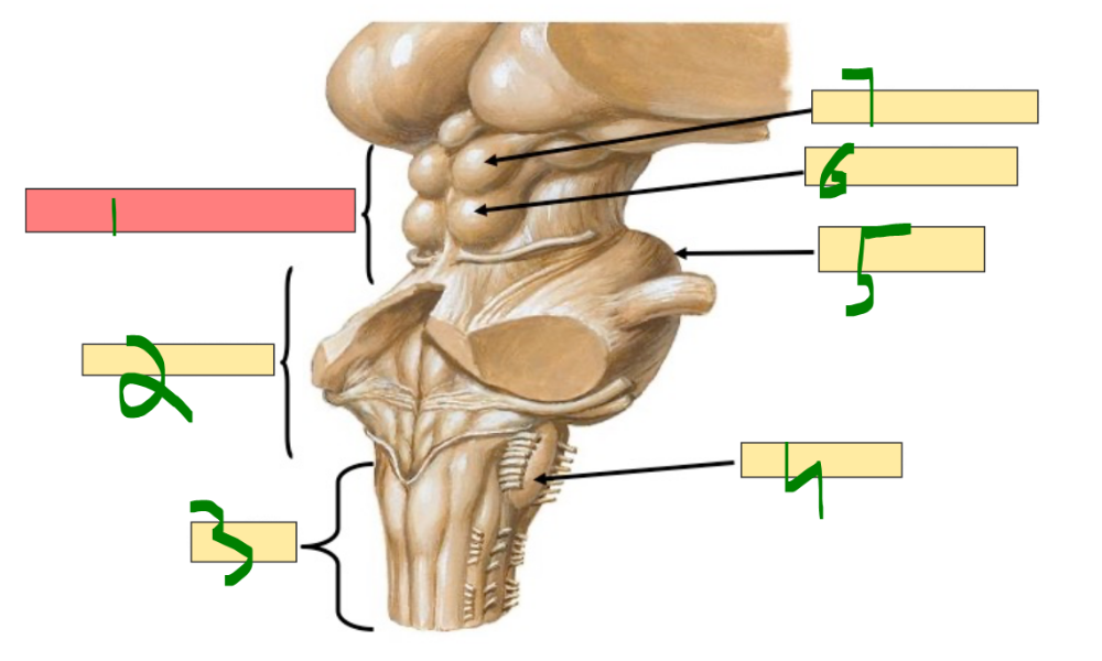 What is 1? | back 1 mesencephalon (midbrain) |
front 2 What is 2? | back 2 metencephalon |
front 3 What is 3? | back 3 medulla |
front 4 What is 4? | back 4 inferior olive |
front 5 What is 5? | back 5 pons |
front 6 What is 6? | back 6 inferior colliculus |
front 7 What is 7? | back 7 superior colliculus |
front 8 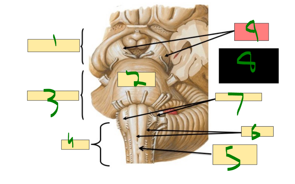 What is 1? | back 8 mesencephalon |
front 9 What is 2? | back 9 pons |
front 10 What is 3? | back 10 metencephalon |
front 11 What is 4? | back 11 medulla |
front 12 What is 5? | back 12 pyramidal decussation |
front 13 What is 6? | back 13 pyramids |
front 14 What is 7? | back 14 inferior olives |
front 15 What is 9? | back 15 cerebral peduncles |
front 16 The brain is derived from the ______ ______ located ______ (cranial) to the ______ pair of somites. | back 16 neural tube, rostral, fourth |
front 17 The three primary brain vesicles are the ______, the ______, and the ______. | back 17 prosencephalon, mesencephalon, rhombencephalon |
front 18 The prosencephalon, or ______, gives rise to the ______ and the ______. | back 18 forebrain, telencephalon, diencephalon |
front 19 The mesencephalon, or ______, remains as the ______ in the secondary vesicle stage. | back 19 midbrain, mesencephalon |
front 20 The rhombencephalon, or ______, gives rise to the ______ and the ______. | back 20 hindbrain, metencephalon, myelencephalon |
front 21 The telencephalon gives rise to the ______ ______ and the ______ ______. | back 21 cerebral cortex, basal ganglia |
front 22 The ______ ______ are the remnant of the telencephalon ______. | back 22 lateral ventricles, vesicle |
front 23 The diencephalon gives rise to the ______, the ______, and the ______ gland. | back 23 thalamus, hypothalamus, pineal |
front 24 The ______ ______ is the remnant of the diencephalon ______. | back 24 third ventricle, vesicle |
front 25 The metencephalon gives rise to the ______ and the ______. | back 25 pons, cerebellum |
front 26 The ______ ______ is the remnant of the metencephalon ______. | back 26 fourth ventricle, vesicle |
front 27 what does the myelencephalon give rise to? ______ ______ | back 27 medulla oblongata |
front 28 The aqueduct of ______, also known as the ______ ______, is a remnant of the ______ vesicle. | back 28 Sylvius, cerebral aqueduct, mesencephalon |
front 29 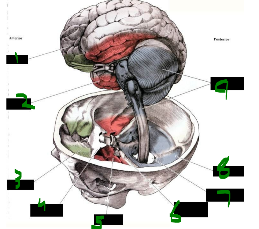 What is 1? | back 29 frontal lobe |
front 30 What is 2? | back 30 temporal lobe |
front 31 What is 3? | back 31 anterior fossa |
front 32 What is 4? | back 32 lesser wing of sphenoid bone |
front 33 What is 5? | back 33 middle fossa |
front 34 What is 6? | back 34 petrous ridge of temporal bone |
front 35 What is 7? | back 35 foramen magnum |
front 36 What is 8? | back 36 posterior fossa |
front 37 What is 9? | back 37 cerebellum and brainstem |
front 38 The ______ ______ contains the ______ ______ cortex responsible for voluntary movement. | back 38 precentral gyrus, primary motor |
front 39 The ______ ______ contains the ______ cortex responsible for processing somatic sensations. | back 39 postcentral gyrus, sensory |
front 40 The ______ gyrus of the ______ lobe is involved in ______ and aspects of memory. | back 40 superior, temporal, audition |
front 41 The ______ lobe, specifically on the banks of the ______ ______, processes ______ information. | back 41 occipital, calcarine fissure, visual |
front 42  What is 1? | back 42 primary motor cortex |
front 43 What is 2? | back 43 central sulcus |
front 44 What is 3? | back 44 primary somatosenosry cortex |
front 45 What is 4? | back 45 parietal lobe |
front 46 What is 5? | back 46 occipital lobe |
front 47 What is 6? | back 47 primary visual cortex |
front 48 What is 7? | back 48 primary auditory cortex |
front 49 What is 8? | back 49 temporal lobe |
front 50 What is 9? | back 50 sylvian fissure |
front 51 What is 10? | back 51 frontal lobe |
front 52 The primary motor cortex is located in the ______ ______ and corresponds to Brodmann's area ______. | back 52 precentral gyrus, 4 |
front 53 The precentral gyrus, where the primary motor cortex resides, is organized ______, meaning specific regions control specific ______ parts. | back 53 somatotopically, body |
front 54 Axons from ______ ______ neurons leave the ______ and descend through the ______ ______. | back 54 upper motor, cortex, internal capsule |
front 55 After descending through the internal capsule, upper motor neuron axons travel through the ______ ______, enter the ______, and reach the ______ where they form the pyramids. | back 55 cerebral peduncle, pons, medulla |
front 56 In the medulla, about ______ percent of fibers cross in the ______ ______ before descending. | back 56 ninety, pyramidal decussation |
front 57 After decussation, fibers descend in the ______ ______ tract and synapse on ______ ______ neurons in the ______ horn. | back 57 lateral corticospinal, alpha motor, ventral |
front 58 from medial to lateral, describe the motor cortex humunculus: ______ , ______ , ______ ______ ,______ ______ , ______ , ______ , ______ , ______ , ______ ______ ______ , ______ , ______ , ______ , ______ , ______ , ______ , ______ | back 58 toes, knee, hip trunk,shoulder arm, elbow, wrist, hand, fingers, thumb neck brow, eye, face, lips, jaw, tongue, pharynx, larynx |
front 59 The primary sensory cortex is located in the ______ ______ and corresponds to Brodmann's areas ______, ______, and ______. | back 59 postcentral gyrus, 3, 1, 2 |
front 60 Like the primary motor cortex, the postcentral gyrus is organized ______, with distinct regions processing input from specific ______ parts. | back 60 somatotopically, body |
front 61 The dorsal column pathway carries ______ ______, ______, and ______ sensations, and it crosses in the ______. | back 61 discriminative touch, proprioception, vibration, brainstem |
front 62 The anterolateral system, also known as the ______ tract, carries ______ ______, ______, and ______ sensations, and it crosses in the ______ ______. | back 62 spinothalamic, crude touch, pain, temperature, spinal cord |
front 63 Both major sensory pathways relay through the ______ before reaching the ______ cortex. | back 63 thalamus, sensory |
front 64 from medial to lateral, describe the sensory cortex humunculus: ______ , ______ , ______ , ______ , ______ , ______ ______ , ______ , ______ , ______ , ______ , ______ ______ , ______ , ______ , ______ , ______ , ______ ______ , ______ , ______ ______ | back 64 genitals, leg, hip, trunk, neck, head arm, elbow, forearm, hand, fingers, thumb eye, nose, face, lips, teeth, gums jaw, tongue, pharynx abdomen |
front 65 An injury to the corticospinal system (pyramidal tract) ______ the pyramidal decussation results in ______ paralysis. | back 65 above, contralateral |
front 66 An injury to the corticospinal system ______ the pyramidal decussation results in ______ paralysis ______ the lesion. | back 66 below, ipsilateral, below |
front 67 A spinal cord lesion causes loss of ______ crude touch, ______, and ______ below the lesion due to damage to the ______ system. | back 67 contralateral, pain, temperature, anterolateral |
front 68 A spinal cord lesion also results in loss of ______ discriminative touch, ______, and ______ below the lesion due to damage to the ______ columns. | back 68 ipsilateral, proprioception, vibration, dorsal |
front 69 The internal carotid artery is a branch of the ______ ______ artery, enters the cranial cavity through the ______ ______, passes through the ______ ______, and primarily supplies the ______, ______, and ______. | back 69 common carotid, carotid canal, cavernous sinus, orbit, eye, brain |
front 70 The vertebral artery is the first branch of the ______ artery, travels through the transverse foramina of ______ to ______, enters the ______ ______, and joins the opposite vertebral artery to form the ______ artery. | back 70 subclavian, C6, C1, foramen magnum, basilar |
front 71 what is the main blood supply of the brain? ______ ______ a. ______ a. | back 71 internal carotid a. vertebral a. |
front 72 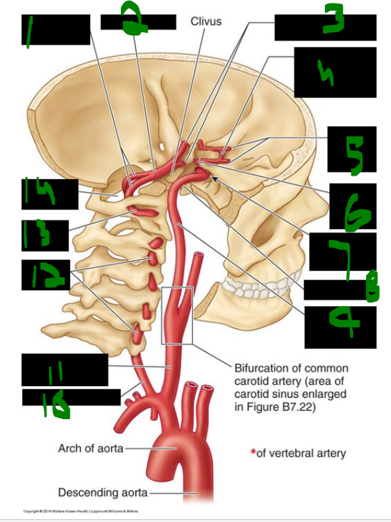 What is 1? | back 72 intracranial parts |
front 73 What is 2? | back 73 basilar artery |
front 74 What is 3? | back 74 posterior cerebral arteries |
front 75 What is 4? | back 75 anterior communicating artery |
front 76 What is 5? | back 76 anterior cerebral arteries |
front 77 What is 6? | back 77 middle cerebral artery |
front 78 What is 7? | back 78 origin of opthalmic artery |
front 79 What is 8? | back 79 carotid canal |
front 80 What is 9? | back 80 internal carotid artery |
front 81 What is 10? | back 81 vertebral artery |
front 82 What is 11? | back 82 common carotid artery |
front 83 What is 12? | back 83 cervical part |
front 84 What is 13? | back 84 atlantic part |
front 85 What is 14? | back 85 foramen magnum |
front 86 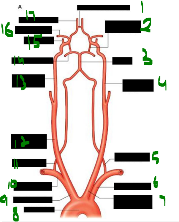 What is 1? | back 86 anterior communicating |
front 87 What is 2? | back 87 posterior communicating |
front 88 What is 3? | back 88 basilar |
front 89 What is 4? | back 89 left internal carotid |
front 90 What is 5? | back 90 left vertebral |
front 91 What is 6? | back 91 left subclavian |
front 92 What is 7? | back 92 left common carotid |
front 93 What is 8? | back 93 aortic arch |
front 94 What is 9? | back 94 brachiocephalic |
front 95 What is 10? | back 95 right subclavian |
front 96 What is 11? | back 96 right vertebral |
front 97 What is 12? | back 97 right common carotid |
front 98 What is 13? | back 98 right internal carotid |
front 99 What is 14? | back 99 posterior cerebral |
front 100 What is 15? | back 100 opthalmic |
front 101 What is 16? | back 101 middle cerebral |
front 102 What is 17? | back 102 anterior cerebral |
front 103 what is the first branch of the internal carotid a.? ____ ____ | back 103 ophthalmic a. |
front 104 what branches arise from the vertebral artery? -____ ____ a. -____ ____ a. -____ ____ ____ a. | back 104 -anterior spinal a. -posterior spinal a. -posterior inferior cerebellar a. "PICA" |
front 105 The basilar artery is formed by the fusion of two ______ arteries and travels rostrally on the anterior aspect of the ______. | back 105 vertebral, pons |
front 106 Branches of the basilar artery, from caudal to rostral, include the ______ ______ cerebellar arteries, about ______ pontine arteries, and the ______ cerebellar arteries. | back 106 anterior inferior, three, superior |
front 107 The basilar artery bifurcates into two ______ ______ arteries. | back 107 posterior cerebral |
front 108 The internal carotid artery enters the cranial cavity via the ______ ______ and passes through the ______ ______. | back 108 carotid canal, cavernous sinus |
front 109 The terminal branches of the internal carotid artery are the ______ ______ artery, the ______ ______ artery, and the ______ ______ artery. | back 109 posterior communicating, middle cerebral, anterior cerebral |
front 110 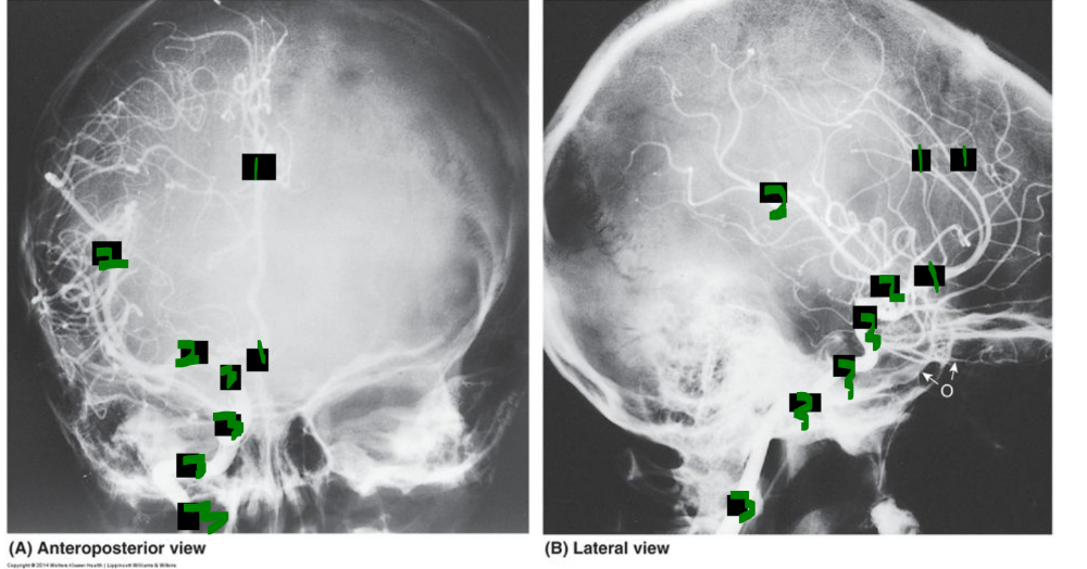 arteriogram | back 110 1= A = anterior cerebral a 2= M = middle cerebral a. 3= I = internal carotid a. |
front 111 The circle of Willis is an anastomosis between the ______ and ______ ______ systems formed at the base of the ______. | back 111 vertebrobasilar, internal carotid, brain |
front 112 The circle of Willis is formed by the ______ ______ artery connecting the left and right ______ ______ arteries. | back 112 anterior communicating, anterior cerebral |
front 113 The ______ ______ arteries, one on each side, connect the internal carotid artery to the ______ ______ artery. | back 113 posterior communicating, posterior cerebral |
front 114 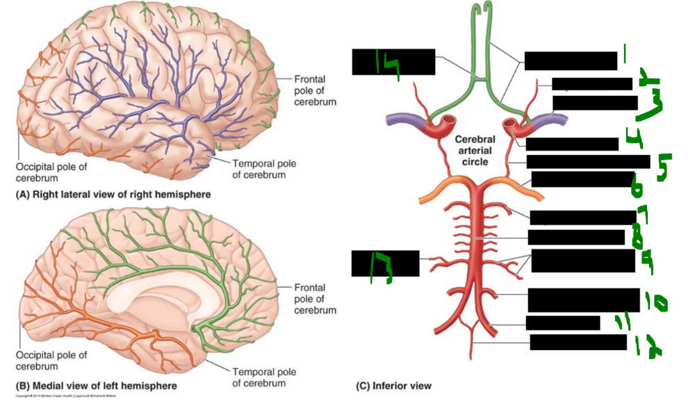 What is 1? | back 114 anterior cerebral |
front 115 What is 2? | back 115 opthalmic |
front 116 What is 3? | back 116 middle cerebral |
front 117 What is 4? | back 117 internal carotid |
front 118 What is 5? | back 118 posterior communicating |
front 119 What is 6? | back 119 posterior cerebral |
front 120 What is 7? | back 120 superior cerebellar |
front 121 What is 8? | back 121 basilar |
front 122 What is 9? | back 122 anterior inferior cerebellar |
front 123 What is 10? | back 123 posterior inferior cerebellar |
front 124 What is 11? | back 124 vertebral |
front 125 What is 12? | back 125 anterior spinal |
front 126 What is 13? | back 126 labyrinthine artery |
front 127 What is 14? | back 127 anteiror communicating |
front 128  What is 1? | back 128 frontal lobe |
front 129 What is 2? | back 129 anterior cerebral artery |
front 130 What is 3? | back 130 anterior communicating artery |
front 131 What is 4? | back 131 anterior cerebral artery |
front 132 What is 5? | back 132 middle cerebral artery |
front 133 What is 6? | back 133 posterior communicating artery |
front 134 What is 7? | back 134 oculomotor nerve (CN 3) |
front 135 What is 8? | back 135 trochlear nerve (CN IV) |
front 136 What is 9? | back 136 trigeminal nerve (CN 5) |
front 137 What is 10? | back 137 basilar artery |
front 138 What is 11? | back 138 labyrinthine artery |
front 139 What is 12? | back 139 anterior inferior posterior inferior (these are the cerebellar arteries) |
front 140 What is 13? | back 140 vertebral artery |
front 141 What is 14? | back 141 anterior spinal artery |
front 142 What is 15? | back 142 hypoglossal nerve (CN 12) |
front 143 What is 16? | back 143 spinal accessory nerve (CN 11) |
front 144 What is 17? | back 144 vagus nerve (CN 10) |
front 145 What is 18? | back 145 glossopharyngeal nerve (cn 9) |
front 146 What is 19? | back 146 vestibulocochlear nerve (CN 8) |
front 147 What is 20? | back 147 facial nerve (CN 7) |
front 148 What is 21? | back 148 abducent nerve (CN 6) |
front 149 What is 22? | back 149 superior cerebellar artery |
front 150 What is 23? | back 150 posterior cerebral artery |
front 151 What is 24? | back 151 temporal lobe |
front 152 What is 25 | back 152 internal carotid artery |
front 153 What is 26? | back 153 optic nerve (CN 2) |
front 154 What is 27? | back 154 corpus callosum |
front 155 What is 28? | back 155 olfactory bulb and tract |
front 156 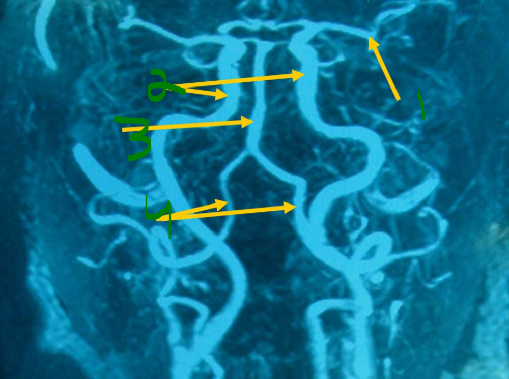 What is 1? | back 156 middle cerebral artery |
front 157 What is 2? | back 157 internal carotid arteries |
front 158 What is 3? | back 158 basilar artery |
front 159 What is 4? | back 159 vertebral arteries |
front 160 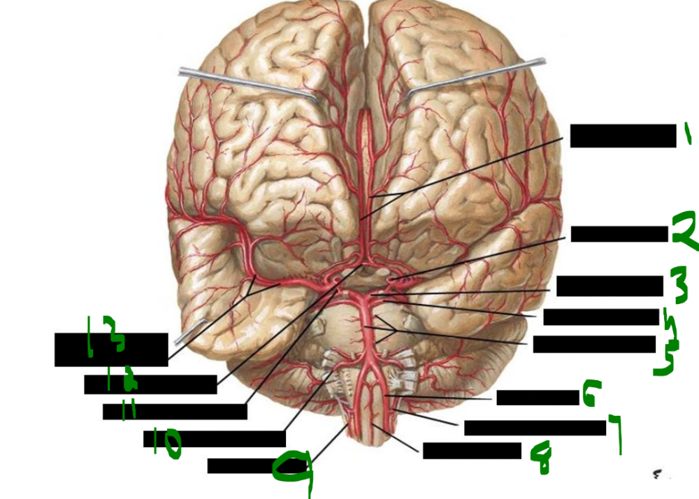 What is 1? | back 160 anterior cerebral arteries |
front 161 What is 2? | back 161 internal carotid artery |
front 162 What is 3? | back 162 posterior cerebral artery |
front 163 What is 4? | back 163 superior cerebellar artery |
front 164 What is 5? | back 164 basilar and pontine arteries |
front 165 What is 6? | back 165 vertebral artery |
front 166 What is 7? | back 166 posteior inferior cerebrellar artery |
front 167 What is 8? | back 167 anteior spinal artery |
front 168 What is 9? | back 168 posterior spinal artery |
front 169 What is 10? | back 169 anterior inferior cerebellar artery |
front 170 What is 11? | back 170 posterior communicating artery |
front 171 What is 12? | back 171 anterior communicating artery |
front 172 What is 13? | back 172 middle cerebral artery and branches |
front 173 an aneurysm to which artery might cause compression/damage to abducens n. (CN VI)? ______ ______ ______ artery | back 173 anterior inferior cerebellar artery |
front 174 aneurysm to which artery might cause compression/damage to oculomotor n (CN III)? ______ ______ or ______ ______ arteries | back 174 posterior cerebral or superior cerebellar arteries |
front 175 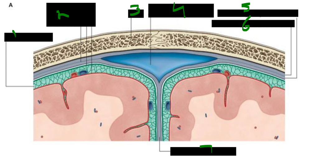 What is 1? | back 175 subarachnoid space |
front 176 What is 2? | back 176 dura mater arachnoid mater pia mater |
front 177 What is 3? | back 177 skull |
front 178 What is 4? | back 178 intracranial venous structure (superior sagittal sinus) |
front 179 What is 5? | back 179 inner meningeal layer of dura mater |
front 180 What is 6? | back 180 outer periosteal layer of dura mater |
front 181 What is 7? | back 181 dura partition (falx cerebri) |
front 182 The falx cerebri is a crescent-shaped ______ specialization that projects downward between the ______ ______. | back 182 dural, cerebral hemispheres |
front 183 Anteriorly, the falx cerebri attaches to the ______ ______ of the ethmoid bone and the ______ ______ of the frontal bone. | back 183 crista galli, frontal crest |
front 184 Posteriorly, the falx cerebri attaches to and blends with the ______ ______. | back 184 tentorium cerebelli |
front 185 what sinuses traverse the falx cerebri? ______ & ______ ______ sinuses | back 185 superior & inferior sagittal sinuses |
front 186 What is 1? | back 186 tentorium cerebelli |
front 187 What is 2? | back 187 falx cerebri |
front 188 What is 3? | back 188 infundibulum |
front 189 What is 4? | back 189 diaphragma sellae |
front 190 What is 5? | back 190 tentorium cerebelli |
front 191 What is 6? | back 191 falx cerebelli |
front 192 What is 7? | back 192 tentorial notch |
front 193 what structure separates the cerebellum from the overlying posterior lobes of the cerebral hemispheres? ______ cerebelli - ______ shelf of ______ dura | back 193 tentorium cerebelli - horizontal shelf of meningeal dura |
front 194 The tentorium cerebelli is a horizontal shelf of ______ ______ that separates the ______ from the overlying posterior lobes of the ______ ______. | back 194 meningeal dura, cerebellum, cerebral hemispheres |
front 195 The tentorium cerebelli attaches posteriorly to the ______ bone, laterally to the superior border of the ______ part of the ______ bone, and anteriorly and medially forms the ______ ______ where the ______ passes through. | back 195 occipital, petrous, temporal, tentorial notch, midbrain |
front 196 what structures pass through the tentorial notch? ______ ______ ______ | back 196 midbrain basilar artery |
front 197 Transtentorial herniation is the herniation of the ______ ______ lobe and ______ through the ______ ______. | back 197 medial temporal, uncus, tentorial notch |
front 198 A common type of transtentorial herniation is ______ herniation. | back 198 uncal |
front 199 Uncal herniation is a common type of ______ herniation involving the ______ ______ lobe and ______ passing through the ______ ______. | back 199 transtentorial, medial temporal, uncus, tentorial notch |
front 200 The clinical triad of uncal herniation includes a ______ pupil due to compression of the ______ complex, ______ from compression of the cerebral ______, and ______ due to distortion of the midbrain ______ system. | back 200 blown, oculomotor nuclear, hemiplegia, peduncles, coma, reticular |
front 201 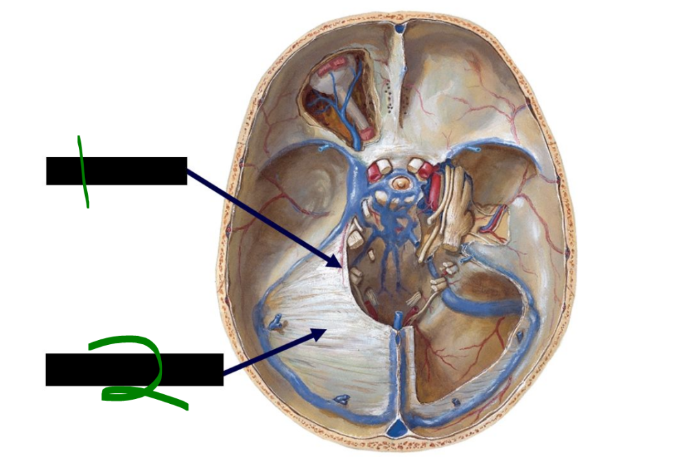 What is 1? | back 201 tentorial notch |
front 202 What is 2? | back 202 tentorium cerebelli |
front 203 The diaphragma sellae is a small, horizontal shelf of ______ ______ that covers the ______ ______ of the ______ ______. | back 203 meningeal dura, hypophyseal fossa, sella turcica |
front 204 The ______ passes through a small opening in the diaphragma sellae within the ______ ______. | back 204 infundibulum, sella turcica |
front 205 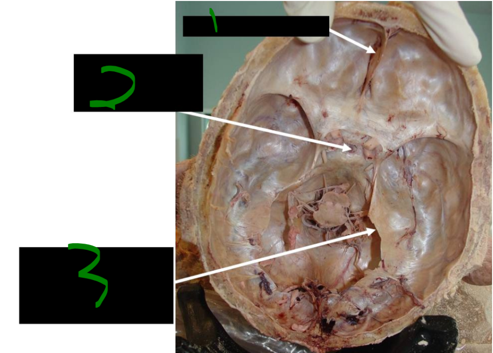 What is 1? | back 205 cut anterior falx cerebri |
front 206  What is 2? | back 206 infundibular stalk projecting through the sella turcica |
front 207 What is 3? | back 207 tentorium cerebelli has been cut to allow the removal of the cerebellum |
front 208 An epidural hemorrhage, also called an ______ hemorrhage, is a ______-shaped accumulation of blood between the ______ of the calvarium and the ______ layer of the dura. | back 208 extradural, lens, bone, periosteal |
front 209 Epidural hemorrhage is usually caused by tearing of the ______ ______ artery, particularly its ______ branch, due to trauma to the ______. | back 209 middle meningeal, anterior, pterion |
front 210 In an epidural hemorrhage, blood slowly separates the ______ ______ from the underlying ______. | back 210 periosteal dura, bone |
front 211 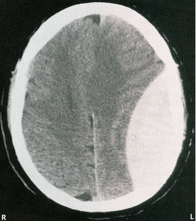 what has happened to this patient? | back 211 epidural/extradural hemorrhage |
front 212 The dura is mostly innervated by the ______ nerve (CN ______), except for the ______ ______. | back 212 trigeminal, V, posterior fossa |
front 213 The posterior fossa of the dura, below the ______, is innervated by cervical nerves ______ and ______, which enter through the ______ ______, ______ canal, and ______ foramen. | back 213 tentorium, C2, C3, foramen magnum, hypoglossal, jugular |
front 214 The brain itself has no ______ or ______ receptors and is therefore ______. | back 214 touch, pain, insensate |
front 215 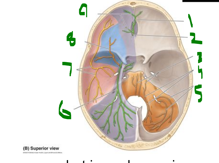 What is 1? | back 215 anterior ethmoidal nerve |
front 216 What is 2? | back 216 posterior ethmoidal nerve |
front 217 What is 3? | back 217 C2,C3 fibers |
front 218 What is 4? | back 218 C2,C3 fibers distributed by CN XII |
front 219 What is 5? | back 219 C2 fibers distributed by CN X |
front 220 What is 6? | back 220 tentorial nerve (recurrent meningeal branch of opthalmic nerve-CN v3) |
front 221 What is 7? | back 221 meningeal branches of mandibular nerve (CN V3) (including nervus spinosus) |
front 222 What is 8? | back 222 meningeal branch of maxillary nerve (CN V2) |
front 223 What is 9? | back 223 anterior meningeal branches of ethmoidal nerve (CN V1) |
front 224 what attaches the arachnoid to pia? ___ ___ - "look like spider webs" | back 224 fine trabeculae - "look like spider webs" |
front 225 within what layer of mater do BVs travel? _____ space - between _____ & _____ _____ | back 225 subarachnoid space - between arachnoid & pia mater |
front 226 A subdural bleed is a crescent-shaped hemorrhage caused by torn ______ veins, filling the potential ______ space between the ______ and ______ mater. | back 226 emissary, subdural, dura, arachnoid |
front 227 Subdural hemorrhage typically occurs in ______ individuals due to brain ______, which increases the space between the brain and ______, straining veins that connect to the ______ ______ sinuses. | back 227 older, atrophy, arachnoid, dural venous |
front 228 The history of a subdural bleed may involve a ______ injury, with or without ______ of ______. | back 228 trivial, loss, consciousness |
front 229 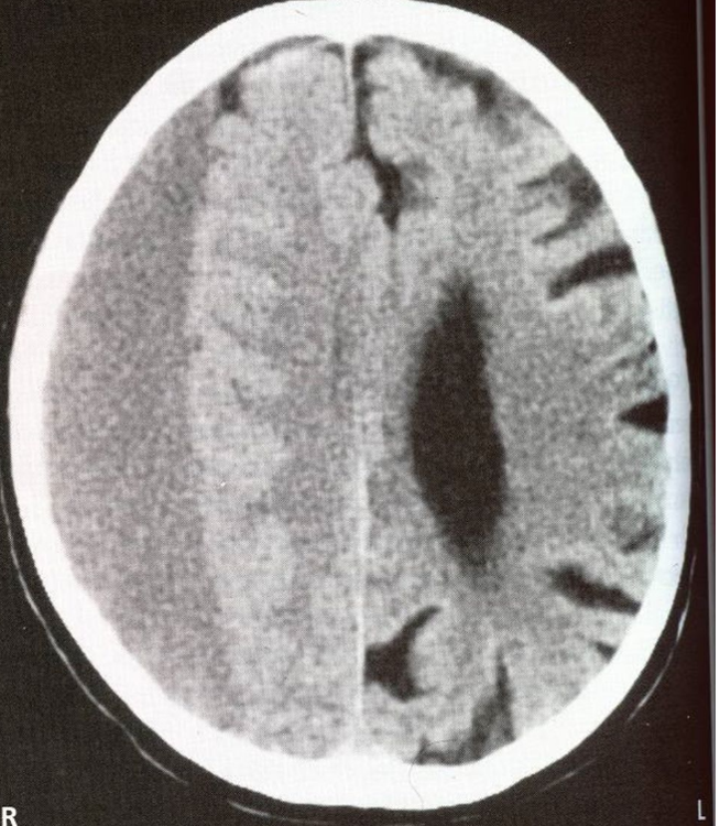 what happened to this patient? | back 229 subdural bleed/hemorrhage |
front 230 subdural vs epidural bleed (shape of pooled blood) subdural - ______ shaped | back 230 cresent lens |
front 231 A subarachnoid bleed is a hemorrhage of ______ blood in the ______ space that flows between ______ of the brain into the ______ and accumulates ______. | back 231 arterial, subarachnoid, gyri, sulci, rapidly |
front 232 Subarachnoid hemorrhage is frequently caused by the bursting of a ______ ______ and may also result from ______ head trauma. | back 232 cerebral aneurysm, significant |
front 233 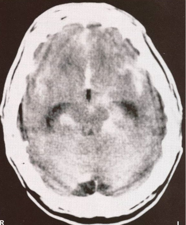 what has happened to this patient? what most likely caused this injury? | back 233 subarachnoid bleed/hemorrhage significant head trauma |
front 234 The four typical sites of brain herniation are: ______ (under the falx cerebri), ______ (downward herniation of the brainstem), ______ (medial temporal lobe and uncus through the tentorial notch), and ______ (cerebellar tonsil through the foramen magnum). | back 234 subfalcine, central, uncal, tonsillar |
front 235 Cushing’s triad is a classic sign of elevated ______ ______ and includes ______, ______, and ______. | back 235 intracranial pressure, hypertension, bradycardia, irregular respiration |
front 236 In Cushing’s triad, hypertension is a reflex to maintain ______ ______, bradycardia is a reflex response to ______, and irregular respiration indicates impaired ______ function. | back 236 cerebral perfusion, hypertension, brainstem |
front 237 Dural venous sinuses are ______-lined spaces located between layers of the ______ that drain blood into the ______ ______ veins via the ______ foramen. | back 237 endothelial, dura, internal jugular, jugular |
front 238 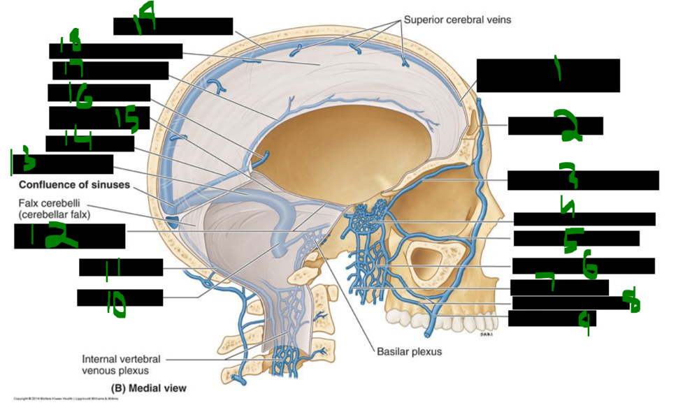 What is 1? | back 238 beginning of superior sagittal sinus |
front 239 What is 2? | back 239 supra-orbital vein |
front 240 What is 3? | back 240 superior opthalmic vein |
front 241 What is 4? | back 241 cavernous sinus |
front 242 What is 5? | back 242 inferior opthalmic vein |
front 243 What is 6? | back 243 pterygoid venous plexus |
front 244 What is 7? | back 244 maxillary vein |
front 245 What is 8? | back 245 deep facial veins |
front 246 What is 9? | back 246 facial vein |
front 247 What is 10? | back 247 sigmoid sinus |
front 248 What is 11? | back 248 occipital sinus |
front 249 What is 12? | back 249 superior and inferior petrosal sinuses |
front 250 What is 13? | back 250 transverse sinus |
front 251 What is 14? | back 251 straight sinus |
front 252 What is 15? | back 252 tentorium cerebelli (inferior surface) |
front 253 What is 16? | back 253 great cerebral vein |
front 254 What is 17? | back 254 inferior sagittal sinus |
front 255 What is 18? | back 255 falx cerebri (cerebral falx) |
front 256 What is 19? | back 256 superior sagittal sinus |
front 257 The straight sinus is formed by the junction of the ______ ______ vein (of Galen) and the ______ ______ sinus. | back 257 great cerebral, inferior sagittal |
front 258 The straight sinus drains into the ______ of sinuses. | back 258 confluence |
front 259 what drains the confluence of sinuses? ______ ______ | back 259 transverse sinuses |
front 260 The sigmoid sinus receives blood from the ______ sinus and the ______ and ______ petrosal sinuses. | back 260 transverse, superior, inferior |
front 261 The sigmoid sinus empties its blood into the ______ ______ vein. | back 261 internal jugular |
front 262 The structures that pass through the cavernous sinus are the ______ ______ artery and the ______ nerve (CN ______). | back 262 internal carotid, abducens, VI |
front 263 The structures that pass along the wall of the cavernous sinus are the ______ nerve (CN ______), the ______ nerve (CN ______), the ______ division (CN ______₁), and the ______ division (CN ______₂) of the trigeminal nerve. | back 263 oculomotor, III, trochlear, IV, ophthalmic, V1, maxillary, V2 |
front 264 what structure lies lateral to the body of the sphenoid bone on either side of the sella turcica? ______ ______ | back 264 cavernous sinus |
front 265 Cavernous sinus syndrome can occur due to metastases from ______, ______, and ______ cancers or from a ______ artery aneurysm in the cavernous sinus. | back 265 breast, prostate, lung, carotid |
front 266 Symptoms of cavernous sinus syndrome include ______ (double vision), painful ______, and possible ______ sensory loss. | back 266 diplopia, ophthalmoplegia, trigeminal |
front 267 what cancers can metastasize to the cavernous sinus? ______ , ______ , & ______ ______ | back 267 breast, prostate, & lung cancer |
front 268 The four ventricles of the brain are CSF-filled spaces including two ______ ventricles, one ______ ventricle, and one ______ ventricle. | back 268 lateral, third, fourth |
front 269 The two lateral ventricles are located in each ______ ______, the third ventricle is between the ______, and the fourth ventricle is in the region of the ______ beneath the ______. | back 269 cerebral hemisphere, diencephalons, pons, cerebellum |
front 270 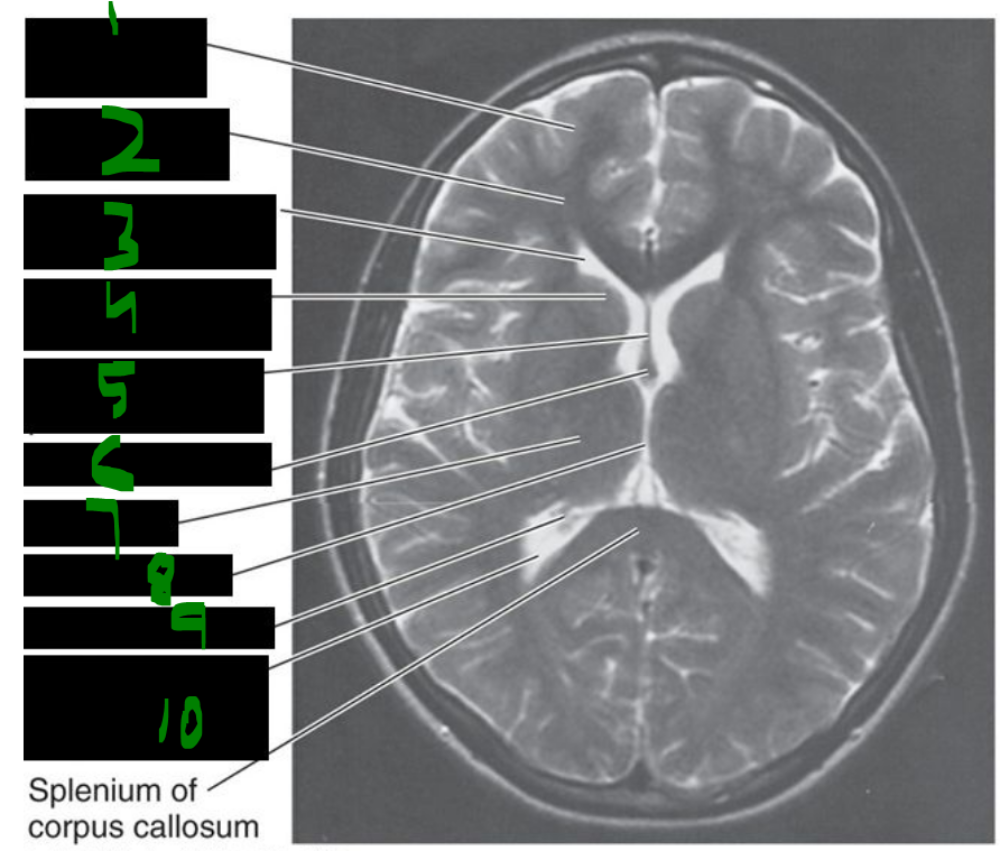 What is 1? | back 270 frontal lobe cortex |
front 271 What is 2? | back 271 frontal lobe (white matter) |
front 272 What is 3? | back 272 anterior horn of lateral ventricle |
front 273 What is 4? | back 273 head of caudate nucleus |
front 274 What is 5? | back 274 septum pellucidum |
front 275 What is 6? | back 275 column of fornix |
front 276 What is 7? | back 276 thalamus |
front 277 What is 8? | back 277 3rd ventricle |
front 278 What is 9? | back 278 choroid plexus |
front 279 What is 10? | back 279 posterior horn of lateral ventricle |
front 280 CSF is produced in the ______ ventricles (~500 cc per day) and flows to the ______ ventricle via the foramina of ______. | back 280 lateral, third, Monro |
front 281 CSF flows from the third ventricle to the fourth ventricle through the aqueduct of ______. | back 281 Sylvius |
front 282 CSF leaves the fourth ventricle via the median foramen of ______ and the two lateral foramina of ______. | back 282 Magendie, Luschka |
front 283 CSF is reabsorbed by the ______ ______ in the ______ ______ sinus. | back 283 arachnoid villi, superior sagittal |
front 284 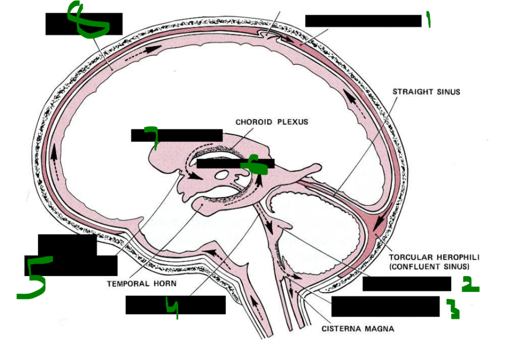 What is 1? | back 284 superior sagittal sinus |
front 285 What is 2? | back 285 fourth ventricle |
front 286 What is 3? | back 286 foramen of magendie (medial foramen) |
front 287  What is 4? | back 287 aqueduct of sylvius (cerebral aqueduct) |
front 288 What is 5? | back 288 foramen of monro (interventricular foramen) |
front 289 What is 6? | back 289 third ventricle |
front 290 What is 7? | back 290 laterla ventricle |
front 291 What is 8? | back 291 subarachnoid space |
front 292 CSF is reabsorbed in the ______ ______ (also called ______) located in the ______ ______ sinus. | back 292 arachnoid villi, granulations, superior sagittal |
front 293 Hydrocephalus is the buildup of ______ in the brain caused by congenital obstruction of the aqueduct of ______ or by ______. | back 293 CSF, Sylvius, tumors |
front 294 In young children, before the skull sutures fuse, hydrocephalus causes ______ swelling and can severely damage ______ tissue. | back 294 head, brain |
front 295 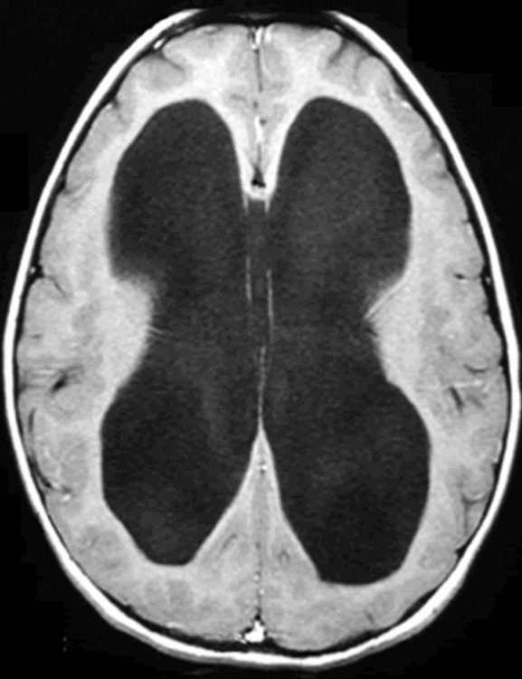 what has happened to this patient? | back 295 hydrocephalus |
front 296 The anterior cranial fossa is supplied by the ______ meningeal arteries, which are branches of the ______ arteries. | back 296 anterior, ethmoidal |
front 297 The middle cranial fossa is supplied by the ______ and ______ meningeal arteries, branches of the ______ artery. | back 297 middle, accessory, maxillary |
front 298 The middle meningeal artery enters the skull through the ______ ______, while the accessory meningeal artery enters through the ______ ______. | back 298 foramen spinosum, foramen ovale |
front 299 The posterior meningeal artery is a branch of the ______ ______ artery. | back 299 ascending pharyngeal |
front 300 The corticobulbar tract is an upper motor neuron tract for the ______ with predominantly ______ projections and some ______ projections. | back 300 head, crossed, bilateral |
front 301 The corticobulbar tract does not synapse in the ______, ______, or ______ nuclei. | back 301 oculomotor, trochlear, abducens |
front 302 The lower part of the ______ nucleus and the ______ nucleus receive crossed corticobulbar tract input. | back 302 facial, hypoglossal |
front 303 The ______ ______, motor nucleus for the pharynx and larynx, receives ______ projections via the corticobulbar tract. | back 303 nucleus ambiguus, bilateral |
front 304 The trigeminothalamic tract is the head equivalent of the ______ tract. Its primary afferent neurons have cell bodies in the ______ ______ and other sensory ganglia. | back 304 anterolateral, trigeminal ganglion |
front 305 In the trigeminothalamic tract, second-order neuron axons ascend and synapse on the ______ ______ nucleus of the ______. | back 305 posteromedial, thalamus |
front 306 Third-order neurons of the trigeminothalamic tract project to the ______ ______. | back 306 sensory cortex |
front 307 The trigeminal lemniscus is the head equivalent of the ______ ______ pathway. | back 307 dorsal column |
front 308 Primary afferent axons for fine touch and vibratory sense synapse in the ______ sensory nucleus of CN V. | back 308 chief |
front 309 Primary afferent axons for proprioception have cell bodies in the ______ nucleus of CN V. | back 309 mesencephalic |
front 310 Second-order neurons of the trigeminal lemniscus synapse in the ______ nucleus of the thalamus. Third-order neurons of the trigeminal lemniscus project to the ______ ______. | back 310 VPM sensory cortex |