Instructions for Side by Side Printing
- Print the notecards
- Fold each page in half along the solid vertical line
- Cut out the notecards by cutting along each horizontal dotted line
- Optional: Glue, tape or staple the ends of each notecard together
Anatomy week 1 - communicating about the body
front 1 regions of body in anatomical position | back 1 no data |
front 2 head | back 2 cephalic / cranial |
front 3 skull | back 3 cranial |
front 4 face | back 4 facial |
front 5 chin | back 5 mental |
front 6 neck | back 6 cervical |
front 7 chest | back 7 pectoral |
front 8 armpit | back 8 axillary |
front 9 arm | back 9 brachial |
front 10 front of elbow | back 10 antecubital / cubital |
front 11 forearm | back 11 antebrachial |
front 12 wrist | back 12 carpal |
front 13 palm | back 13 palmar |
front 14 fingers | back 14 digital / phalangeal |
front 15 thigh | back 15 femoral |
front 16 anterior surface of knee | back 16 patellar |
front 17 forehead | back 17 frontal |
front 18 eye | back 18 orbital |
front 19 cheek | back 19 buccal |
front 20 ear | back 20 otic |
front 21 nose | back 21 nasal |
front 22 mouth | back 22 oral |
front 23 breastbone | back 23 sternal |
front 24 breast | back 24 mammary |
front 25 naval | back 25 umbilical |
front 26 hip | back 26 coxal |
front 27 groin | back 27 inguinal |
front 28 hand | back 28 manual |
front 29 pubis | back 29 pubic |
front 30 chest | back 30 thoracic |
front 31 abdomen | back 31 abdominal |
front 32 breastbone to pelvis | back 32 trunk |
front 33 base of skull | back 33 occipital |
front 34 shoulder | back 34 acromial |
front 35 shoulder blade | back 35 scapular |
front 36 spinal column | back 36 vertebral |
front 37 back of elbow | back 37 olecranal |
front 38 between hips | back 38 sacral |
front 39 buttock | back 39 gluteal |
front 40 hollow behind knee | back 40 popliteal |
front 41 calf | back 41 sural |
front 42 sole | back 42 plantar |
front 43 heel | back 43 calcaneal |
front 44 hips to heel | back 44 lower limb |
front 45 back of hand | back 45 dorsal |
front 46 upper limb | back 46 hand to shoulder |
front 47 loin | back 47 lumbar |
front 48 Standard anatomical position | back 48 SAP - palms up - forearm underside is ANTERIOR |
front 49 anterior | back 49 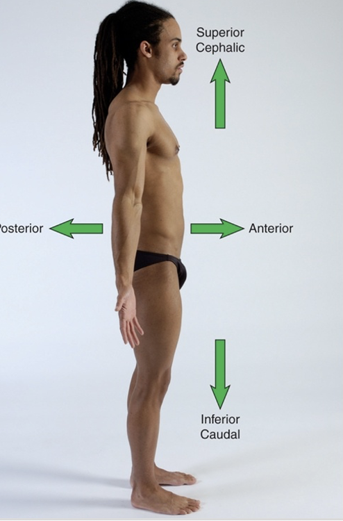 towards the front |
front 50 superior | back 50 cephalic (upper body direction) |
front 51 posterior | back 51 towards the back |
front 52 inferior | back 52 caudal (lower body direction) |
front 53 proximal | back 53 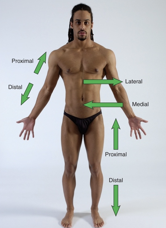 proximal means closer to the origin of the body part of the point of attachement of a limb to the body trunk |
front 54 distal | back 54 further away from origin of a body part or the point of attachment of a limb to the body trunk |
front 55 lateral | back 55 Lateral - to be farther away from midline of body, in direction of either side from midline of body OR a structure away from the midline of the body |
front 56 medial | back 56 Medial - to be closer to the midline of the body or a structure (internal opposed to external) toward the midline of the body |
front 57 sagittal plane of movement and motions | back 57 cuts body into right vs. left motions: flexion and extension |
front 58 frontal plane of movement and motions | back 58 cuts body into anterior vs. posterior motions: abduction and adduction |
front 59 transverse plane and motions | back 59 cuts body into superior and inferior motions: rotation |
front 60 axes | back 60 imaginary line at right angles to the plane at which it rotates or spins each plane perpendicular to its axis |
front 61 frontal axis | back 61 line thru side of body plane spinning: sagittal plane |
front 62 sagittal axis | back 62 line anterior - posterior of body plane: frontal axis |
front 63 longitudinal axis | back 63 line head/heels plane: transverse |
front 64 tissue types in body | back 64
|
front 65 epithelial tissue location | back 65
|
front 66 epithelial tissue function and functional categories | back 66 protect, absorb, filter, secretes substances in body functional categories:
regenerate fast, constantly repair/replace |
front 67 connective tissue locations | back 67 bones, tendons, ligaments, fascia, cartilage, adipose (fat), blood |
front 68 connective tissue types differ by | back 68 density |
front 69 connective tissue components | back 69 Extracellular matrix (EXM): various fibers suspended in fluid Ground substance: fluid portion of extracellular matrix Three fiber types: collagen, reticular, elastic |
front 70 exm vs ground substance | back 70 no data |
front 71 fibroblasts | back 71 in connective tissue cells that secrete proteins that make up fibers in matrix include osteoblasts (bone) and chondroblasts (cartilage) |
front 72 macrophages | back 72 part of connective tissue respond to injury / infection |
front 73 fat cells | back 73 part of connective tissue adipose |
front 74 types of connective tissues | back 74
|
front 75 muscle tissue | back 75 network of muscle cells containing myofibrils |
front 76 myofibrils | back 76 contractile protein structures stimulated by nervous system to contract / shorten = movement force generated by shortening myofibrils -> transmitted into surrounding myofascia -> force drives internal + external human movement |
front 77 myofibril unit | back 77 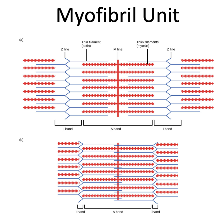 sarcomere, actin, myosin, I band, A band, Z line, H zone, M line |
front 78 sarcomere | back 78 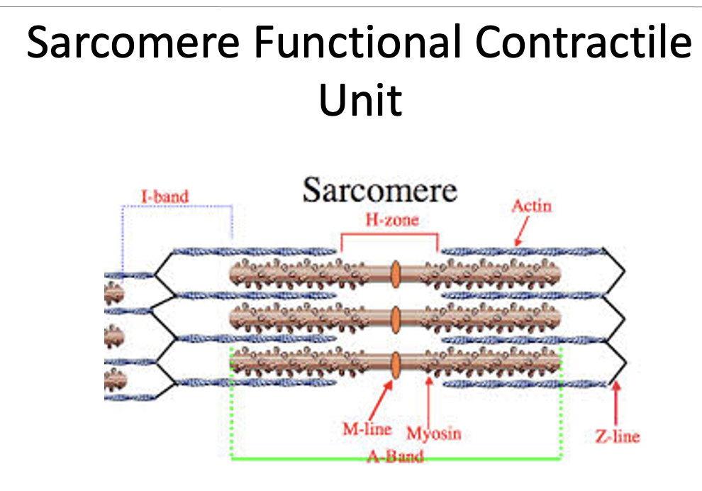 basic contractile unit of a myofibril |
front 79 actin | back 79 thin filament |
front 80 myosin | back 80 thick filament |
front 81 I band | back 81 primarily contains actin (thin) filaments |
front 82 A band | back 82 primarily contains myosin (thick) filaments |
front 83 Z line | back 83 defines boundary of sarcomere and anchors the actin filament |
front 84 H zone | back 84 zone of myosin filaments with no actin filaments |
front 85 M line | back 85 runs down the middle of the H zone |
front 86 nervous tissue | back 86 network of neurons (nerve cells) can be stimulated, conduct stimulus, respond to stimulus electrical impulses travel in btw neurons + btw them and other cells impulses allow communication btw nervous system + other tissues nervous system monitor + regulate body functions |
front 87 sensory vs. motor | back 87 sensory
motor
|
front 88 structures involved in movement | back 88
|
front 89 bone characteristics | back 89
|
front 90 epiphysis | back 90 epiphysis are bone ends with only ossify once skeleton matures as an adult |
front 91 diaphysis | back 91 shaft of bone |
front 92 bone anatomy | back 92 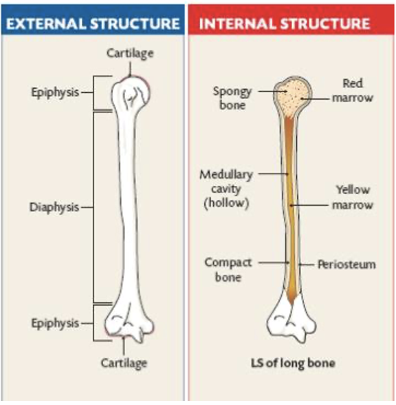 |
front 93 long bone shape | back 93 shaft in middle w/ bumpy ends |
front 94 small bone shape | back 94 cube-shaped; allows fine, gliding movements in hand + foot |
front 95 flat bone shape | back 95 sternum, lilium |
front 96 irregular bone shape | back 96 unique; vertebrae + fascial bones |
front 97 sesamoid | back 97 encased in tendon; helps improve leverage + strength of muscle that cross it (patella) |
front 98 bone functions | back 98
|
front 99 ligaments | back 99 fibrous structures made of dense connective tissue that connects bones to each other prevent movements at joints + help stabilize joints (static stabilizers) poorly vascularized |
front 100 structure of ligaments | back 100 complex networks of collagen fibers that resist stress in multiple directions (grisltly feel) present at both ends of bones, help form joints, joint capsules interosseous membrane - broad sheet of dense connective tissue, thinner than ligaments; connects bones along length of shaft |
front 101 types of muscle | back 101 smooth
cardiac
skeletal muscle
|
front 102 smooth muscle | back 102
|
front 103 cardiac muscle | back 103
|
front 104 skeletal muscle | back 104
|
front 105 muscle features | back 105
|
front 106 tendon | back 106 connect muscle to bone abundant in collagen fibers = strength + elasticity change shape as moves smother than muscle |
front 107 fascia | back 107
|
front 108 fascia layers | back 108
|
front 109 superficial fascia | back 109 under dermis of skin stores fat + water, passage for nerves + vessels |
front 110 deep fascia | back 110 form network around muscles + internal structures help muscle movement, make passageways for nerves + vessels, make muscle attachment sites, cushion muscle |
front 111 subserous fascia | back 111 separates deep fascia from membranes, allow movement of internal organs |
front 112 skin functions | back 112 protect vs environment help regulate internal temp excretes waste |
front 113 skin layers | back 113 epidermis dermis hypodermis |
front 114 epidermis | back 114 epithelial tissue; thin layers of cells cont. keratin, melanin, defensive cells |
front 115 dermis | back 115 dense connective tissue; contains hair follicles, glands, nerves, blood vessels, tiny muscles |
front 116 hypodermis | back 116 loose connective tissue, lying under dermis; contains adipose cells -> cushion + protect structures |
front 117 burns | back 117 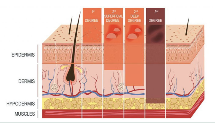 1st degree - superficial thickness 2nd degree - partial thickness burn 3rd degree - full thickness burn 4th degree - burn to bone / muscle, result in loss of limb / part |
front 118 blood vessel functions | back 118 path blood flow deliver oxygen + nutrients to tissue removes waste |
front 119 types blood vessels | back 119
|
front 120 lymph vessels and node functions | back 120 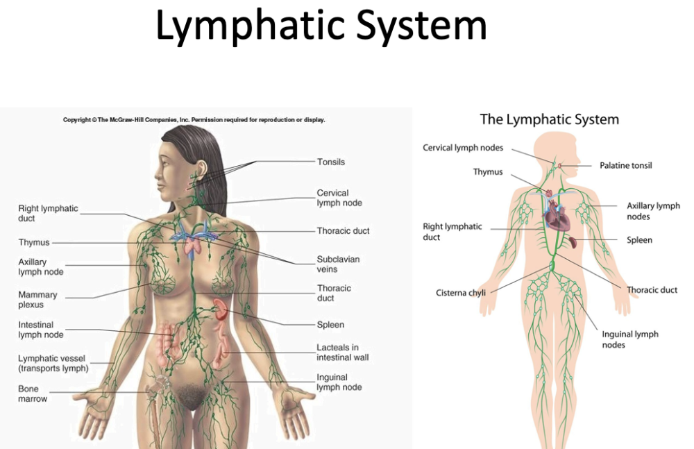
|
front 121 circulation of lymphatic system | back 121 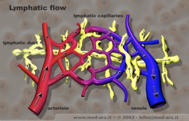
|
front 122 nerves | back 122
= cell body (nucleus), dendrites, axon |
front 123 nerve types | back 123
|
front 124 cartilage | back 124 supporting connective tissue
|
front 125 cartilage types | back 125
|
front 126 labrum | back 126 fibrocartilaginous ring deepen concavity of joint attachment site for many joint ligaments ex: in shoulder + hip joints |
front 127 bursae | back 127 small/flat sacs w/ synovial fluid in shoulder, elbow, hip, knee become big + swollen when exposed to excess friction (bursitis) |
front 128 synovial fluid | back 128 lubricant helps decrease friction + create gliding movement btw structures |