Instructions for Side by Side Printing
- Print the notecards
- Fold each page in half along the solid vertical line
- Cut out the notecards by cutting along each horizontal dotted line
- Optional: Glue, tape or staple the ends of each notecard together
A&P Chapter 13
front 1 Which layer of protective connective tissue covers a nerve and fuses with the outer menix layer? | back 1 Epineurium |
front 2 Sensory information travels to the brain via | back 2 Ascending tracts |
front 3 Which structure listed contains cerebrospinal fluid? | back 3 subarachnoid space |
front 4 Spinal nerves | back 4 1, 2, 3, 4 |
front 5 Which statements describe dermatomes? Select all that apply. | back 5 Can be used clinically to determine area of spinal cord damage Complete anesthesia of a single dermatome often requires blocking three adjacent spinal nerves The dermatome serving the face is supplied by the trigeminal cranial nerve Dermatomes are designated based on the cranial or spinal nerve that serves that area of the skin |
front 6 Which of the following lists the connective tissue coverings of the axons, fascicles, and entire nerve in the correct order? | back 6 endoneurium, perineurium, epineurium |
front 7  In this diagram which portion of the spinal cord contains cell bodies and axons of interneurons as well as incoming axons of sensory neurons? | back 7 C |
front 8 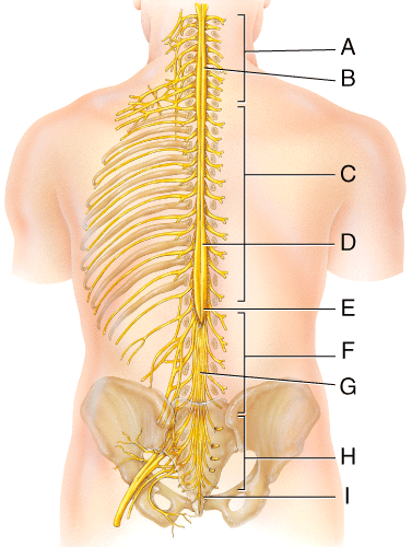 Which of the following structures travels from the area labeled D in the diagram? | back 8 obturator nerve |
front 9 Which of the following parts of a reflex arc would involve the posterior root ganglion? | back 9 Sensory neuron |
front 10 Which branch of a spinal nerve serves the deep muscles and skin of the posterior surface of the trunk? | back 10 Posterior ramus |
front 11 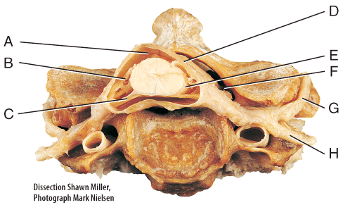 If you had a rash on the anterior surface of your trunk, what labeled structure would serve the muscles and skin of that area? | back 11 H |
front 12 On what labeled structure would a ganglion be found? | back 12 C |
front 13 A nerve impulse initiated at a muscle spindle has to travel through which of the following structures to enter the integrating center? | back 13 Posterior root of spinal nerve |
front 14 When the quadriceps muscle extends the lower leg in the patella reflex, what is occurring in the motor neuron to produce an action potential? | back 14 Influx of sodium and efflux of potassium |
front 15 In response to a muscle being stretched, a muscle spindle initiates a
somatic spinal reflex that causes | back 15 1 and 2 |
front 16  In this diagram, which layer is the thickest protective covering? | back 16 I |
front 17 In this diagram, where is the space that contains a buoyant liquid to form a hydraulic cushion that absorbs shock? | back 17 E |
front 18 A severed obturator nerve will lead into paralysis of which region of the body? | back 18 Thigh |
front 19 The nerves that supply the thigh and calf of leg emerge from the | back 19 lumbar enlargement |
front 20 A gymnast has damaged their hip flexor muscles. The nerve that innervates these muscles arise from which group of spinal nerves in this diagram? | back 20 F |
front 21 Excitation of the quadriceps femoris group and inhibition of the hamstring group is termed _______ innervation. | back 21 reciprocal |
front 22 Which of the following spinal nerves does not travel through an intervertebral foramen to reach its destination? | back 22 cervical spinal nerve 1 |
front 23 Which type of descending motor pathway conveys nerve impulses that originate in the cerebral cortex and allows you to use your fingers for sewing and sign language? | back 23 corticospinal tract |
front 24 In this diagram where are tracts located? Select all that apply. | back 24 B, D, I |
front 25 Intercostal nerves can be described by what statements below? Select all that apply | back 25 are also known as thoracic nerves do not enter into a plexus directly connect to the structures they supply |
front 26 An individual suffers a stroke and looses control over their left arm. The individual is affected with __________ paralysis. | back 26 Monoplegia |
front 27 In this diagram where would the medial reticulospinal tract be located? | back 27 I |
front 28 In this diagram, where is the avascular structure located between the subdural and subarachnoid space? | back 28 H |
front 29 Which of the nerves in this diagram would be involved with ‘flexing’ the chest wall to bring the ribs together? | back 29 D |
front 30 In this diagram which structure is the conus medullaris? | back 30 E |
front 31 Place the reflex arc components in order from detecting a stimulus to
response. | back 31 5, 3, 4, 1, 2 |
front 32 A patient who has loss of pin prick sensation starting at the level of the nipple and downward on the right side would have a lesion in the spinal cord at which level? | back 32 T6-T7 |
front 33 Which of the following parts of a reflex arc would be represented by the contraction of the gastrocnemius muscle or the radial iris muscles? | back 33 Effector |
front 34 In this diagram, where is the structure that adheres to the spinal cord and brain? | back 34 G |
front 35 In this diagram, where would the axons of unipolar neurons be located? | back 35 C |
front 36 An individual with shingles presents with pain and lesions in the mouth, ears, and pharynx. Which dermatomal region would be sending the afferent signals? | back 36 Trigeminal |
front 37 In this diagram which portion of the spinal cord contains cell bodies that regulate cardiac muscle, smooth muscle and glands? | back 37 F |
front 38 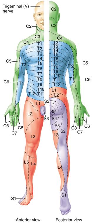 A child was in a diving accident. When brought into the emergency room, the doctor used sharp and blunt objects to determine if there was any sensation on the bottom of the foot and the lateral aspect of their palm. The child stated they could not feel the stimulus in either area. What nerves were most likely damaged? | back 38 S1 and C6 |
front 39 The area of the skin that provides sensory input to the CNS via one pair of spinal nerves is called | back 39 a dermatome. |
front 40 Which of the labeled structures is composed of squamous to cuboidal cells and is very vascular? | back 40 E |
front 41 In reciprocal innervation of the tendon reflex stimulation of afferent fibers causes | back 41 excitation of antagonist muscles |
front 42 Which of the nerves in this diagram only have anterior and posterior rami branches and do not form plexuses? | back 42 D |
front 43 In this diagram which area is continuous with the 4thventricle of the brain? | back 43 G |
front 44 In the diagram which structure contains nerve roots that arise from the lumbar, sacral, and coccygeal region of the spinal cord? | back 44 G |
front 45 If these structures were severed, the nerve impulses from pain and stretch receptors would not reach the CNS. | back 45 posterior root of spinal nerves |
front 46 Which statements describe the spinothalamic tract? Select all that apply | back 46 Begins in the spinal cord Terminates in the thalamus Carries information for pain, temperature and tickle |
front 47 Which plexus arises from the structure labeled B in the diagram? | back 47 brachial plexus |
front 48 Which spinal meninges is composed of collagen fibers arranged in an irregular pattern that is very strong? | back 48 Dura mater |
front 49 A typical spinal nerve has how many connections to the spinal cord? | back 49 2 |
front 50 Which of the labeled structures allows doctors to safely introduce antibiotics or anesthetics and measure CSF pressure? | back 50 A |
front 51 Which of the following parts of a reflex arc can be monosynaptic or polysynaptic? | back 51 Integrating center |
front 52 The spinal cord ends | back 52 between L1 and L2 |
front 53 In the 400 meter relay, runners handoff the baton to the person in front of them. The second runner in the relay team had to be replaced. They were unable to pronate their forearm to reach behind for the baton because of injury to their brachial plexus. What is the most likely site of their injury? | back 53 Median nerve |
front 54 Which of the nerves in this diagram innervates muscles that extend the toes and if damaged results in foot drop (plantar flexion)? | back 54 F |
front 55 Which type of descending motor pathway excites antigravity muscles in order to exert control over postural changes necessary to compensate for tilts and movements of the body? | back 55 vestibulospinal tract |
front 56 Which are functions of the spinal reflexes that use muscle spindles and tendon organs as sensors? Select all that apply. | back 56 Awareness of muscle tension in body Prevention of damage to muscles Awareness of muscle length Prevention of damage to tendons Maintenance of muscle tone |
front 57 Which region of the spinal cord allows a dancer to be aware of their movements and body position? | back 57 Posterior white column |
front 58 In this diagram where would somatic motor nuclei be located? | back 58 H |
front 59 What parts of a neuron within a nerve is/are wrapped in a protective
endoneurium? | back 59 Both 1 & 2 |
front 60 The ______ nerve would be involved with a dancer performing pointe (standing on their toes). Select all that apply | back 60 obturator nerve tibial nerve |
front 61 A patient is being examined for muscle weakness. To assess their condition, the doctor tests their knee-jerk reflex by tapping the patella tendon with a reflex hammer and tapping his lower jaw with your finger. Both pathways are considered monosynaptic. What do they have in common? | back 61 They both a two neuron pathway, sensory to motor |
front 62 In this diagram, where is the space that contains interstitial fluid? | back 62 F |
front 63 Why is a spinal tap performed below L2 of the spinal cord? | back 63 The spinal meninges and spaces extend passed the proper spinal cord, allowing fluid to be withdrawn with a minimal risk of damage to the spinal cord. |
front 64 A pupillary reflex is integrated in the brain stem so it is classified as a ________ reflex. The smooth muscle of the iris is the effector, so this is classified as a(n) ___________. | back 64 Cranial; autonomic |
front 65 In response to a tendon being stretched excessively, a tendon organ
initiates a somatic spinal reflex that causes | back 65 3 and 4 |
front 66 Which of the labeled structures contains both sensory and motor axons? Select all that apply. | back 66 G & H |
front 67 Which of the following parts of a reflex arc monitors the rectus femoris muscle stretching? | back 67 Sensory receptor |
front 68 Which nerve innervates the muscle that abducts the arm at the shoulder and the skin overlying the deltoid? | back 68 axillary nerve |
front 69 Which of the labeled structures would receive injections to cause loss of sensation to control pain in childbirth? | back 69 F |
front 70 Christopher Reeve, a famous actor and competitive equestrian, suffered fractures to the top two vertebra and could not breathe without the help of a respirator. Which of the nerves in this diagram were likely damaged? | back 70 A |
front 71 In this diagram, respiratory distress can occur if damage occurs to which group of spinal nerves? | back 71 A |