Anatomy of the Digestive System (Exercise 38)

1
Mouth (oral cavity)

2
Tongue

3
Esophagus

4
Liver

5
Gallbladder

6
Small intestine

7
Duodenum

8
Jejunum

9
Ileum

10
Anus

11
Anal canal

12
Appendix

13
Rectum

14
Sigmoid colon

15
Cecum

16
Ascending Colon

17
Descending Colon

18
Transverse colon

19
Large Intestine

20
Spleen

21
Pancreas

22
Stomach

23
Pharynx
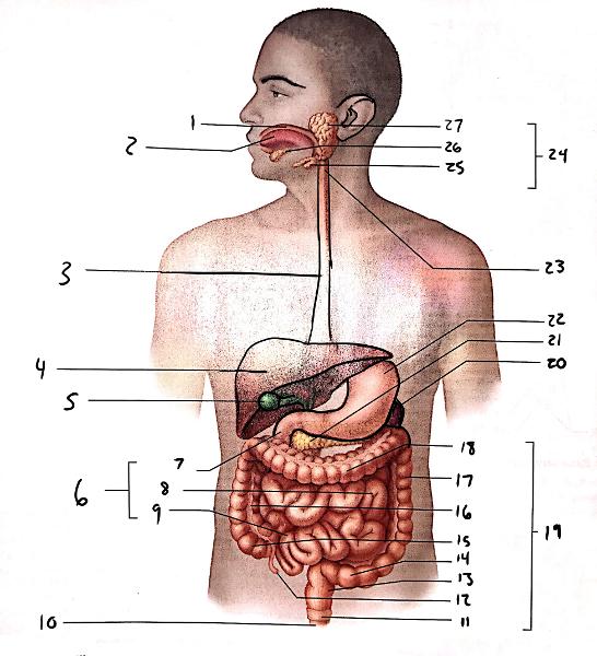
24
Salivary glands

25
Submandibular gland

26
Sublingual gland

27
Parotid gland

1
Esophagus

2
Longitudinal layer

3
circular layer

4
Oblique layer

5
Muscularis externa
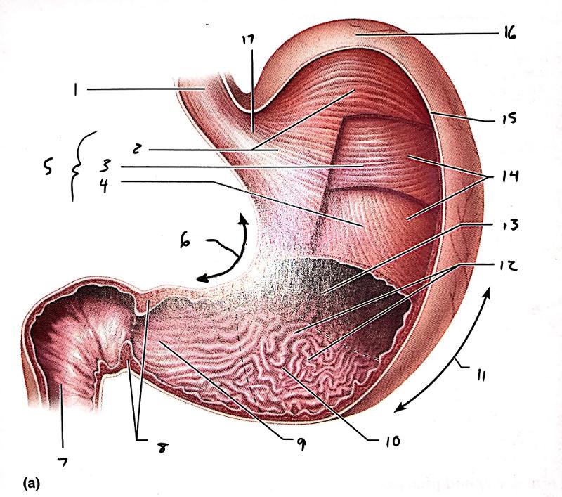
6
Lesser curvature

7
Duodenum

8
Pyloric sphincter at pylorus

9
Pyloric canal

10
Pyloric antrum

11
Greater curvature

12
Rugae of mucosa

13
Lumen

14
Body

15
Serosa

16
Fundus

17
Cardial part

1
Fundus
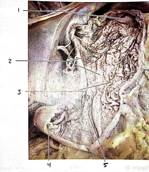
2
Body

3
Rugae of mucosa

4
Pyloric sphincter

5
Pyloric antrum
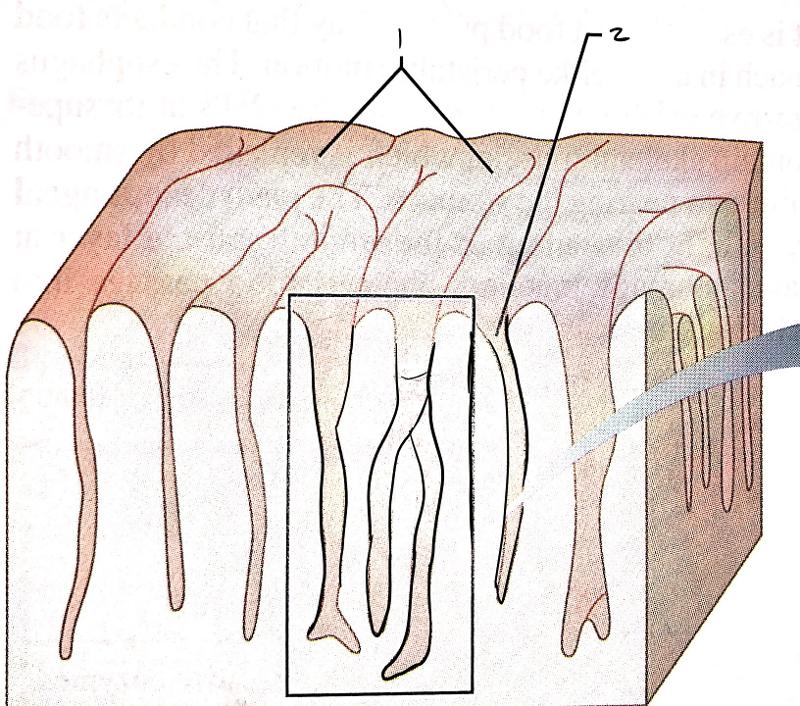
1
Rugae

2
Gastric pit

1
Gastric pit

2
Gastric gland

3
Enteroendocrine cell

4
Chief cell
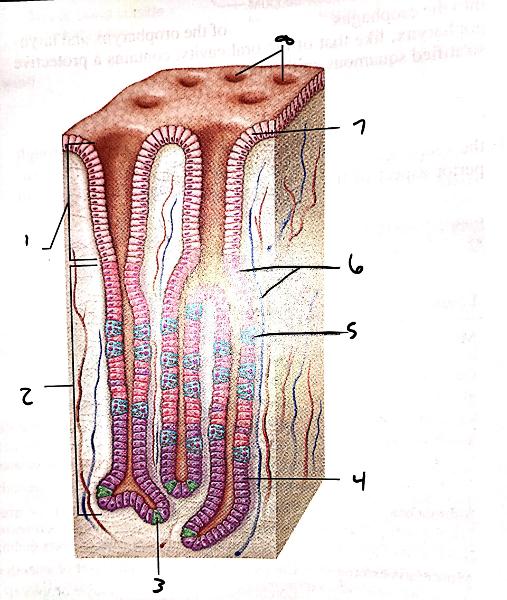
5
Parietal cell

6
Mucous neck cells

7
Surface epithelium (mucous cells)

8
Gastric pits

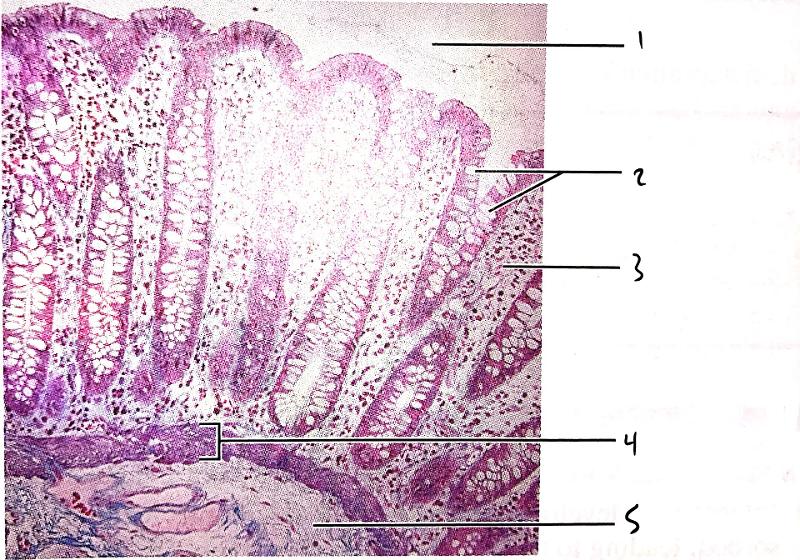
1
Gastric glands

2
Muscularis mucosae
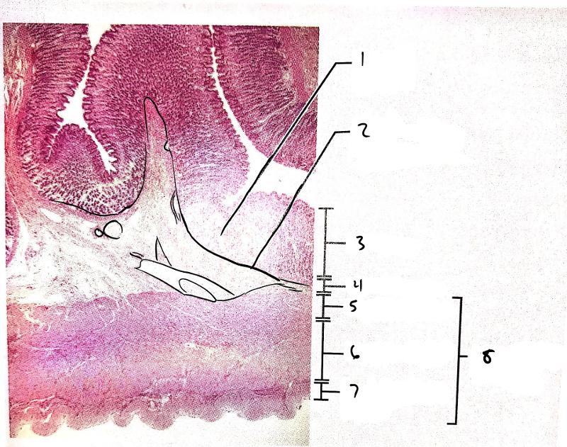
3
mucosa

4
submucosa

5
oblique layer

6
circular layer

7
longitudinal layer

8
muscularis externa

1
Simple columnar epithelium

2
lamina propria
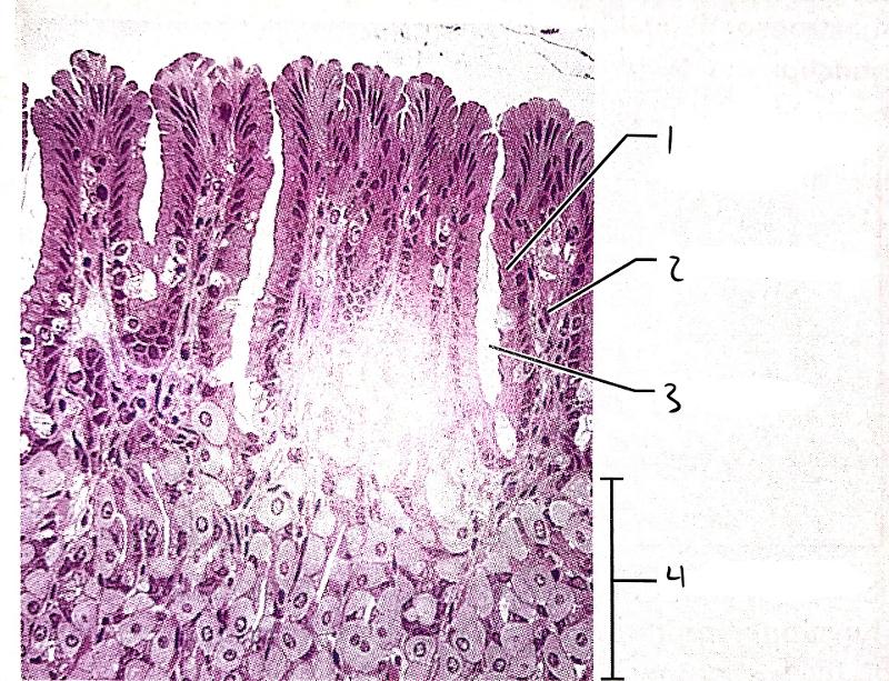
3
gastric pit

4
gastric glands

1
Stratified squamous epithelium of esophagus

2
gastroesophageal junction

3
simple columnar epithelium of stomach
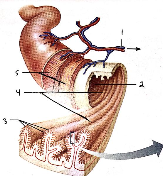
1
Vein carrying blood to hepatic portal vessel

2
Lumen

3
Villi

4
Circular folds

5
muscle layers

1
Absorptive cells

2
lacteal

3
Goblet cell

4
Blood capillaries

5
Mucosa-associated lymphoid tissue

6
intestinal crypt

7
muscularis mucosae

8
Duodenal gland
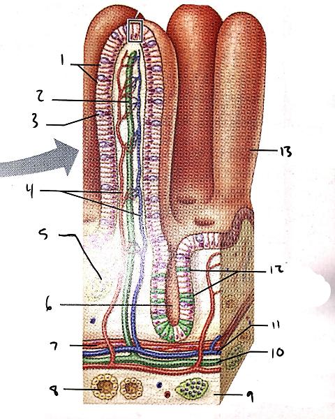
9
submucosa

10
lymphatic vessel

11
venule

12
entero-endocrine cells

13
villus
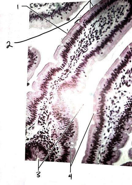
1
Villus

2
Absorptive cells

3
Intestinal crypt

4
Goblet cells

1
Villus

2
Simple columnar epithelium

3
lamina propria

4
Intestinal crypt

5
Muscularis mucosae
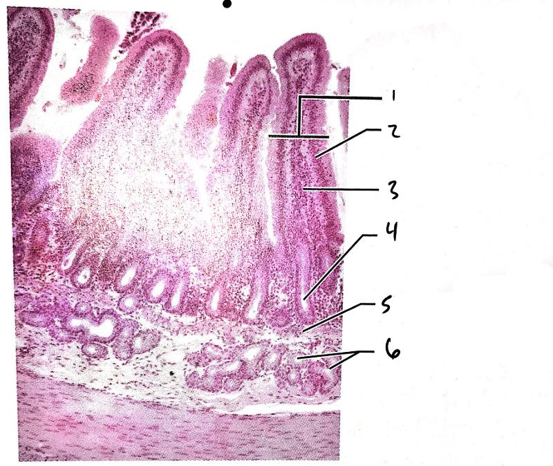
6
Duodenal glands
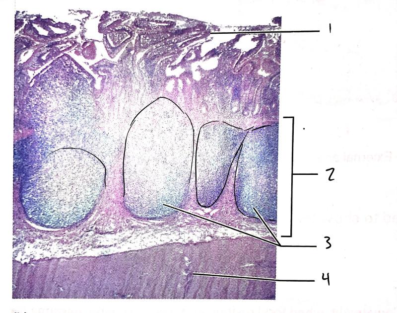
1
Villus

2
Submucosa

3
Peyer's patches

4
Muscularis externa
1
Lumen

2
Goblet cells in epithelium

3
Lamina propria

4
Muscularis mucosae

5
Submucosa

1
Right colic (hepatic) flexure

2
Transverse colon

3
Superior mesenteric artery

4
Haustrum

5
Ascending colon

6
Ileum

7
Ileocecal valve

8
Cecum

9
Appendix

10
Rectum

11
Anal canal

12
External anal sphincter
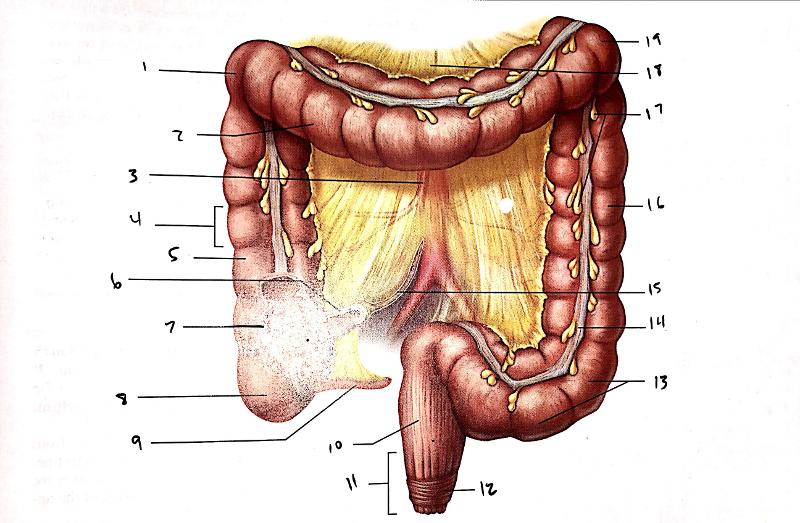
13
Sigmoid colon

14
tenia coli

15
Mesentery (cut edge)

16
Descending colon

17
Epiploic appendages

18
Transverse mesocolon

19
Left colic (splenic) flexure