Exercise 11: The Appendicular Skeleton
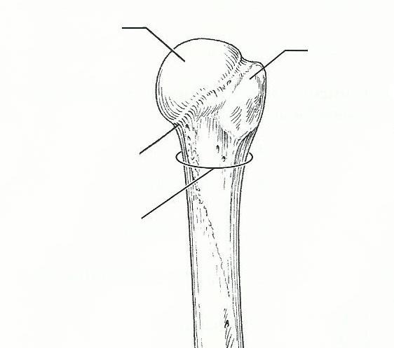
deltoid tuberosity
raised area on lateral surface of humerus to which deltoid muscle attaches
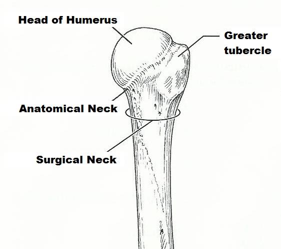
humerus
arm bone
scapula, clavicle
bones of the shoulder girdle
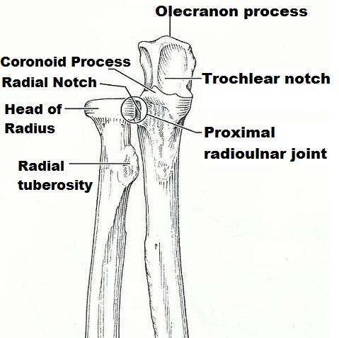
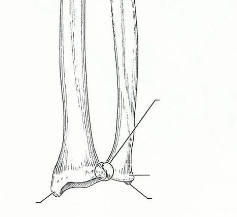
radius, ulna
forearm bones
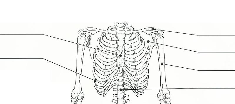
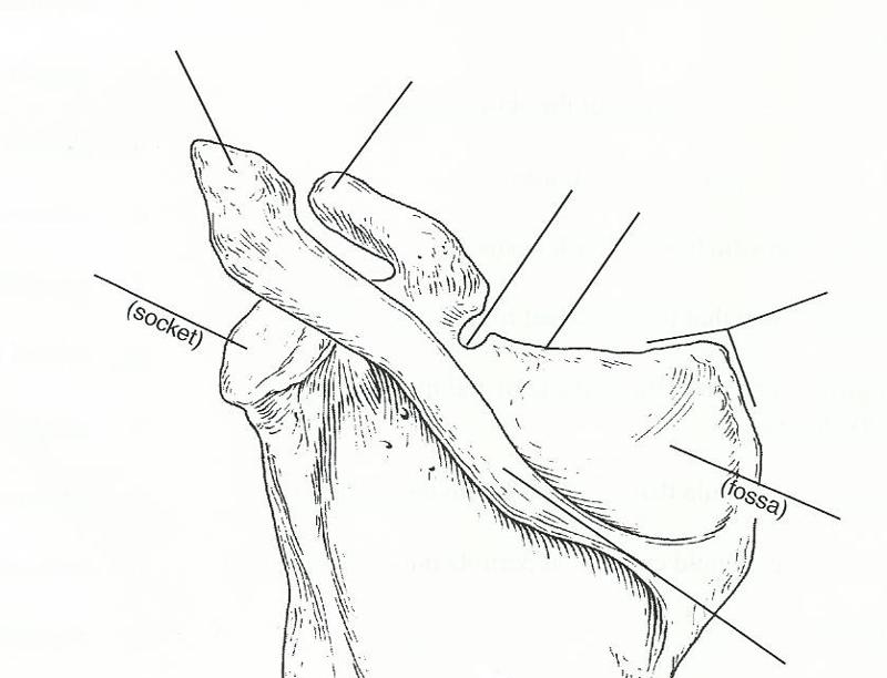
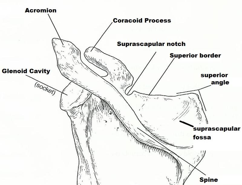
acromion
scapular region to which the clavicle connects
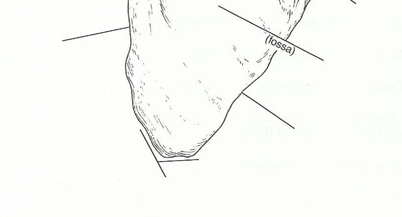
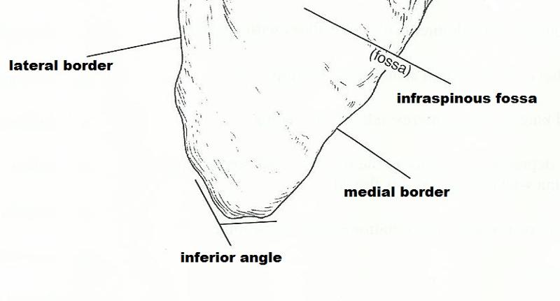
scapula
shoulder girdle bone that is unattached to the axial skeleton
clavicle
shoulder girdle bone that articulates with and transmits forces to the bony thorax
glenoid cavity
depression in the scapula that articulates with the humerous
coracoid process
process above the glenoid cavity that permits muscle attachment
clavicle
the "collarbone"
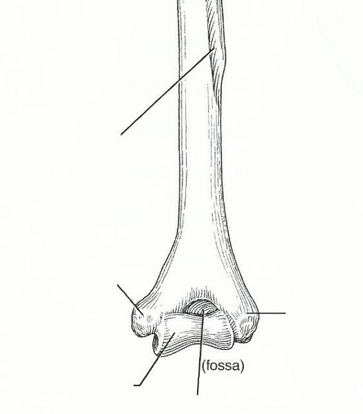
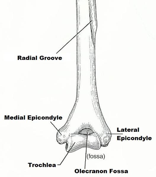
trochlea
distal condyle of the humerus that articulates with the ulna
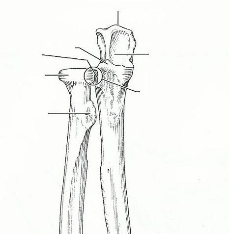
ulna
medial bone of forearm in anatomical position
capitulium
rounded knob on the humerus; adjoins the radius
coronoid fossa
anteriar depression, superior to the trochlea, which receives part of the ulna when the forearm is flexed
ulna
forearm bone involved in formation of the elbow joint
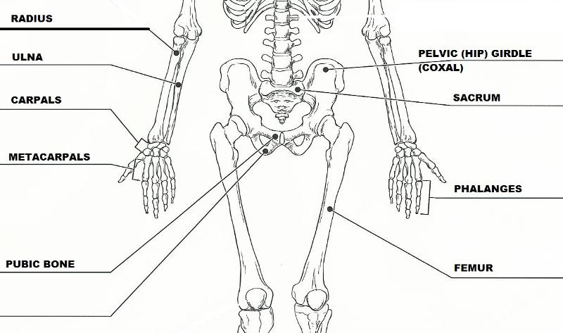
carpals
wrist bones
phalanges
finger bones
metacarpals
heads of these bones form the knuckles
scapula, sternum
bones that articulate with the clavicle
How is the arm held clear of the widest dimension of the thoracic cage?
Clavicle serves as an anterior base or strut to hold the arm away from the top of the thorax
What is the total number of phalanges in the hand?
14
What is the total number of carpals in the wrist?
8
Name the carpals (medial to lateral) in the proximal row.
pisiform-triquetral-lunate-scaphoid
In the distal row, they are (medial to lateral)
trapezium-trapezoid-capitate-hamate

The humerus is a (right/left) bone in (an anterior/a posterior) view.
right; Posterior
The radius and ulna are (right/left) bones in (an anterior/a posterior) view.
left; Anterior
Pectoral
flexibility most important; lightweight; insecure axaial and limb attachments
Pelvic
massive; secure axail and limb attachments; weight-bearing most important
What organs are protected, at least in part, by the pelvic girdle?
Uterus, bladdar, rectum, small intestine, and reproductive organs
What is the difference between the true pelvis and the false pelvis?
The true pelvis is small bowl-like shape containing the unrinary bladder and sexual organs. The false pelvis is the area been the alla or wings of the pelvis. This is a much larger area and contains some of the abdominal organs like part of the small intestine and part of the colon.
This is a (female/male) pelvis because?
Female; It is adapted for childbearing, defines birth canal, Farther apart then a males, the sacrum is wider, shorter and less curved then a males. The pelvic inlet (brim) is wider; oval from side to side.
Deduce why the pelvis bones of a four-legged animal such as a cat or pig are less massive than those of the human.
The pelvic bones of a two-legged animal such as a human have to carry his whole weight, divided by two. A four-legged animal such as a pig divides its weight over four legs, so each leg and each pelvic bone has to bear only a fourth of the animal's weight. As the strain on the bones is a half than in the two-legged case, the bones can be less massive.
A person instinctively curls over his abdominal area in times of danger. Why?
To prevent a smaller target and also to protect the internal organs and genitalia.
For what anatomical reason do many women appear to be slightly knock-kneed?
Biologically, women have wider pelvises than men. The angle of their hips turn the legs slightly inward, making some to actually touch at the knees.
How might this anatomical arrangement contribute to knee injuries in female athletes?
they can be more prone to knee related injuries, such as ACL tears due to high intensity straight knee landing or impact since the knee is already in a weak position.
What does fallen arches mean?
It mean flat feet or flat footed
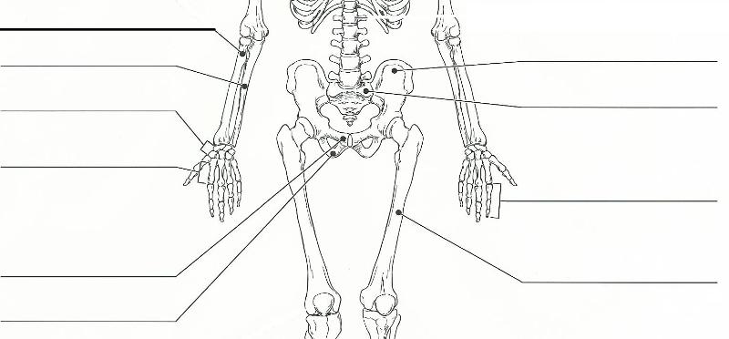
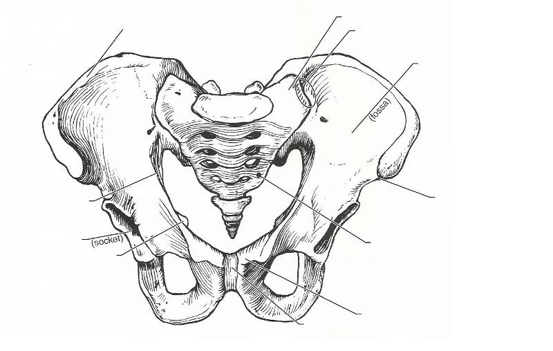
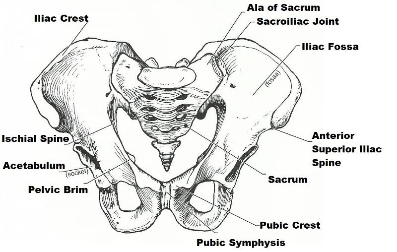
illium, ischium, pubis
fuse to form the coxal bone
ischium
"sit-down" bone of the coxal bone
pubic symphysis
point where the coxal bones join anteriorly
iliac crest
superiormost margin of the coxal bone
acetabulum
deep socket in the coxal bone that receives the head of the thigh bone
sacroiliac joint
joint between axaial skeleton and pelvic girdle
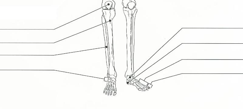
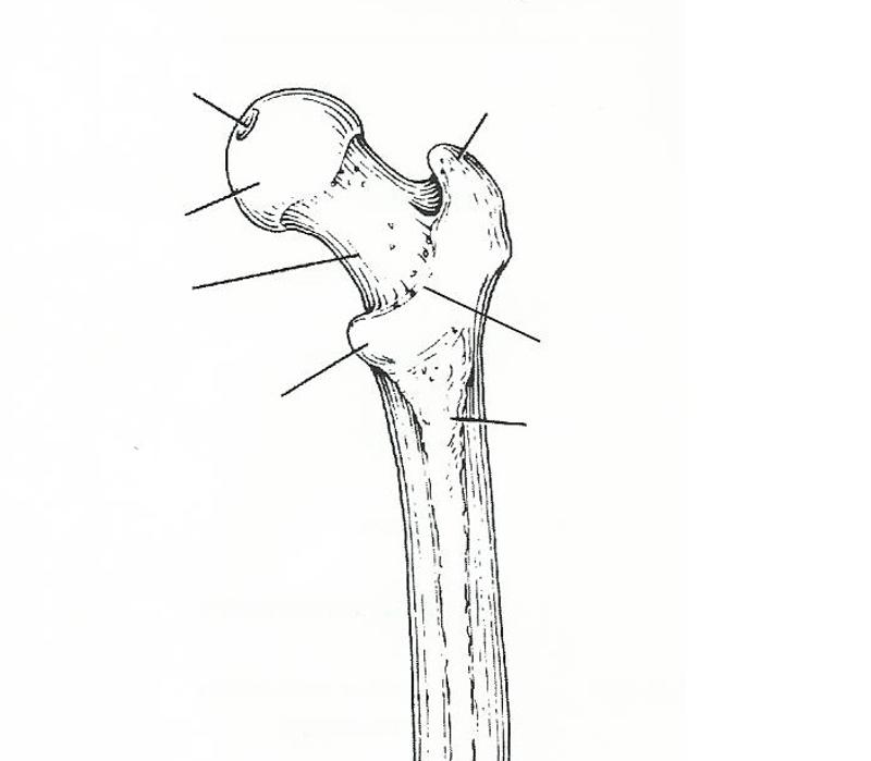
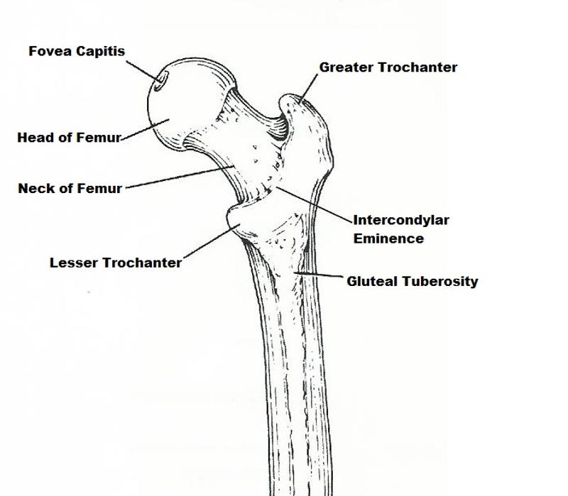
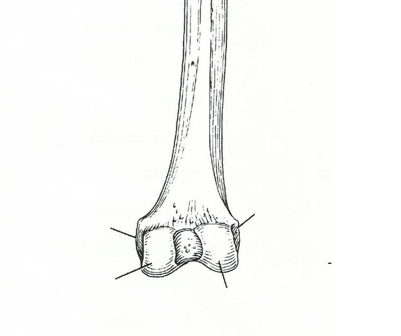
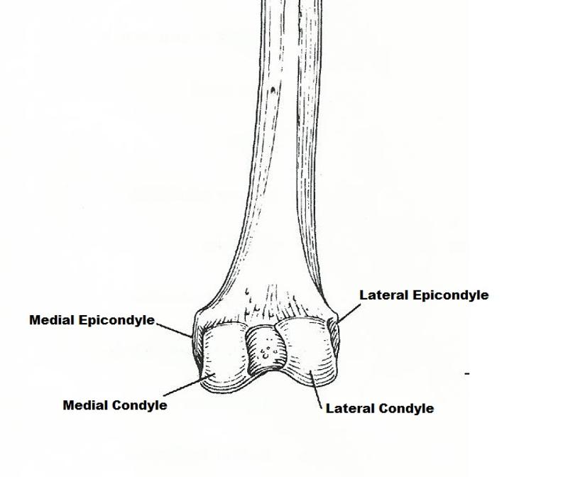
femur
longest, strongest bone in the body
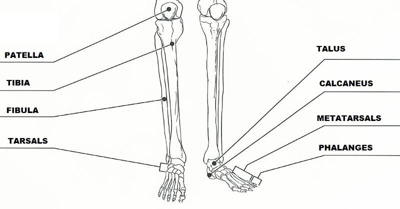
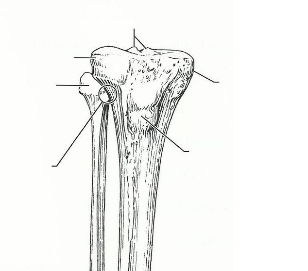
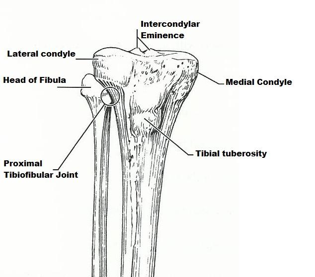
fibula
thin lateral leg bone
tibia
heavy medial leg bone
femur, tibia, patella
bones forming knee joint
tibial tuberosity
point where the patellar ligament attaches
patella
kneecap
tibia
shinbone
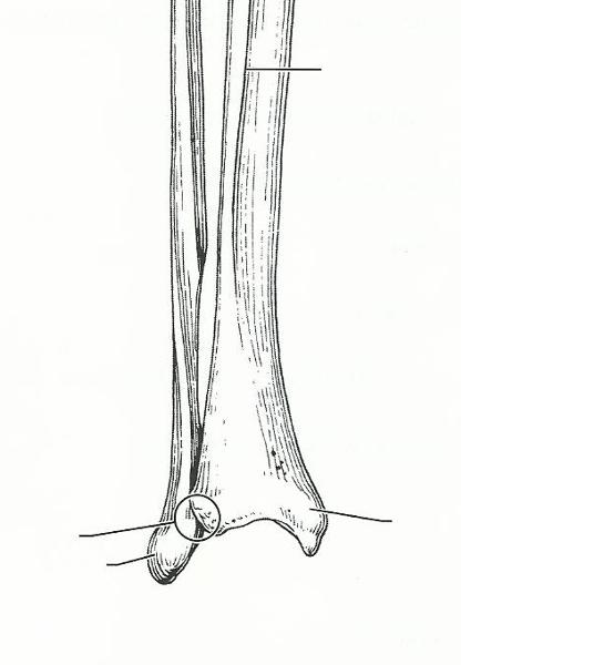
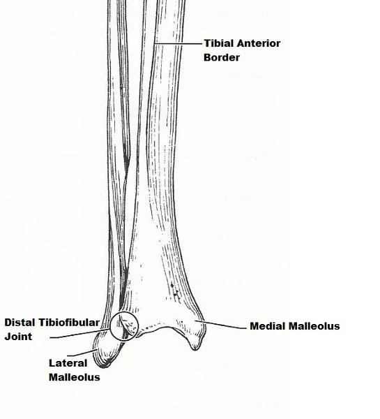
medial malleolus
medial ankle projection
lateral malleolus
lateral ankle projection
calcaneus
largest tarsal bone
tarsals
ankle bones
metatarsals
bones forming the instep of the foot
obturator foramen
opening in hip bone formed by the pubic and ischial rami
gluteal tuberosity, greater and lesser trochanters
sites of muscle attachment on the proximal femur
talus
tarsal bone that "sits" on the calcaneus
tibia
weight-bearing bone of the leg
talus
tarsal bone that articulates with the tibia
The femur is a (right/left) bone in (an anterior/a posterior) view.
right; posterior
The tibia and fibula are (right/left) bones in (an anterior/a posterior) view.
right; anterior