A&P II Lab Practical 3 Review
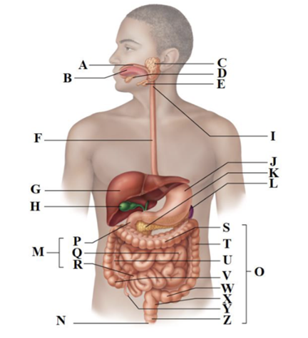
A
Mouth (oral cavity)
B
Tongue
C
Parotid gland
D
Sublingual gland
E
Submandibular gland
F
Esophagus
G
Liver
H
Gallbladder
I
Pharynx
J
Stomach
K
Pancreas
L
Spleen
M
Small intestine
N
Anus
O
Large intestine
P
Duodenum
Q
Jejunum
R
Ileum
S
Transverse colon
T
Descending colon
U
Ascending colon
V
Cecum
W
Sigmoid colon
X
Rectum
Y
Appendix
Z
Anal canal
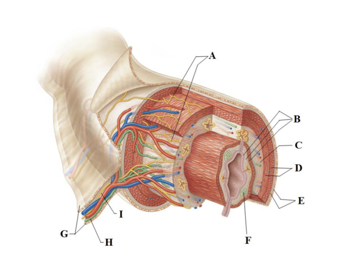
A
Intrinsic nerve plexuses
B
mucosa
C
submucosa
D
muscularis externa
E
serosa
F
mucosa-associated lymphoid tissue
H
lymphatic vessel
I
nerve
G
mesentery
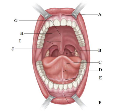
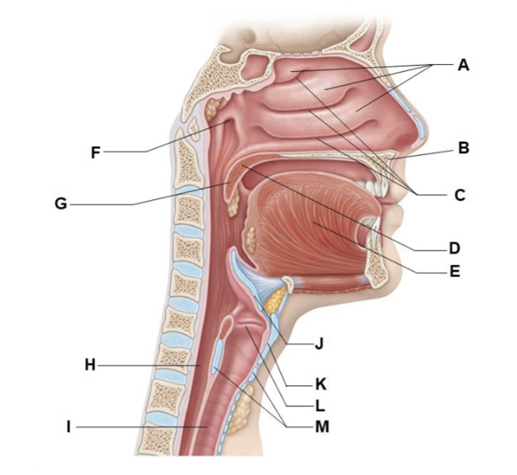
A
Superior labial frenulum
B
posterior wall of oropharynx
C
tongue
D
lingual frenulum
E
opening of submandibular duct
F
inferior labial frenulum
G
palatine raphe
H
hard palate
I
soft palate
J
uvula
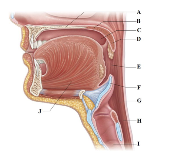
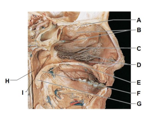
A
hard palate
B
soft palate
C
oral cavity
D
uvula
E
oropharynx
F
epiglottis
G
laryngopharynx
H
esophagus
I
trachea
J
tongue
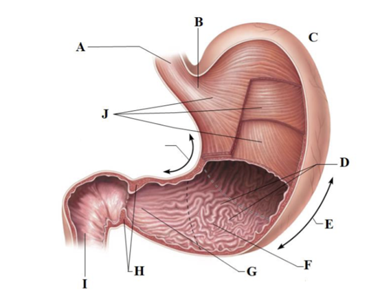
A
esophagus
B
cardial part
C
fundus
D
rugae of mucosa
E
greater curvature
F
pyloric antrum
G
pyloric canal
H
pyloric sphincter
I
duodenum
J
muscularis externa 3 layers
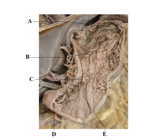
A
fundus
B
body
C
rugae of mucosa
D
pyloric sphincter
E
pyloric antrum
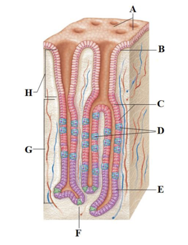
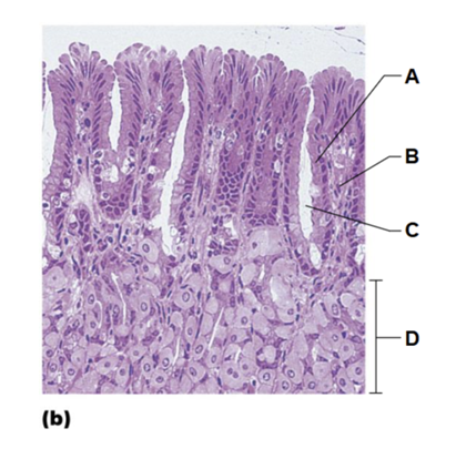
A
gastric pits
B
surface epithelium (mucous cells)
C
mucous neck cells
D
parietal cell
E
chief cell
F
enteroendocrine cell
G
gastric gland
H
gastric pit
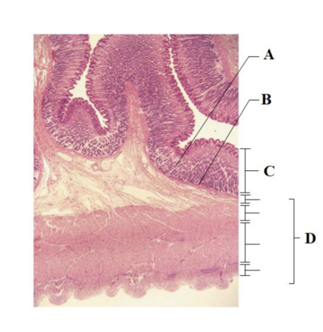
A
gastric glands
B
muscularis mucosae
C
mucosa
D
muscularis externa
A
simple columnar epithelium
B
lamina propria
C
gastric pit
D
gastric glands
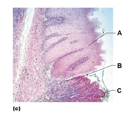
A
stratified squamous epithelium of esophagus
B
gastroesophageal junction
C
simple columnar epithelium of stomach
A
falciform ligament
B
liver
C
gallbladder
D
spleen
E
stomach
F
greater omentum
G
small intestine
H
cecum
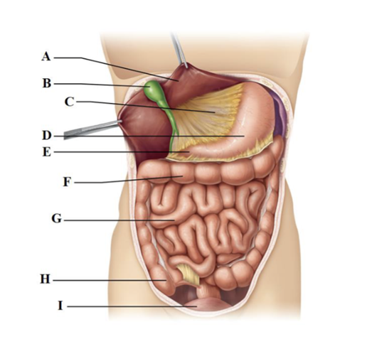
A
liver
B
gallbladder
C
lesser omentum
D
stomach
E
duodenum
F
transverse colon
G
small intestine
H
cecum
I
rectum
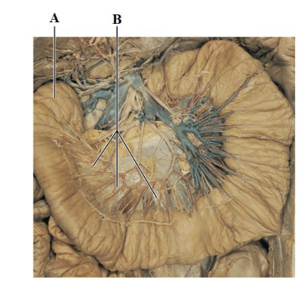
A
small intestine
B
spread mesentery
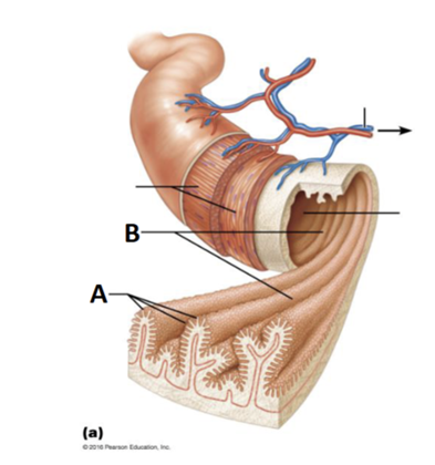
A
villi
B
circular folds
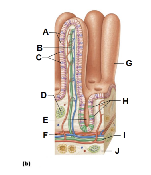
A
absorptive cells
B
lacteal
C
goblet cell
D
mucosa-associated lymphoid tissue
E
intestinal crypt
F
muscularis mucosae
G
villus
H
entero-endocrine cells
I
lymphatic vessel
J
submucosa
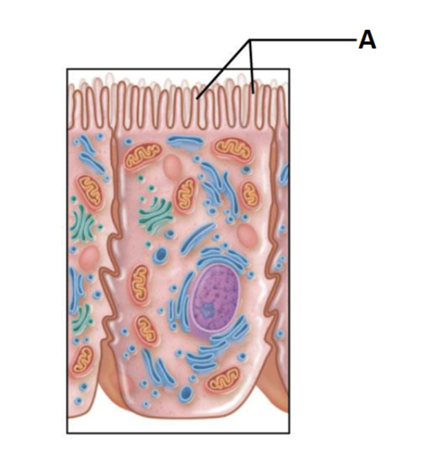
A
microvilli (brush border)
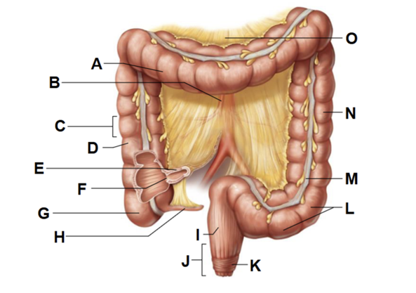
A
transverse colon
B
superior mesenteric artery
C
haustrum
D
ascending colon
E
ileum
F
ileocecal valve
G
cecum
H
appendix
I
rectum
J
anal canal
K
external anal sphincter
O
transverse mesocolon
N
descending colon
M
tenia coli
L
sigmoid colon
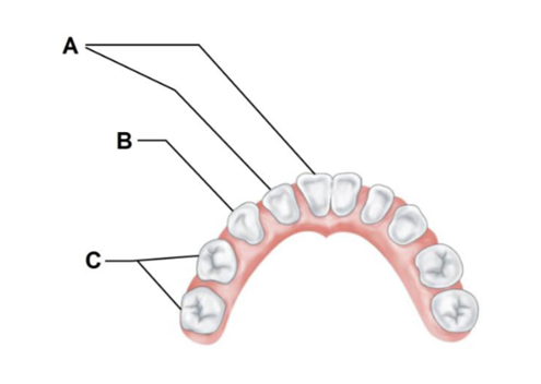
A
incisors
B
canine (eyetooth)
C
molars
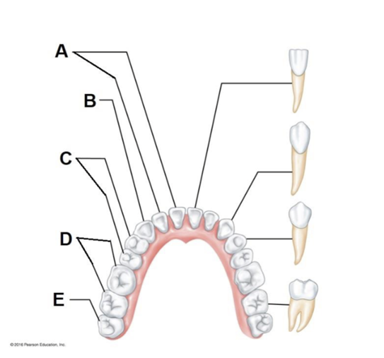
A
incisors
B
canine (eyetooth)
C
premolars (bicuspids)
D
molars
E
third molar (wisdom tooth)
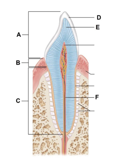
A
crown
B
neck
C
root
D
enamel
E
dentin
F
root canal
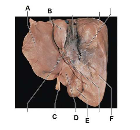
A
left lobe of liver
B
caudate lobe of liver
C
ligamentum
D
quadrate lobe of liver
E
gallbladder
F
right lobe of liver
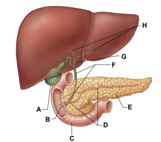
A
gallbladder
B
major duodenal papilla
C
duodenum
D
main pancreatic duct and sphincter
E
pancreas
F
bile duct and sphincter
G
common hepatic duct
H
right and left hepatic ducts of liver
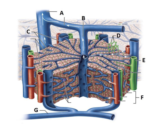
A
interlobular veins
B
central vein
C
sinusoids
D
bile canaliculi
E
bile duct
F
portal triad
G
portal vein
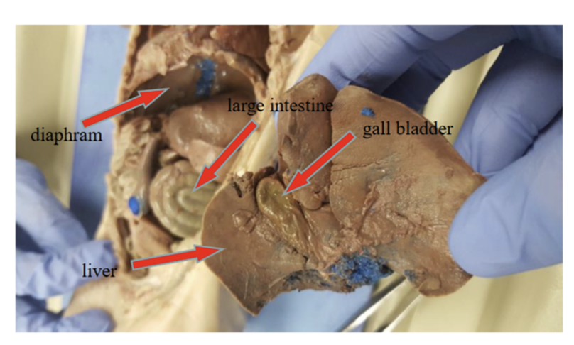
Find the gallbladder
GALLBLADDER of pig
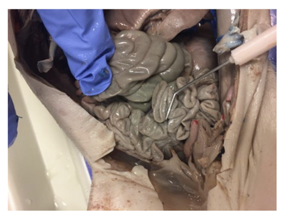
Find the large intestine
LARGE INTESTINE of pig
Find the liver
LIVER of pig
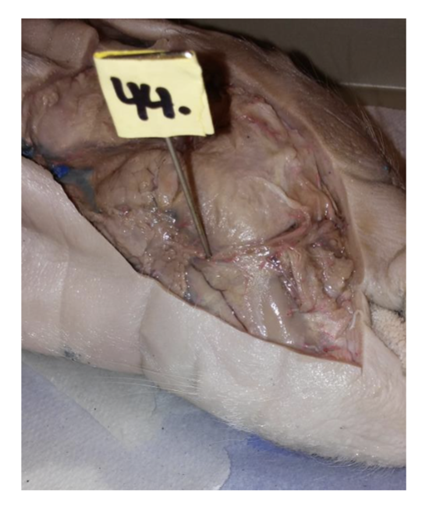
Find the diaphragm
DIAPHRAGM of pig
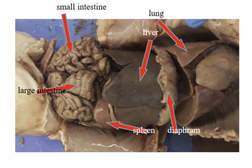
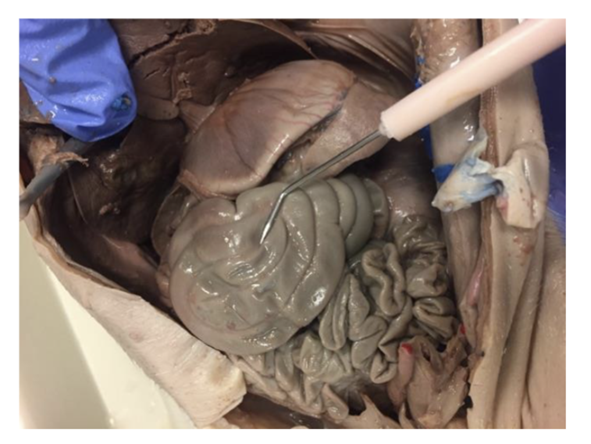
find the small intestine
SMALL INTESTINE of pig
find the lungs
LUNGS of pig
find the diaphragm
DIAPHRAGM of pig
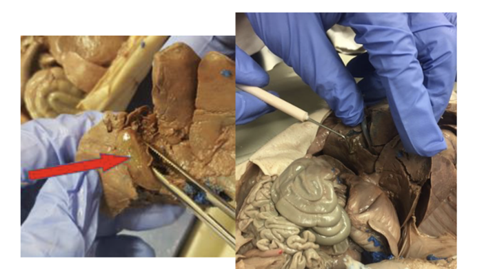
find the spleen
SPLEEN of pig
find the large intestine
LARGE INTESTINE of pig
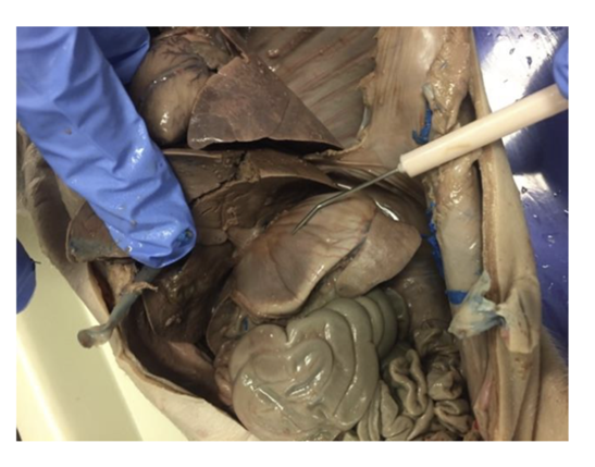
What is the pointer pointing to?
stomach
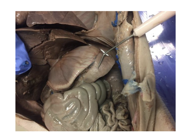
What is the pointer pointing to?
spleen
What is the pointer pointing to?
pancreases
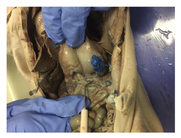
What is the pointer pointing to?
gallbladder
What is the pointer pointing to?
large intestine
What is the pointer pointing to?
small intestine
What is the pointer pointing to?
masseter muscle or submandibular gland
A
nasal conchae
B
hard palate
C
nasal meatuses
D
soft palate
E
tongue
F
opening of pharyngotympanic tube
G
uvula
H
esophagus
I
trachea
J
epiglottis
K
thyroid cartilage
L
vocal fold
M
cricoid cartilage
A
olfactory nerves
B
superior nasal concha and superior nasal meatus
C
middle nasal concha and middle nasal meatus
D
inferior nasal concha and inferior nasal meatus
E
hard palate
F
soft palate
G
uvula
H
nasopharynx
I
pharyngotympanic tube
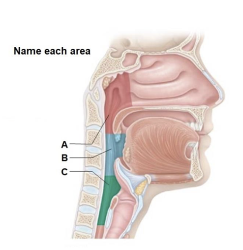
A
Nasopharynx
B
oropharynx
C
laryngopharynx
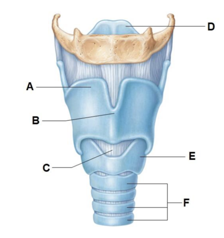
A
thyroid cartilage
B
laryngeal prominence (Adam's apple)
C
Cricothyroid ligament
D
epiglottis
E
cricoid cartilage
F
tracheal cartilages
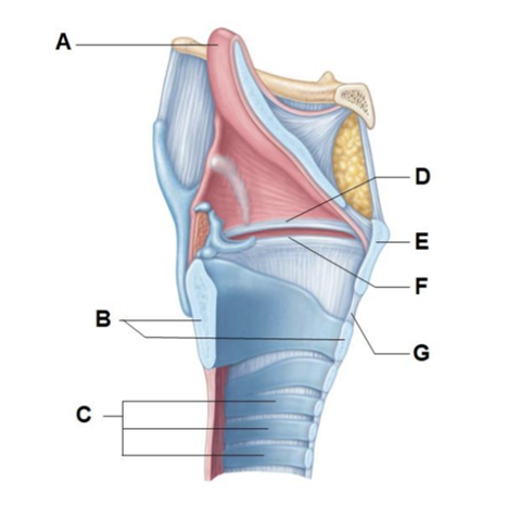
A
epiglottis
B
cricoid cartilage
C
tracheal cartilages
D
vestibular fold (false vocal cord)
E
thyroid cartilage
F
vocal fold (true vocal cord)
G
cricothyroid ligament
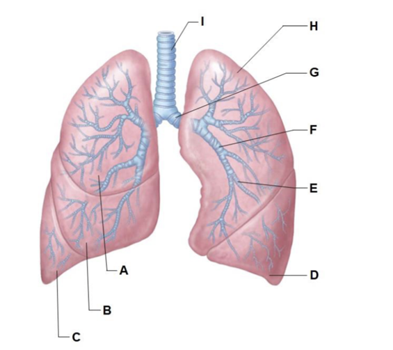
A
superior lobe of right lung
B
middle lobe of right lung
C
inferior lobe of right lung
D
inferior lobe of left lung
E
segmental (tertiary) bronchus
F
lobar (secondary) bronchus
G
left main (primary) bronchus
H
superior lobe of left lung
I
trachea
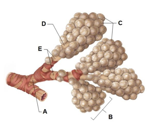
A
terminal bronchiole
B
alveolar sac
C
alveoli
D
alveolar duct
E
respiratory bronchioles
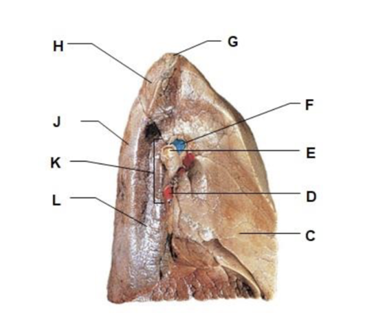
C
cardiac impression
D
pulmonary vein
E
left main bronchus
F
pulmonary artery
G
apex of lung
H
left superior lobe
J
left inferior lobe
K
hilum of lung
L
aortic impression
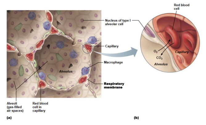
UNDERSTAND THIS IMAGE AND GAS EXCHANGE AS A WHOLE
UNDERSTAND THIS IMAGE AND GAS EXCHANGE AS A WHOLE
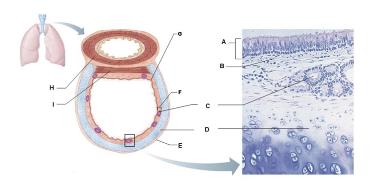
A
pseudostratified ciliated columnar epithelium
B
lamina propria (connective tissue)
C
seromucous gland in submucosa
D
hyaline cartilage
E
adventitia
F
submucosa
G
mucosa
H
esophagus
I
trachealis muscle
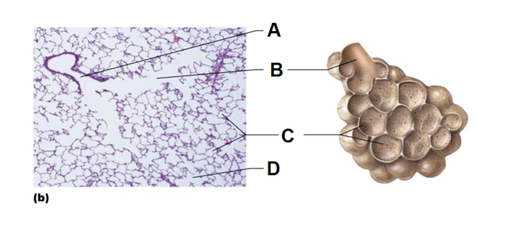
A
respiratory bronchiole
B
alveolar duct
C
alveoli
D
alveolar sac
LEFT PHOTO
the vocal fold (true vocal cord)
RIGHT PHOTO
Vestibular fold (false vocal cord)
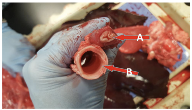
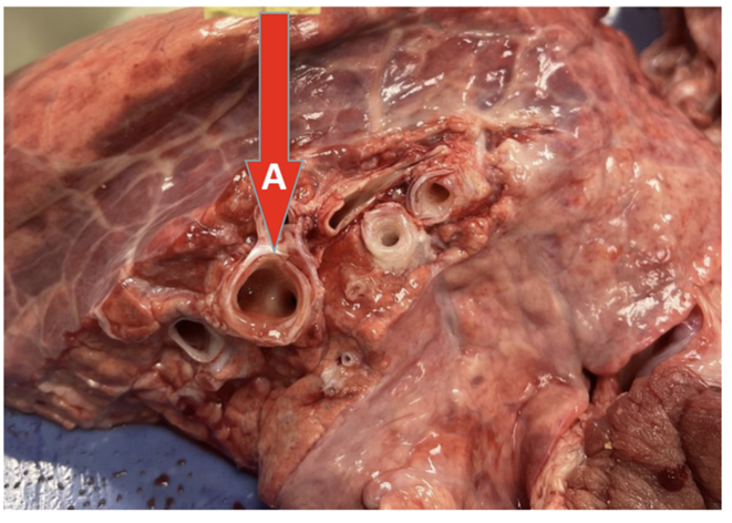
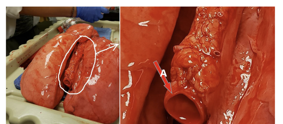
Where does A lead?
esophagus and stretches from the pharynx to the stomach
Where does B lead?
trachea and stretched from the pharynx to the lungs
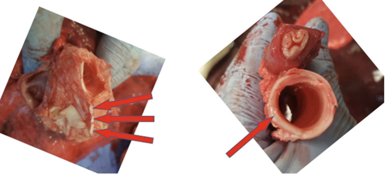
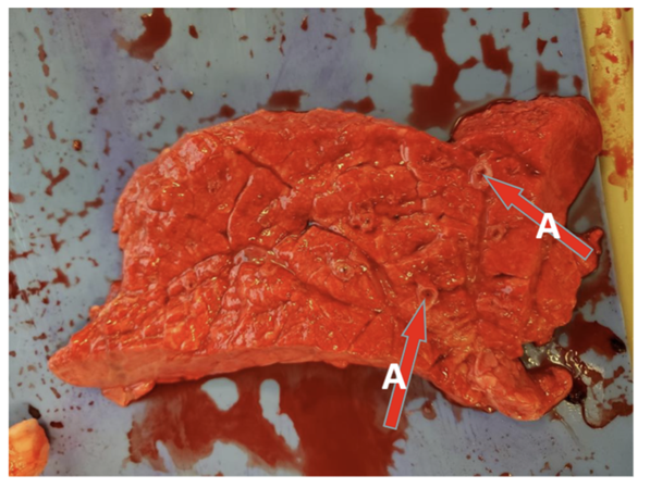
What are all the red arrows pointing at?
trachea
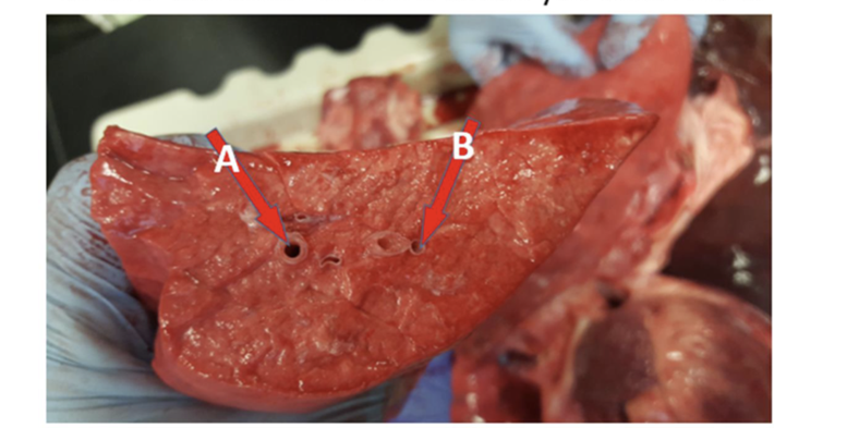
A
lobar
B
main/primary bronchus
C
trachea
A
trachea
B
carina
C
lobar/secondary bronchus
D
main/primary bronchus
A
primary bronchus
A
segmental/tertiary bronchus in lung tissue
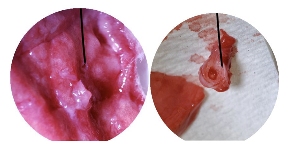
What is this under dissecting microscope
terminal bronchiole
A
tertiary bronchus
B
blood vessel
A
abdominal aorta