Instructions for Side by Side Printing
- Print the notecards
- Fold each page in half along the solid vertical line
- Cut out the notecards by cutting along each horizontal dotted line
- Optional: Glue, tape or staple the ends of each notecard together
Anatomy-Chapter 11-Nervous System
front 1 Two basic types of nerve cells | back 1 Neurons-transmit electrical signals Neuroglia (glial cells)-support the neurons |
front 2 4 types of neuroglia-CNS | back 2 Astrocytes Microglial cells Ependymal cells Oligodendrocytes |
front 3 2 types of neuroglia-PNS | back 3 Satellite cells Schwann cells |
front 4 Neuroglia compared to Neurons | back 4 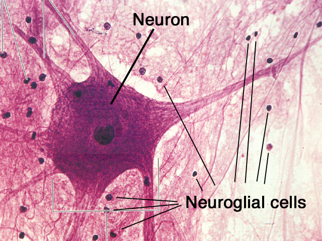 Neuroglia are smaller, darker then neurons they outnumber neurons 10 to 1 making up at least 50% of brain and spinal cord mass |
front 5 Astrocytes-CNS | back 5  "star-shaped" most abundant and versatile Jobs: support and brace neurons, guide young neurons, synapse formation, adjust capillary permeability, adjust "chemical environment" by absorbing ions, information processing |
front 6 Microglial cells-CNS | back 6  oval cells with long, thorny processes Jobs: monitor health of neurons, transform into a macrophage (**because cells of immune system are denied access to CNS) |
front 7 Ependymal cells-CNS | back 7  can be squamous, cuboid, or columnar, most are ciliated Jobs: line central cavities of brain and spinal cord, cilia help to circulate CSF |
front 8 Oligodendrocytes-CNS | back 8  Job: wrap around neurons of CNS, insulating them and forming myelin sheath **cannot regenerate like schwann cells *does not wrap around Nodes of Ranvier |
front 9 Satellite cells-PNS | back 9  *surrounds neuron cell bodies of PNS Job: is thought to help guide young neurons like the astrocytes |
front 10 Schwann cells-PNS | back 10  AKA neurolemmocytes Jobs: wrap around nerve fibers in PNS forming myelin sheath; similar to oligodendrocytes of CNS; regenerate damaged peripheral fibers |
front 11 Neurons (nerve cells) characteristics | back 11 conduct nerve impulses, in CNS and PNS, last a lifetime, have high metabolic rate (O2) **Amitotic-once they reach maturity, they lose ability to divide; except the olfactory epithelium and some hippocampal regions that have stem cells |
front 12 Nerve cell anatomy parts | back 12 Neuron cell body Nissl body (rough ER) Microtubules and Neurofibrils Inclusions Dendrites and Axons |
front 13 Neuron Cell body | back 13 AKA (ALSO KNOWN AS) perikaryon or soma do not have centrioles, which is why they are amitotic Most are in CNS, in receptive region |
front 14 Nissl Bodies | back 14  AKA Rough ER AKA chromatophilic substance (because it stains darkly with basic dyes) |
front 15 Microtubules and Neurofibrils | back 15 help maintain shape and integrity of cell |
front 16 Inclusions | back 16  little packages of metabolic byproducts that accumulate in the cell Some are pigments: melanin and lipofuscin |
front 17 Melanin inside inclusions | back 17 red iron-containing pigment |
front 18 Lipofuscin inside inclusions | back 18 golden-brown pigment that accumulates with age AKA "aging pigment" |
front 19 Nuclei | back 19 clusters of nerve cell bodies in CNS |
front 20 Ganglia | back 20 clusters of nerve cell bodies in PNS |
front 21 Processes of nerve cell (neuron) anatomy | back 21 CNS contains both neuron cell bodies and their processes; tracts PNS contains mostly neuron processes; nerves |
front 22 Bundles of neuron processes in the CNS | back 22 tracts |
front 23 Bundles of neuron processes in the PNS | back 23 nerves |
front 24 2 Types of Nerve cell processes | back 24 Dendrites Axons |
front 25 Dendrites | back 25 short, branching main receptive or input regions with graded potential **Motor neurons have 100's--dendritic spines are points of synapses with other neurons |
front 26 Graded potential | back 26 NOT action potentials, but are a type of short-distance signal |
front 27 Axons | back 27 **Conducting region of neuron that sends action potentials 1 per neuron, can have many branches starts with axon hillock, a funnel-shaped region by cell body long ones (3-4 ft in leg) are called nerve fibers branches are called axon collaterals ends in thousands of terminal branches (telodendria) with knob-like end called an axon terminal |
front 28 What happens when axons get cut | back 28 axon contains the same organelles found in the dendrites and cell body with 2 exceptions (no nissl bodies AKA rough ER, no golgi apparatus) **so axons will quickly decay if cut |
front 29 Axonal Transport | back 29 single bidirectional transport system that brings stuff up and down axons *motor protein that uses ATP *goes along microtubules at 15 inches per day directions: retrograde and anterograde |
front 30 Retrograde | back 30 transport back to the cell body **polio, herpes simplex and tetanus toxin use this to read cell body |
front 31 Anterograde | back 31 toward axon terminals |
front 32 Myelin Sheath and Neurolemma | back 32 Jobs: whitish fat covers long nerve fibers, protects and electrically insulates nerve fiber, increases speed of nerve impulse transmission *dendrites always unmyelated, axons either way |
front 33 Myelin sheath and neurolemma composition-PNS | back 33 Schwann cells make it up, Nodes of Ranvier (gaps in between these cells), can wrap around 15 nerve cell axons |
front 34 Gray Matter-CNS | back 34 contains mostly nerve cell bodies and unmyelinated fibers |
front 35 Myelin sheath and neurolemma composition-CNS | back 35 Oligodendrocytes make it up, can wrap around 60 nerve cell axons with widely spaced nodes of ranvier |
front 36 White Matter-CNS | back 36 dense collections of myelinated fibers |
front 37 Classification of Neurons-Structural | back 37 Multipolar neurons Bipolar neurons Unipolar neurons |
front 38 Multipolar neurons | back 38 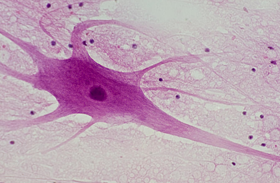 have 3 or more processes *most common type! (about 99%) |
front 39 Bipolar neurons | back 39  2 processes-cell body in middle and dendrite *rare (retina of eye and olfactory mucosa) |
front 40 Unipolar neurons | back 40 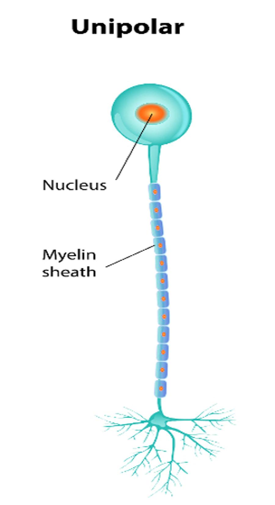 1 short process emerging from cell body and divides into peripheral and central processes *found in ganglia or PNS used as sensory neurons |
front 41 Classification of Neurons-Functional | back 41 Sensory (afferent) neurons Motor (efferent) neurons Interneurons or association neurons |
front 42 Sensory (Afferent) neurons | back 42 transmit impulse into CNS unipolar located in PNS...ganglia |
front 43 Motor (Efferent) neurons | back 43 carry impulse away from CNS multipolar cell bodies in CNS...nuclei |
front 44 Interneurons or Association neurons | back 44 between motor and sensory neurons multipolar and in CNS |
front 45 Neurophysiology Components | back 45 Voltage Current Resistence |
front 46 Voltage | back 46 "potential energy" from separation of charges measured in volts or with tiny stuff like a nerve, millivolts |
front 47 Current | back 47 flow of electrical charge from one point to another used to do work |
front 48 Resistance | back 48 hindrance to charge flow |
front 49 Ohm's Law | back 49 Current (I) = Voltage (V) / Resistance (R) Electrical currents reflect flow of ions across cell membranes Plasma membranes maintaining membrane potential use ion channels **Open channels allow ions to move along their electrochemical gradients |
front 50 Types of Charged Channels | back 50 Chemically (Ligand) gated ion channels Voltage gated ion channels Mechanically gated ion channels Leak channels |
front 51  Chemical (Ligand) gated ion channel | back 51 **found on nerve dendrites and cell bodies transmembrane--open or close in response to a chemical neurotransmitters like ligands in neurons, help react quickly to messages **uses lock and key fit, site far from channel ion permeability changes, let potassium, sodium, calcium pass through to evoke intracellular electrical signal |
front 52 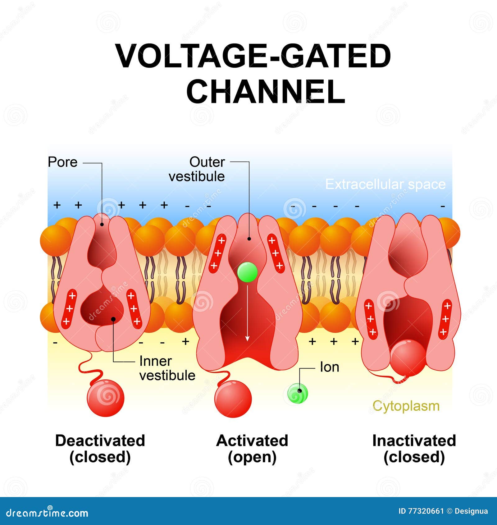 Voltage gated ion channels | back 52 **Found on nerve axons and axon hillock **rely on difference in membrane potential Change potential to let Action potentials occur Starts at a resting potential with Sodium-potassium ATPase *reverses resting membrane potential, potassium leaves, which removes positive charges closes channel by ball-and-chain method RESULT: more negative cell and an action potential |
front 53 Resting Membrane Potential | back 53 *graded potential + and - charges inside and outside the nerve cell, greater negative charge inside *leak channels allow ions to diffuse down concentration gradients--many more potassium leak channels than sodium leak channels--so more permeable to potassium ions sodium and potassium gradients, more sodium ions outside, more potassium ions inside **measured using a voltmeter, on average the charge is -70 millivolts |
front 54 Depolarization and Hyperpolarization | back 54 *graded potentials for short distances *action potentials for long distances anything that produces a change in ion permeability can change membrane potential |
front 55 Depolarization | back 55 loss of difference in charge in a nerve cell where sodium ions enter cell |
front 56 Hyperpolarization | back 56 increase in potential difference |
front 57 Spread and Decay of graded potentials | back 57  |
front 58 Action Potential | back 58 **send signals over long distances AP transmission and generation in skeletal muscle cells and neurons are the SAME. **To have this occur, enough voltage gated channels need to open, graded potentials at the axon hillock transition into action potentials **NO dissipation over distance, unlike graded potentials **ALL of these of from -70mV to +30mV |
front 59 Depolarization of an Action Potential | back 59 restores resting electrical conditions, but does NOT restore resting ionic conditions by Sodium-potassium pump Amounts of sodium needed for Action potential is a 0.012% change in intracellular Na+ concentrations |
front 60 Saltatory Conduction | back 60 in myelinated nerves and motor neurons action potential is propagated along axon's entire length from each node of ranvier and is self-propagating 30x faster than continuous conduction seen in unmyelinated axons |
front 61 Propagation of an Action Potential | back 61 reversal of charges across a membrane, positive charge from axon hillock to the next segment of axon to trigger action potential in the next axon, then axon hillock returns to resting membrane potential ---action potential travels from one axon to the next --with action potential also moving from one segment of an axon to the next |
front 62 Graded Potential | back 62 positive charge dissipates over distance, Action potentials go from -70mV to a range of values up to +30mV |
front 63 Refractory Period of an action potential propagation | back 63 **Cannot trigger an action potential going backwards on the gradient --the ball-and-chain method where the ball closes off the channel so no charge can go through it again...until they reset for another action potential |
front 64 Relationship between stimulus strength and action potential frequency | back 64 **any voltage that is NOT strong enough to open enough sodium channels will NOT generate an action potential *increasing axon width will also increase action potential speed *once threshold is reached for an action potential, the stronger the stimulus, the more frequently the action potentials are generated |
front 65 Absolute Refractory Period in Action Potentials | back 65 **when sodium channels open until sodium channels reset *this patch of membrane can NOT respond to another stimulus |
front 66 Relative Refractory Period in Action Potentials | back 66 **most of the sodium channels have closed *still repolarizing, but a STRONG stimulus can cause an action potential |
front 67 Action Potential in Bare Plasma Membrane | back 67 no data |
front 68 Action Potential in Unmyelinated Axons | back 68 https://www.youtube.com/watch?v=pbg5E9GCNVE |
front 69 Action Potential in Myelinated Axons | back 69 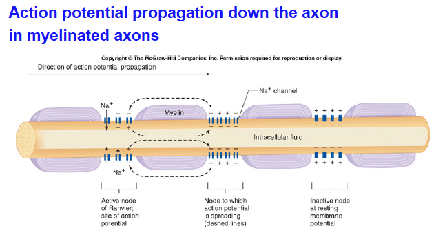 |
front 70 MS-Multiple Sclerosis | back 70 autoimmune disease-due to myelin sheath in CNS being gradually destroyed and hardened (sclerosis) axons are not affected though onset in young adults! 1st sign is visual (temporary blindness) problems controlling muscles (weak, clumsy)-from peripheral motor nerve demyelination |
front 71 Synapse (tiny gaps) Types | back 71 Electrical Chemical |
front 72 Electrical Synapse | back 72 **less common neurons joined like this are electrically coupled action potential transmission is very rapid unidirectional OR bidirectional |
front 73 Chemical Synapse | back 73 transmit action potentials to postsynaptic neurons by specific chemicals called neurotransmitters *inside synaptic vesicles at axon terminals neurotransmitters shoot across a tiny gap called a synaptic cleft (30 to 50nm wide) *only unidirectional |
front 74 Excitatory Post Synaptic Potentials | back 74 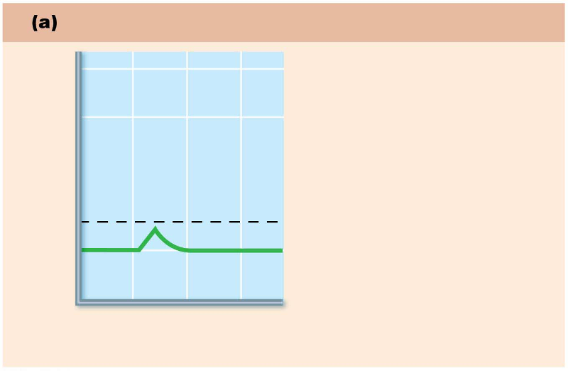 |
front 75 Inhibitory Post Synaptic Potentials | back 75 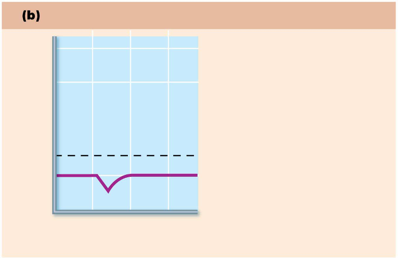 |
front 76 Action potential vs. Graded potential!! | back 76 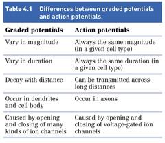 VERY important for test! |
front 77 Types of Neurotransmitters | back 77 Acetylcholine Norepinephrine Dopamine Serotonin |
front 78 Acetylcholine | back 78 nicotinic and muscarinic subtypes *when prolonged, you can get titanic muscle spasms because acetylcholinesterase is blocked (sarin nerve gas and insecticides can do this)--also inhibited by the botulism toxin |
front 79 Alzheimers and Acetylcholine connection | back 79 less overall acetylcholine with people who have this disease |
front 80 Norepinephrine | back 80 release enhanced by amphetamines removal from synapse blocked by cocaine and antidepressants **low levels in depression |
front 81 Dopamine | back 81 release and removal similar to Norepinephrine **low levels in Parkinson's disease |
front 82 Serotonin | back 82 acts like a brake on a bicycle blocked by LSD and Prozac (treats depression) blocking an inhibitor makes it activate! |
front 83 Differences between action potentials and graded potentials | back 83  |