Instructions for Side by Side Printing
- Print the notecards
- Fold each page in half along the solid vertical line
- Cut out the notecards by cutting along each horizontal dotted line
- Optional: Glue, tape or staple the ends of each notecard together
Activity 3: Coronary Circulation
front 1 Does the contained blood inside the heart chambers nourish the myocardium? | back 1 No |
front 2 What are the 6 coronary arteries? | back 2 1. Right coronary artery (RCA) 2. Right marginal artery 3. Posterior interventricular artery 4. Left coronary artery (LCA) 5. Anterior interventricular artery 6. Circumflex artery |
front 3 Where does the functional blood supply of the heart come from then? (Hint: 2 sets of arteries) | back 3 Right and left coronary arteries |
front 4 What are the 2 branches of the right coronary artery? | back 4 1. Right marginal artery 2. Posterior interventricular artery |
front 5 The right marginal artery supplies what side of the heart? | back 5 Lateral right side of the heart |
front 6 The posterior interventricular artery supplies what 2 regions of the heart? | back 6 1. Posterior walls of the ventricles 2. Posterior portion of the interventricular septum |
front 7 What are the 2 branches of the left coronary artery? | back 7 1. Anterior interventricular artery 2. Circumflex artery |
front 8 The anterior interventricular artery supplies what 2 regions of the heart? | back 8 1. Anterior portion of the interventricular spetum 2. Anterior walls of both ventricles |
front 9 The circumflex artery supplies what 2 regions of the heart? | back 9 1. Left atrium 2. Posterior portion of the left ventricle |
front 10 What are the 5 cardiac veins? | back 10 1. Great cardiac vein 2. Middle cardiac vein 3. Small cardiac vein 4. Coronary sinus 5. Anterior cardiac veins |
front 11 The great cardiac vein drains what 2 anterior regions of the heart? | back 11 1. Anterior left ventricle 2. Anterior right ventricle |
front 12 The middle cardiac vein drains what 2 posterior regions of the heart? | back 12 1. Posterior right ventricle 2. Posterior left ventricle |
front 13 The small cardiac vein drains what part of the heart? | back 13 Lateral right ventricle |
front 14 The great, middle, and small cardiac veins all drain into which coronary vein? | back 14 Coronary sinus |
front 15 The coronary sinus drains what part of the heart? | back 15 The entire heart |
front 16 The anterior cardiac veins drain directly into which chamber of the heart? | back 16 Right atrium |
front 17 The auricles cover what 2 parts of the heart? | back 17 Right and left atria |
front 18 What tissue is the balance of the heart muscle? | back 18 Ventricular tissue |
front 19 Why is the left ventricle thicker? | back 19 There is greater demand on the left ventricle. It must pump blood through the much longer systemic circuit, a pathway with much higher resistance than the pulmonary circuit. |
front 20 What is the first branching of the aorta? | back 20 Brachiocephalic trunk |
front 21 The ligamentum arteriosum is a cordlike remnant of what structure? | back 21 Ductus arteriosus |
front 22 What is the function of the ductus arteriosus in the fetus? | back 22 In the fetus, it allows blood to pass from the pulmonary trunk to the aorta by bypassing the nonfunctional fetal lungs. |
front 23 What is the cordlike ridges of muscle throughout most of the right atrium called? | back 23 Pectinate muscle |
front 24 The fossa ovalis marks the site of what opening in the fetal heart? | back 24 Foramen ovale |
front 25 What is the function of the foramen ovale in the fetal heart? | back 25 It allows blood to pass from the right to the left atrium, thus bypassing the fetal lungs. |
front 26 The ridged and pitted muscle located in the ventricles are called what? | back 26 Trabeculae carneae |
front 27 The moderator band connects what 2 structures of the heart? | back 27 1. Interventricular septum 2. Anterior papillary muscles |
front 28 The moderator band is a bundle of what type of fibers? | back 28 Cardiac muscle fibers |
front 29 What is the function of the moderator band? | back 29 Coordinate contraction of the ventricle. |
front 30 Is the myocardium of the left ventricle thicker than that of the right's? | back 30 Yes |
front 31 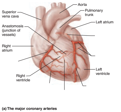 Identify the blanks. | back 31 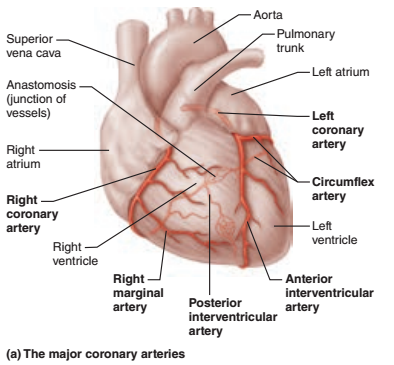 |
front 32 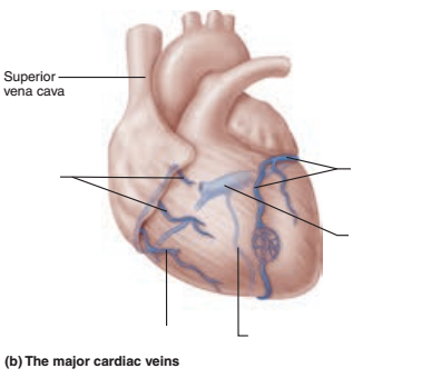 Identify the blanks. | back 32 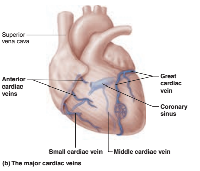 |