Instructions for Side by Side Printing
- Print the notecards
- Fold each page in half along the solid vertical line
- Cut out the notecards by cutting along each horizontal dotted line
- Optional: Glue, tape or staple the ends of each notecard together
The spinal cord and tracts
front 1 Cell bodies of interneurons can be found in which of the following locations? Ventral horns of the spinal cord Anterior horns of the spinal cord White matter of the spinal cord Dorsal root ganglia Dorsal horns of the spinal cord | back 1 Dorsal horns of the spinal cord |
front 2 __________ line the central canal of the spinal cord and help to circulate CSF. | back 2 ependymal cells |
front 3 The cell bodies of sensory neurons whose axons travel in spinal nerves are located within the _________. | back 3 dorsal root ganglia |
front 4 ________ roots the spinal cord to the coccyx | back 4 filum terminale |
front 5 There are __________ pairs of spinal nerves. | back 5 31 |
front 6 A spinal nerve is formed from the union of a dorsal _______ and a ventral _______. What word fills in both blanks? anterior root has only ______ posterior root has only ____ | back 6 root motor axons sensory axons |
front 7 distal tapered end of the spinal cord | back 7 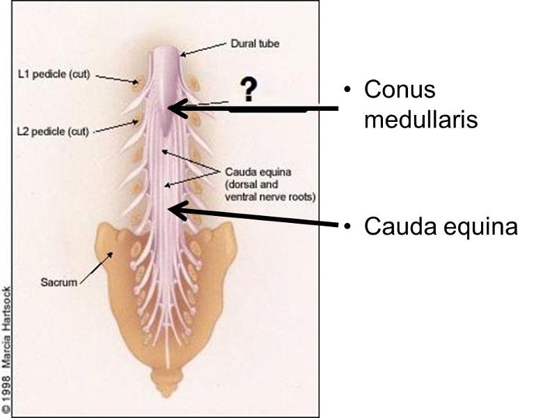 conus medullaris |
front 8 the spinal cord runs inferiorly from ______ to ________- | back 8 the foramen magnum; L1 or L2 |
front 9 collection of spinal nerve roots extend inferior to spinal cord | back 9 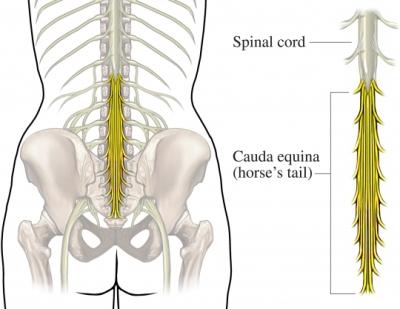 cauda equina |
front 10 extension of pia mater from the conus medullaris anchoring to the posterior surface of the coccyx | back 10 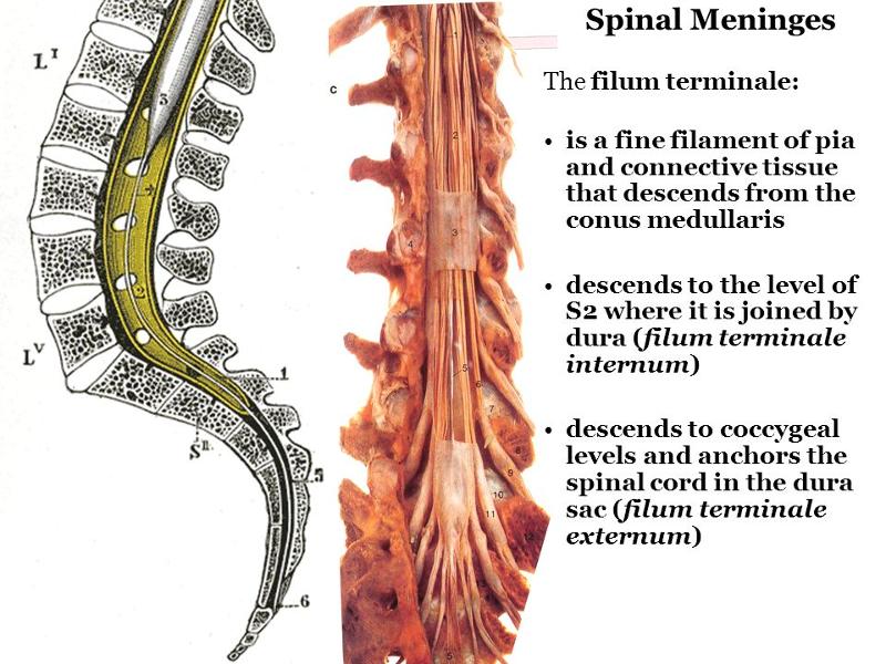 filum terminale |
front 11 what are the two major enlargements of the spinal cord | back 11 cervical enlargement and lumbrosacral enlargement |
front 12 what group of nerves is found near the cervical enlargement of the spine? | back 12 brachial plexus |
front 13 which vertebrae do the cervical and lumbrosacral enlargements cover? | back 13 c5-T1 and L1-S3 |
front 14 what protects the spinal cord? | back 14 vertebrae, meninges, ligaments, muscles CSF |
front 15 what is the function of a denticulate ligament and what are they made of? | back 15  help anchor the spinal cord out laterally up and down the spinal column. made of pia mater |
front 16 what are the meningeal spaces of the spinal cord and brain and are they potential or actual | back 16 brain spinal cord epidural potential actual subdural potential potential subarachnoid actual actual |
front 17 which epidural space is continuous between brain and spinal cord? | back 17 subarachnoid |
front 18 where is an epidural administered? why? | back 18 in the epidural space between L3 and L4 o4 L4 and L5. The epidural space in the spinal cord is not continuous with the space in the brain. you don't want anesthetic in the brain |
front 19 where is a spinal tap administered AKA LUMBAR PUNCTURE? | back 19 into the subarachnoid space - used to remove CSF to dx meningitis |
front 20 through which fissure does the anterior spinal artery travel? | back 20 the anterior median fissure |
front 21 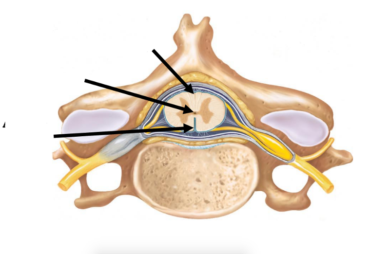 | back 21 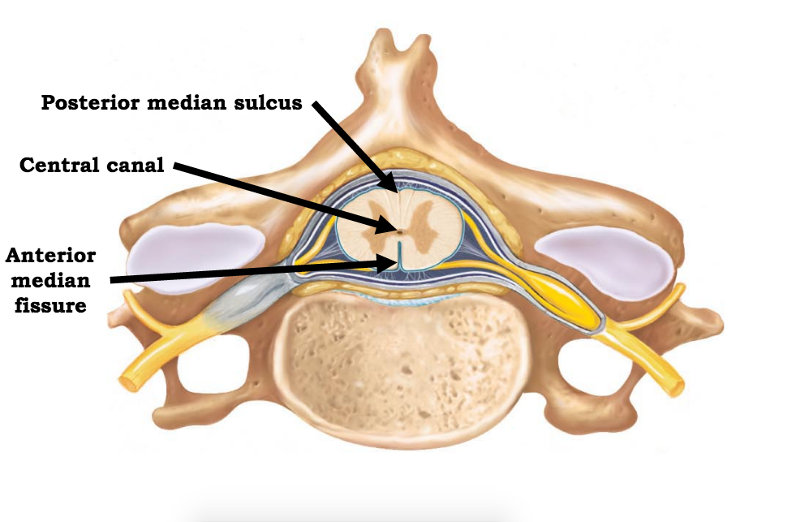 |
front 22 what is more deep, the posterior median sulcus or anterior median fissure? | back 22 anterior median fissure is deep to posterior median sulcus |
front 23 where are cell bodies of motor neurons found? | back 23  in the ventral horn |
front 24 where are cell bodies of sensory neurons found? | back 24 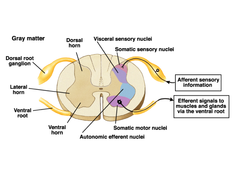 in the dorsal root ganglia |
front 25  | back 25 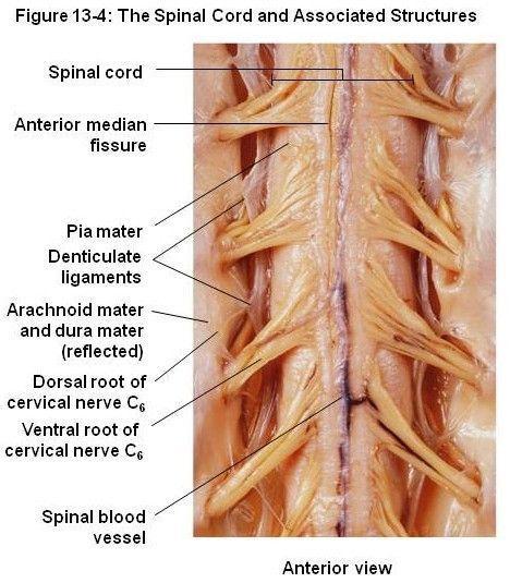 |
front 26 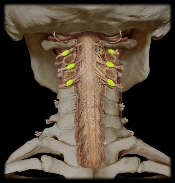 identify yellow structures | back 26 dorsal root ganglion |