Instructions for Side by Side Printing
- Print the notecards
- Fold each page in half along the solid vertical line
- Cut out the notecards by cutting along each horizontal dotted line
- Optional: Glue, tape or staple the ends of each notecard together
Chapter 20: Lymphatic System (Mastering)
front 1 Which of the following are primary lymphoid organs? lymph nodes and tonsils bone marrow and thymus appendix and spleen spleen and thymus | back 1 bone marrow and thymus |
front 2 Which of the following areas in a secondary lymphoid organ allows intimate contact between blood and the lymphocytes? germinal centers of the lymph nodes white pulp of the spleen red pulp of the spleen Hassall’s corpuscles of the thymus | back 2 white pulp of the spleen |
front 3 Where in the lymph node do the T cells first encounter antigens presented by dendritic cells? medullary cords in the medulla lymphoid follicles of the outer cortex germinal centers of the cortex deep in the cortex | back 3 deep in the cortex |
front 4 Collections of lymphoid tissues, called MALT, are strategically placed throughout the respiratory, digestive, and genitourinary systems. Which one of these is located at the end of the small intestine? Peyer’s patches appendix tonsils | back 4 Peyer’s patches |
front 5 There is a decrease in our ability to fight infection as we age. Which lymphoid organ may have a role in this decline? spleen thymus lymph nodes | back 5 thymus |
front 6 Besides lymph nodes, where would you expect to find proliferating (dividing) B cells? in the brain in the thyroid in the skin in the spleen | back 6 in the spleen |
front 7 Which of the following mechanisms is NOT used to propel lymph through lymphatic vessels? small heart-like pumps gravity the milking action of muscles pulmonary motion | back 7 small heart-like pumps |
front 8 Adjacent cells in lymphatic capillaries overlap each other loosely. What is the unique structural modification that increases their permeability? lacteals minivalves fibroblasts trabeculae | back 8 minivalves |
front 9 Which of the following promotes closure of the minivalves associated with lymph capillaries? increasing pressure in the interstitial space anchoring of endothelial cells to adjacent structures by collagen fibers increasing pressure inside the lymph capillary inflammation of tissues surrounding lymphatic capillaries | back 9 increasing pressure inside the lymph capillary |
front 10 Lymph from what regions of the body is drained into the right lymphatic duct? the left upper limb, the left side of the head and thorax, and both lower limbs the right upper limb, the right side of the head, and the thorax the right upper limb, the right side of the head and thorax, and the right lower limb the digestive organs and lower limbs | back 10 the right upper limb, the right side of the head, and the thorax |
front 11 What is the name of the enlarged sac to which the lumbar trunks and the intestinal trunk return lymph? cisterna chyli thoracic duct lacteals right lymphatic duct | back 11 cisterna chyli |
front 12 Where are the three large clusters of superficial lymph nodes? the cervical, acromial, and mammary regions the axillary, brachial, and subclavian regions the lumbar, inguinal, and femoral regions the cervical, inguinal, and axillary regions | back 12 the cervical, inguinal, and axillary regions |
front 13 Once collected, lymph ultimately drains into __________. lymph nodes arterial circulation venous circulation the liver for detoxification | back 13 venous circulation |
front 14 Which statement describes the origin of lymph fluid? Lymph is collected from atrial to venous anastomoses. Lymph is secreted into the lymph vessels. Lymph is collected from fluid that accumulates in veins as blood slowly circulates back toward the heart. Lymph is excess fluid formed from plasma that accumulates in the tissues as interstitial fluid. | back 14 Lymph is excess fluid formed from plasma that accumulates in the tissues as interstitial fluid. |
front 15 Art-based Question Which lymphoid organ extracts aged and defective blood cells and platelets from the blood in addition to storing some of the breakdown products for later reuse? A B C D | back 15 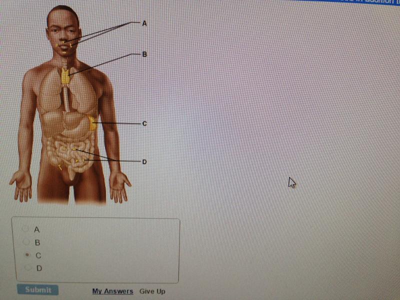 C |
front 16 Which lymph cells produce antibodies? dendritic cells macrophages plasma cells reticular cells | back 16 plasma cells |
front 17 What region of the lymph node contains follicles filled with dividing B cells? hilus cortex subcapsular sinus medulla | back 17 cortex |
front 18 Which of the following is a role of lymph nodes? They produce red blood cells. They return lymph to circulation. They filter lymph. They produce lymph. | back 18 They filter lymph. |
front 19 Which part of the spleen is the site of immune function? splenic sinusoids red pulp white pulp splenic cords | back 19 white pulp |
front 20 After surgical removal of the spleen (i.e., a splenectomy), some other organs take over most of its functions. Which of the following spleen functions in the adult can not be performed by bone marrow? immune surveillance erythropoiesis removal of aged and damaged red blood cells from the blood storage of platelets | back 20 removal of aged and damaged red blood cells from the blood |
front 21 Which of the following lymph organs is NOT matched with its function? thymus: mature T cells Peyer's patches: mature B cells bone marrow: form lymphocytes spleen: remove red blood cells | back 21 Peyer's patches: mature B cells |
front 22 Peyer's patches are mucosa-associated lymph tissue located in the __________. wall of the small intestine spleen wall of the colon liver | back 22 wall of the small intestine |