Instructions for Side by Side Printing
- Print the notecards
- Fold each page in half along the solid vertical line
- Cut out the notecards by cutting along each horizontal dotted line
- Optional: Glue, tape or staple the ends of each notecard together
Multichoice: the Skeletal System: the Appendicular skeleton
front 1 This is the anterior bone that articulates with the manubrium of the sternum at the sternoclavicular joint. a) Scapula b) Clavicle c) Xiphoid d) Rib e) Thoracic vertebra | back 1 b |
front 2 Which of the following bones articulates with the scapula?
| back 2 b |
front 3 Why does a fracture of the clavicle usually occur in the mid-region of the bone? a) Due to the medial pressure from the inflated lungs b) Due to the ligament-reinforced strength of the acromial joint c) Due to weakness at the junction of the two curves of clavicle d) Due to position of the clavicle relative to the humerus e) Due to fusion of the ends of the clavicle to the sternum and scapula | back 3 c |
front 4  Which part of the clavicle articulates with the manubrium?
| back 4 d |
front 5 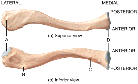 Which is the only part of the clavicle that articulates with the scapula?
| back 5 a |
front 6 Which site labeled on the diagram is considered the weakest point of the clavicle?
| back 6 e |
front 7 Which of the following bones is located in the posterior thorax between the levels of second and seventh vertebrae?
| back 7 d |
front 8 Which bone articulates with the scapula at the glenoid cavity?
| back 8 e |
front 9 This is the thick edge of the scapula that is closer to the arm. a) Axillary border b) Medial border c) Infraspinous fossa d) Coracoid process e) Acromion | back 9 a |
front 10 Which of the following bone markings on the scapula is an indentation into which the head of the humerus fits? a) Coracoid process b) Glenoid cavity c) Acromion d) Scapular notch e) Supraspinous fossa | back 10 b |
front 11 Which of the following statements is TRUE with regard to the human hand? a) There are 5 carpals, 8 metacarpals and 14 phalanges. b) There are 8 carpals, 6 metacarpals and 14 phalanges c) There are 8 carpals, 5 metacarpals and 15 phalanges d) There are 8 carpals, 5 metacarpals and 14 phalanges e) There are 5 carpals, 8 metacarpals and 14 phalanges | back 11 d |
front 12 The glenohumeral joint is formed by articulation of the a) humerus, radius and ulna. b) humerus and radius. c) humerus and clavicle. d) humerus and ulna. e) humerus and scapula. | back 12 e |
front 13 The epiphyseal line on the proximal end of the humerus is found in the
| back 13 a |
front 14 The capitulum of the humerus articulates with the
| back 14 a |
front 15 This is a spool-shaped process on distal end of the humerus that is found medial to the capitulum and articulates with the ulna. a) Coronoid fossa b) Trochlea c) Medial epicondyle d) Lateral epicondyle e) Lesser tubercle | back 15 b |
front 16 The medial and lateral epicondyles are found on the distal end of the humerus and are used for a) formation of the elbow joint. b) tendon attachment. c) passage of nerves and blood vessels through the bone into the marrow cavity. d) Both formation of the elbow joint and tendon attachment. e) All of these choices are correct. | back 16 b |
front 17 Which of the following structures on the ulna receives the trochlea of the humerus?
| back 17 c |
front 18 What is the function of the interosseous membrane between the ulna and radius? a) Joins the shafts of two bones b) Tendon attachment c) Site of bone repair d) Both site of tendon attachment and bone repair e) None of these choices are correct. | back 18 a |
front 19 The distal end of the radius articulates with how many bones of the wrist? a) 1 b) 2 c) 3 d) 4 e) 5 | back 19 c |
front 20 Which of the following carpal bones is named for its large hook-shaped projection on its anterior surface?
| back 20 d |
front 21 What is included in the carpal tunnel?
| back 21 e |
front 22 The carpometacarpal joint consists of the a) base of metacarpal bones and distal row of carpal bones. b) base of metacarpal bones and proximal row of carpal bones. c) head of metacarpal bones and distal row of carpal bones. d) head of metacarpal bones and proximal row of carpal bones. e) None of these choices are correct. | back 22 a |
front 23 How many phalanges are in each hand? a) 10 b) 12 c) 14 d) 16 e) 20 | back 23 c |
front 24 The coxal bones unite anteriorly at a joint called the a) pubic symphysis. b) sacroiliac joint. c) hip. d) acetabulum. e) None of these choices are correct. | back 24 a |
front 25 What is the function of the pelvic girdle? a) Support for vertebral column b) Attachment site for lower limbs c) Attachment site for large pectoral muscles. d) Attachment site for lower limbs and for large pectoral muscles. e) All of these choices are correct. | back 25 d |
front 26 In the standard anatomical position, the _____ is the bone of the pelvis found the most superior.
| back 26 a |
front 27 Which of the following is the largest foramen in the human skeleton?
| back 27 b |
front 28 Which projection extends superiorly and laterally along the superior ramus of the pubis eventually merging with the arcuate line of the ilium? a) Pectineal line b) Ischial tuberosity c) Anterior gluteal line d) Inferior gluteal line e) Greater sciatic notch | back 28 b |
front 29 The hip joint is the joint found between a) the femur and tibia. b) the pelvis and sacrum. c) the pelvis and tibia. d) the femur and patella. e) the pelvis and femur. | back 29 e |
front 30 The portion of the bony pelvis that is found inferior to the pelvic brim is called a) the false pelvis. b) the greater pelvis. c) the true pelvis. d) both the false pelvis and the greater pelvis. e) all of these choices are correct. | back 30 c |
front 31 In comparison to the male pelvis, the female pelvis is NOT a) wider. b) shallower. c) larger in the pelvic inlet. d) larger in the pelvic outlet. e) larger in the acetabulum. | back 31 e |
front 32 32) Each lower limb has a) 30 bones found in 3 locations. b) 30 bones found in 4 locations. c) 32 bones found in 3 locations. d) 32 bones found in 4 locations. e) 34 bones found in 4 locations | back 32 b |
front 33 Which process on the femur serves as an attachment point for tendons of several thigh muscles?
| back 33 d |
front 34 Which of the following markings is located on the medial side of the femur? a) Lesser trochanter b) Greater trochanter c) Gluteal tuberosity d) Lateral epicondyle e) Linea aspera | back 34 a |
front 35 Which bone develops in the tendon of the quadriceps femoris muscle and protects the knee joint?
| back 35 d |
front 36 The medial and lateral condyles of the femur fit into what part of the patella? a) Articular facets b) Base of the patella c) Tibiofemoral crest d) Apex of the patella e) None of these choices are correct. | back 36 a |
front 37 Which of the structures listed below is NOT part of the knee joint? a) Lateral condyle of the femur b) Medial condyle of the femur c) Lateral malleolus of the fibula d) Condyles of the tibia e) Patella | back 37 c |
front 38 The hard sharp ridge of the shin that can easily be felt below the skin is the a) anterior border (crest) of the tibia. b) tibial tuberosity. c) medial condyle of the tibia. d) tibiofemoral joint. e) intercondylar eminence. | back 38 a |
front 39 The lateral malleolus is found on the distal end of what bone?
| back 39 b |
front 40 Which of following bones is NOT a tarsal bone?
| back 40 e |
front 41 Which of the followings structures is not found in the foot? a) Pollex b) Hallux c) Talus d) Longitudinal arch e) Transverse arch | back 41 a |
front 42  Which of the labeled structures in the diagram serve as attachment sites for tendons of the shoulder muscles?
| back 42 e |
front 43  43) Which of the labeled structures in the diagram is the coracoid process?
| back 43 a |
front 44 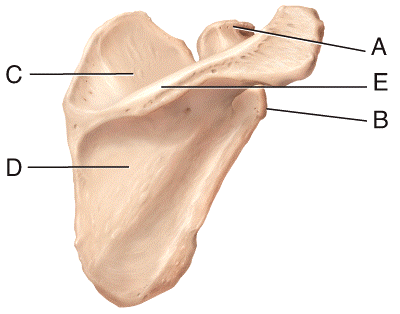 Which of the labeled structures in the diagram is the supraspinous fossa?
| back 44 c |
front 45  In the diagram of the humerus, which is the lateral epicondyle?
| back 45 b |
front 46  In the diagram of the humerus, this structure receives the head of the radius when the forearm is flexed.
| back 46 e |
front 47  In the diagram of the humerus, where is the trochlea?
| back 47 c |
front 48 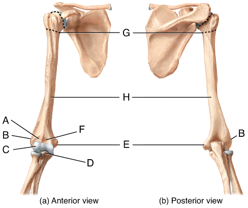 In the diagram of the ulna and radius, where is the styloid process of the radius?
| back 48 c |
front 49  In the diagram of the ulna and radius, where is the radial tuberosity?
| back 49 b |
front 50 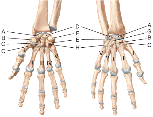 In the diagram of the wrist and hand, where is the capitate bone?
| back 50 d |
front 51 In the diagram of the wrist and hand, where is the trapezoid bone?
| back 51 c |
front 52  Which structure in the pelvis is where the longest nerve in the body passes? a) C b) D c) F d) G e) H | back 52 c |
front 53  In the diagrams of the pelvis, where is the ischial tuberosity?
| back 53 d |
front 54 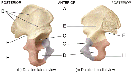 Which labeled structure in the diagrams of the pelvis terminates anteriorly as the anterior superior iliac spine?
| back 54 a |
front 55 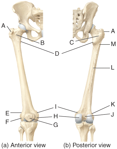 Which of the labeled structures of the femur serve as points of attachment for the tendons of thigh and buttocks muscles?
| back 55 c |
front 56  In the diagram of the femur, where is the medial condyle?
| back 56 d |
front 57  In the diagram of the tibia and fibula, where is the tibial tuberosity?
| back 57 b |
front 58 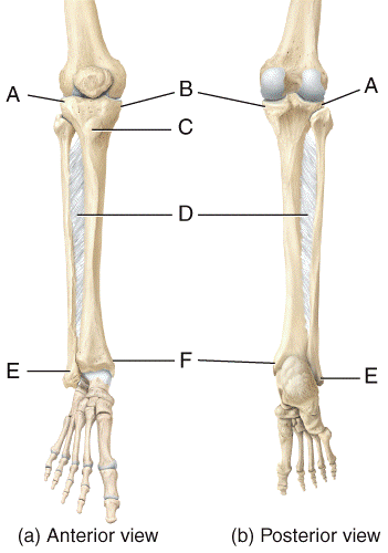 In the diagram of the tibia and fibula, this structure articulates with the talus and forms a protrusion on the medial surface of the ankle.
| back 58 b |
front 59  In the diagram of the foot, where is the navicular?
| back 59 c |
front 60  Which labeled bone in the diagram of the foot is the largest and strongest tarsal bone?
| back 60 a |
front 61 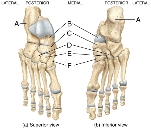 Which labeled bone in the diagram of the foot is the only bone of the foot that articulates with the fibula and tibia?
| back 61 b |
front 62 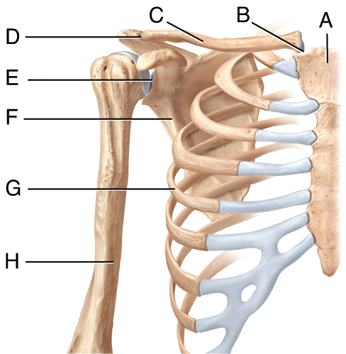 The pectoral girdle consists of 2 bones labeled _____ and _____ in the diagram.
| back 62 c |
front 63 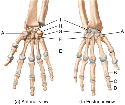 Where on the diagram is a metacarpal bone? a) A b) B c) D d) E e) I | back 63 d |
front 64 This is a common condition experienced by runners, which is caused by the kneecap tracking laterally as well as inferiorly and superiorly. a) Patellofemoral stress syndrome b) Metatarsal microfracture c) Bunions d) Hallux valgus e) Plantar fasciitis | back 64 a |
front 65 Which of the following is a condition where the medial longitudinal arch of the foot is decreased, resulting in fallen arches? a) Patellofemoral stress syndrome b) Bunions c) Flatfoot d) Clawfoot e) Clubfoot | back 65 c |
front 66 Which of the following is a condition where the foot is twisted inferiorly and medially, and the angle of the arch is increased? a) Patellofemoral stress syndrome b) Bunions c) Flatfoot d) Clawfoot e) Clubfoot | back 66 e |
front 67 During embryonic and fetal develop, most skeletal tissues arise from a) the neurocranium. b) the notochord. c) mesenchymal cells. d) endoderm. e) none of these choices are correct. | back 67 c |
front 68 The neurocranium gives rise to bones of the
| back 68 e |
front 69 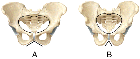 Which pelvis in the diagram shows the characteristics of a female pelvis?
c) Both are male. | back 69 a |
front 70 The boundary between the true pelvis and the false pelvis is the
| back 70 d |
front 71 The boundary between the true pelvis and the false pelvis is the
| back 71 d |
front 72 Which of the following is NOT a way that the skeletal system contributes to homeostasis? a) Provides support and protection for internal organs. b) Stores and releases sodium ions. c) Houses blood forming tissue. d) Protects the brain and spinal cord. e) Serves as attachment and leverage points for muscles. | back 72 b |