Instructions for Side by Side Printing
- Print the notecards
- Fold each page in half along the solid vertical line
- Cut out the notecards by cutting along each horizontal dotted line
- Optional: Glue, tape or staple the ends of each notecard together
Bio 201 Peripheral Nervous System
front 1 What makes up the Peripheral Nervous System and the Central Nervous System? Be sure to include ALL of the subdivisions. | back 1 Central Nervous System (CNS) – –Brain and spinal cord Peripheral Nervous System (PNS) – –Made up of nerves that attach to CNS arms, legs, hands, and feet Subdivisions of the PNS include the Somatic nervous system and the autonomic nervous system. |
front 2 Review the arrangement of sensory, motor, and association neurons. | back 2 Sensory – transmit impulses from skin or other organs toward CNS Motor – carry impulses away from CNS to effector organs Association (interneurons) – lie between motor and sensory neurons |
front 3 Compare and contrast afferent and efferent transmission. | back 3 no data |
front 4 Compare and contrast the somatic and autonomic nervous system in terms of structure AND function. Somatic Nervous System | back 4 Controls voluntary and involuntary skeletal muscle contractions Involuntary skeletal contraction – reflex Single neuron system |
front 5 Compare and contrast the somatic and autonomic nervous system in terms of structure AND function. Autonomic Nervous System | back 5 Controls activity of smooth and cardiac muscles 2 neuron system
|
front 6 What is the name given to the branch of the nervous system that is responsible for the fight or flight response mechanism? | back 6 Sympathetic division |
front 7 What is the name given to the branch of the nervous system that is responsible for resting and digesting? | back 7 Parasympathetic division |
front 8 Describe the main differences between the sympathetic nervous system and parasympathetic nervous system. List multiple examples of what each system does. Sympathetic Nervous System | back 8 “ Fight or flight” response Release adrenaline and noradrenaline Increases heart rate and blood pressure Increases blood flow to skeletal muscles Sweat gland activated Inhibits digestive functions |
front 9 Describe the main differences between the sympathetic nervous system and parasympathetic nervous system. List multiple examples of what each system does. Parasympathetic Nervous System | back 9 “ Rest and digest ” system Salivary and digestive glands activated Urination and defecation stimulated Calms the rest of the body to conserve and maintain energy Lowers heartbeat, breathing rate, blood pressure |
front 10 Diagram AND label the arrangement of a preganglionic fiber, ganglion cell, postganglionic fiber, and effector organ. Make sure to include the synapses! | back 10 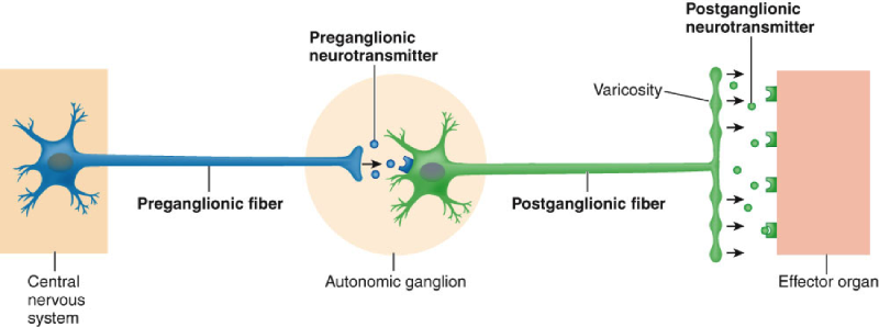 |
front 11 Describe the arrangement, including lengths, of preganglionic and postganglionic fibers in the sympathetic nervous system. | back 11 Preganglionic fibers are SHORT Postganglionic fibers are LONG Preganglionic neurons located between segments T1 and L2 of spinal cord |
front 12 Compare and contrast the locations, targets, AND functions of sympathetic chain ganglia, collateral ganglia, and ganglia found in the adrenal medulla. | back 12 The Sympathetic chain ganglia innervates visceral effectors via spinal nerves and also innervates visceral organs in the thoracic cavity via sympathetic nerves. Target organs are visceral effectors in the thoracic cavity, head, body wall, and limbs. |
front 13 Compare and contrast the locations, targets, AND functions of sympathetic chain ganglia, collateral ganglia, and ganglia found in the adrenal medulla. | back 13 The collateral ganglia innervates visceral effectors in the abdominal cavity. |
front 14 Compare and contrast the locations, targets, AND functions of sympathetic chain ganglia, collateral ganglia, and ganglia found in the adrenal medulla. | back 14 Targets are organs and systems throughout the body. It secretes neurotransmitters into general circulation. |
front 15 Describe the arrangement, including lengths, of preganglionic and postganglionic fibers in the parasympathetic nervous system. | back 15 Preganglionic fibers are LONG Postganglionic fibers are SHORT Preganglionic fibers originate in brain stem and sacral segments of spinal cord |
front 16 Compare and contrast the locations, targets, AND functions of the ganglia in the parasympathetic nervous system. | back 16 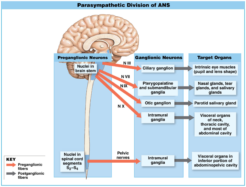 |
front 17 What is the neurotransmitter involved at the synapse between the preganglionic and postganglionic cells of BOTH the sympathetic and parasympathetic nervous system? | back 17 Ach |
front 18 Diagram the neurotransmitters involved between the postganglionic cell and the target organ/gland in BOTH the sympathetic and parasympathetic nervous system. | back 18 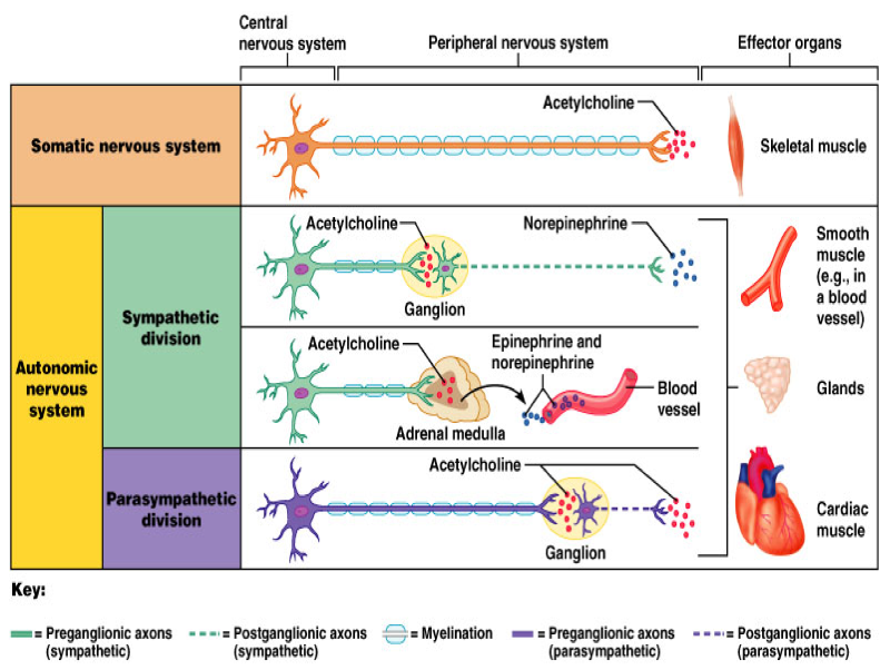 |
front 19 Compare and contrast the locations and functions of nicotinic and muscarinic acetylcholine receptors. | back 19 ***Nicotinic R eceptors –On surfaces of ALL ganglion cells (sympathetic AND parasympathetic) –ALSO found at the NEUROMUSCULAR JUNCTION in the SOMATIC NERVOUS SYSTEM ***Muscarinic Receptors –At the neuromuscular or neuroglandular junctions (parasympathetic) –At few cholinergic junctions (sweat glands innervated by the sympathetic nervous system) |
front 20 Describe the pathophysiology of BOTH nicotine poisoning and muscarine poisoning. Ask yourself WHY a person would experience the symptoms associated with each type of poisoning. Think of what is happening at the receptors! NICOTINE POISONING | back 20 Nicotine: –binds to nicotinic receptors –targets autonomic ganglia and skeletal neuromuscular junctions Symptoms: –vomiting, diarrhea, high blood pressure, rapid heart rate, sweating, profuse salivation, convulsions
|
front 21 Describe the pathophysiology of BOTH nicotine poisoning and muscarine poisoning. Ask yourself WHY a person would experience the symptoms associated with each type of poisoning. Think of what is happening at the receptors! MUSCARINE POISONING | back 21 Muscarine: –binds to muscarinic receptors –targets parasympathetic neuromuscular or neuroglandular junctions Symptoms: –salivation, nausea, vomiting, diarrhea, constriction of respiratory passages, low blood pressure, slow heart rate (bradycardia) |
front 22 What does adrenergic mean? | back 22 Aka SYMPATHOMIMETIC |
front 23 Compare and contrast the following receptors, in detail:
| back 23 Alpha-1 ( a 1 ) –More common type of alpha receptor –Releases intracellular calcium ions from reserves in endoplasmic reticulum –What will this do to the smooth muscle cells in the wall of an artery? Vasoconstrict Alpha-2 ( a 2 ) –Suppresses release of epinephrine –Can lead to vasodilation of blood vessels |
front 24 Compare and contrast the following receptors, in detail:
| back 24 Affect membranes in many organs (skeletal muscles, lungs, heart, and liver) Beta-1 ( b 1 ) –Increase in heart rate (chronotropy) and strength of contractions (ionotropy) Beta-2 ( b 2 ) –Triggers relaxation of smooth muscles along respiratory tract and wall of uterus Beta-3 ( b 3 ) –Is found in adipose tissue –Leads to lipolysis, the breakdown of triglycerides in adipocytes –Releases fatty acids into circulation |
front 25 Why does Primack’s wife carry an epi-pen with her? Hint: Bees! Why would a person with heart disease (a damaged heart muscle) take Atenolol (a b-1 Antagonist) – commonly called a Beta Blocker? Why else might someone be prescribed Atenolol on a PRN (as needed) basis? Why would a person having an asthma attack take Albuterol (a b-2 Agonist)? Why does their heart race increase when taking this drug????????? Would you expect this to happen? Why or why not? Why would a female be administered terbutaline (a b-2 Agonist) during pregnancy? Come up with two different ways to pharmacologically treat hypertension using drugs which target adrenergic receptors | back 25 Review |
front 26 Autonomic Innervation: The Heart | back 26 Receives dual innervation 2 divisions have opposing effects: –parasympathetic division:
–sympathetic division:
–autonomic tone is present –releases small amounts of both neurotransmitters continuously –Parasympathetic innervation dominates under resting conditions Crisis accelerates heart rate by: –stimulation of sympathetic innervation inhibition of parasympathetic innervation |
front 27 Autonomic Innervation: Blood Vessel Dilation | back 27 Blood vessel dilates and blood flow increases Blood vessel constricts and blood flow reduced Sympathetic postganglionic fibers release NE: –innervate smooth muscle cells in walls of peripheral vessels Background sympathetic tone keeps muscles partially contracted To increase blood flow: –rate of NE release decreases –smooth muscle cells relax –vessels dilate and blood flow increases |