Instructions for Side by Side Printing
- Print the notecards
- Fold each page in half along the solid vertical line
- Cut out the notecards by cutting along each horizontal dotted line
- Optional: Glue, tape or staple the ends of each notecard together
Muscles Part 9
front 1 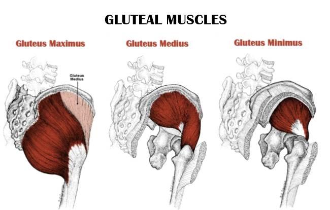 Give the origin, insertion, and action of the gluteus maximus | back 1 O: Iliac crest, lateral surface of the ilium; sacrum, coccyx I: Iliotibial tract and gluteal tuberosity of femur A: Extension and lateral rotation at hip |
front 2  Give the origin, insertion, and action of the gluteus medius and minimus | back 2 O: Anterior iliac crest of ilium/lateral surface of ilium I: Greater trochanter of femur A: Abduction and medial rotation at hip |
front 3 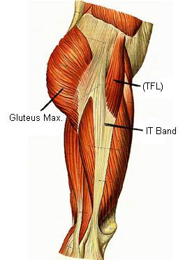 Give the origin, insertion, and action of the tensor fascia latae | back 3 O: Iliac crest and lateral surface of anterior superior iliac spine I: Iliotibial tract A: Flexion and medial rotation at hip; laterally supports the knee (fascia lata) |
front 4  Give the origin, insertion, and action of the Iliacus (iliopsoas group) | back 4 O: Iliac fossa of ilium I: Femur distal to lesser trochanter A: Flexion at hip |
front 5 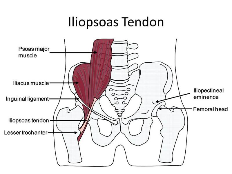 Give the origin, insertion, and action of the psoas major (iliopsoas group) | back 5 O: Anterior surfaces and transverse processes of vertebrae (T12-L5) I: Lesser trochanter in company with iliacus A: Flexion at hip or lumbar intervertebral joints |
front 6 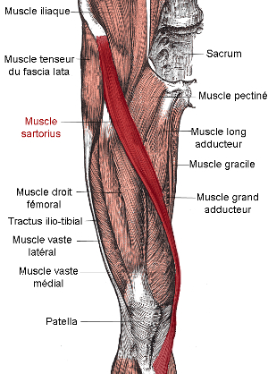 Give the origin, insertion, and action of the sartorius | back 6 O: Anterior superior iliac spine I: Medial surface of tibia near tibial tuberosity A: Flexion at knee; flexion and lateral rotation at hip |
front 7 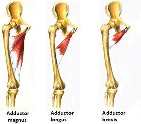 Give the origin, insertion, and action of the adductors | back 7 O: Inferior ramus of pubis I: Linea aspera of femur A: Adduction at hip |
front 8 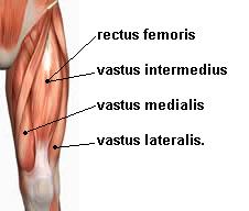 Give the origin, insertion, and action of the rectus femoris | back 8 O: Anterior inferior iliac spine and superior acetabular rim of ilium I: Tibial tuberosity via patellar ligament A: Extension at knee; flexion at hip |
front 9  Give the origin, insertion, and action of the vastus medialis | back 9 O: Entire length of linea aspera of femur I: Tibial tuberosity via patellar ligament A: Extension at knee |
front 10  Give the origin, insertion, and action of the vastus intermedius | back 10 O: Anterolateral surface of femur and line aspera I: Tibial tuberosity via patellar ligaments A: Extension at knee |
front 11  Give the origin, insertion, and action of the vastus lateralis | back 11 O: Anterior and inferior to greater trochanter of femur and along linea aspera (proximal half) I: Tibial tuberosity via patellar ligament A: Extension at knee |
front 12 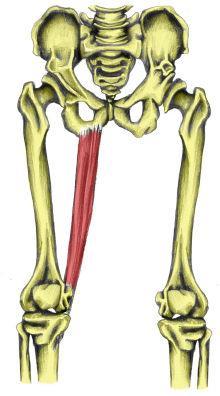 Give the origin, insertion, and action of the gracilis | back 12 O: Inferior ramus of pubis I: Medial surface of tibia inferior to medial condyle A: Flexion at knee; adduction and medial rotation at hip |
front 13 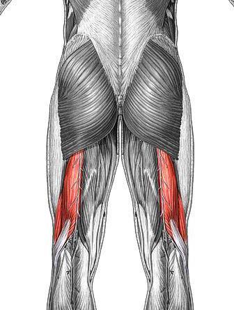 Give the origin, insertion, and action of the biceps femoris | back 13 O: Ischial tuberosity and linea aspera of femur I: Head of fibula, lateral condyle of tibia A: Flexion at knee; extension and lateral rotation at hip |
front 14 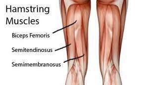 Give the origin, insertion, and action of the semimembranosus | back 14 O: Ischial tuberosity I: Posterior surface of medial condyle of tibia A: Flexion at knee; extension and medial rotation at hip |
front 15  Give the origin, insertion, and action of the semitendinosis | back 15 O: Ischial tuberosity I: Proximal, medial surface of tibia near insertion of gracilis A: Flexion at knee; extension and medial rotation at hip |
front 16 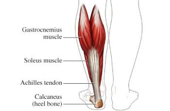 Give the origin, insertion, and action of the gastrocnemius | back 16 O: Femoral condyles I: Calcaneous via calcaneal tendon A: Extension (plantar flexion) at ankle; inversion of foot; flexion at knee |
front 17 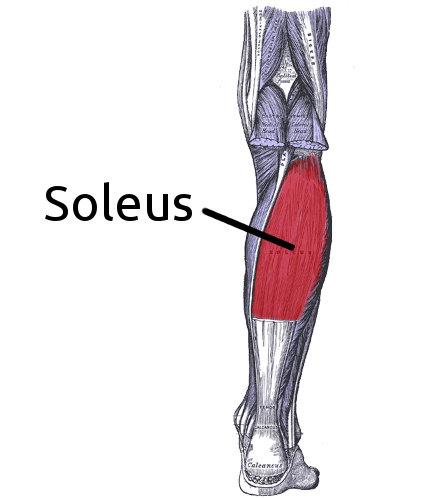 Give the origin, insertion, and action of the soleus | back 17 O: Head and proximal shaft of fibula and adjacent posteromedial shaft of tibia I: Calcaneous via calcaneal tendon (with gastrocnemius) A: Extension (plantar flexion) at ankle |
front 18 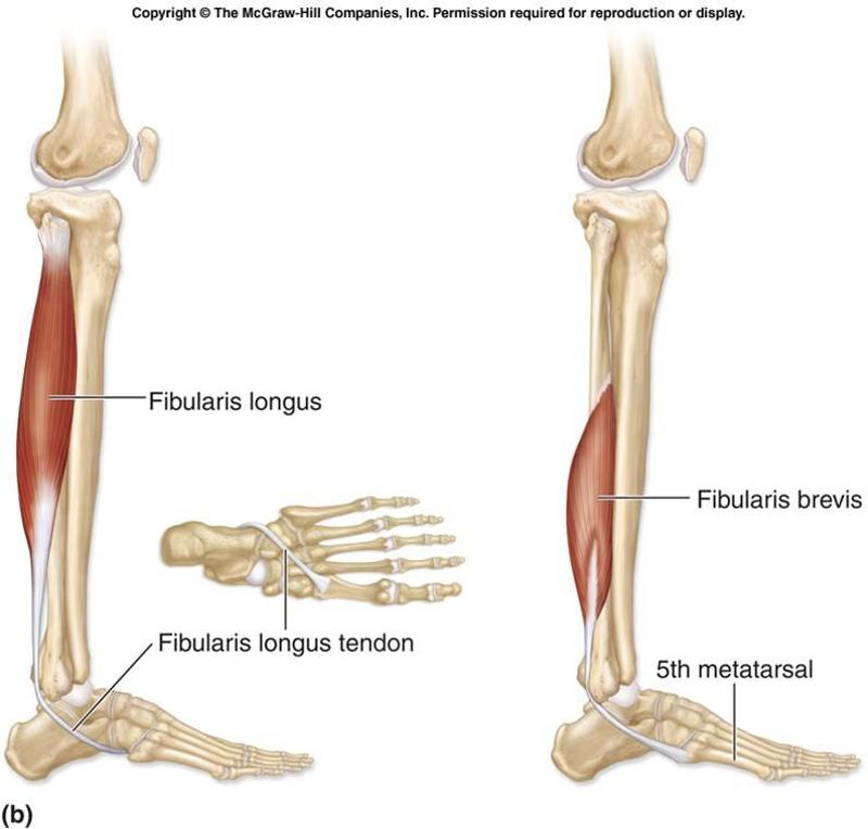 Give the origin, insertion, and action of the fibularis brevis | back 18 O: Midlateral margin of fibula I: Base of fifth metatarsal bone A: Eversion of foot and extension (plantar flexion) at ankle; supports longitudinal arch |
front 19  Give the origin, insertion, and action of the fibularis longus | back 19 O: Lateral condyle of tibia, head and proximal shaft of fibula I: Base of fifth metatarsal bone and medial uniform bone A: Eversion of foot and extension (plantar flexion) at ankle; supports longitudinal arch |
front 20 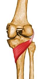 Give the origin, insertion, and action of the popliteus | back 20 O: Lateral condyle of femur I: Posterior surface of proximal tibial shaft A: Medial rotation of tibia (or lateral rotation of femur); flexion at knee |
front 21 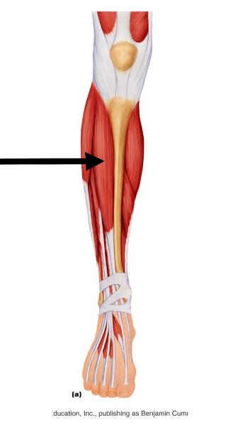 Give the origin, insertion, and action of the tibialis anterior | back 21 O: Lateral condyle and proximal shaft of tibia I: Base of first metatarsal bone and medial uniform bone A: Flexion at ankle; inversion of foot |
front 22 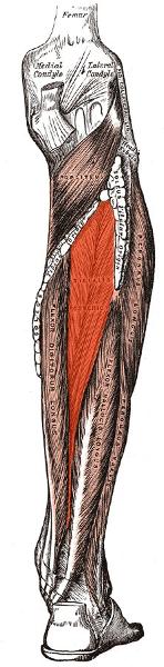 Give the origin, insertion, and action of the tibialis posterior | back 22 O: Interosseous membrane and adjacent shafts of tibia and fibula I: Tarsal and metatarsal bones A: Adduction and inversion of foot; extension (plantar flexion) at ankle |
front 23 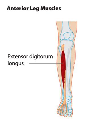 Give the origin, insertion, and action of the extensor digitorus | back 23 O: Anterior surface of fibula I: Superior surface, distal phalanx of great toe A: Extension at joints of great toe |